Article

The Development of Crassulacean Acid Metabolism (CAM) Photosynthesis in Cotyledons of the C4 Species, Portulaca grandiflora (Portulacaceae)

Department of Biology, Roger Williams University, One Old Ferry Rd., Bristol, RI 02809, USA
*
Author to whom correspondence should be addressed.
Plants 2020, 9(1), 55; https://doi.org/10.3390/plants9010055
Submission received: 10 December 2019 / Revised: 26 December 2019 / Accepted: 27 December 2019 / Published: 2 January 2020
(This article belongs to the Special Issue Regulation of Central Carbon and Amino Acid Metabolism in Plants)

Abstract

:
Portulaca grandiflora simultaneously utilizes both the C4 and Crassulacean acid metabolism (CAM) photosynthetic pathways. Our goal was to determine whether CAM developed and was functional simultaneously with the C4 pathway in cotyledons of P. grandiflora. We studied during development whether CAM would be induced with water stress by monitoring the enzyme activity, leaf structure, JO2 (rate of O2 evolution calculated by fluorescence analysis), and the changes in titratable acidity of 10 and 25 days old cotyledons. In the 10 days old cotyledons, C4 and CAM anatomy were evident within the leaf tissue. The cotyledons showed high titratable acid levels but a small CAM induction. In the 25 days old cotyledons, there was a significant acid fluctuation under 7 days of water stress. The overall enzyme activity was reduced in the 10 days old plants, while in the 25 days old plants CAM activity increased under water-stressed conditions. In addition to CAM, the research showed the presence of glycine decarboxylase in the CAM tissue. Thus, it appears both pathways develop simultaneously in the cotyledons but the CAM pathway, due to anatomical constraints, may be slower to develop than the C4 pathway. Cotyledons showed the ancestral Atriplicoid leaf anatomy, which leads to the question: Could a CAM cell be the precursor to the C4 pathway? Further study of this may lead to understanding into the evolution of C4 photosynthesis in the Portulaca.

1. Introduction

CO2 concentrating mechanisms have evolved in terrestrial plants in response to changing environmental conditions. Two different mechanisms have evolved that involve a similar suite of enzymes utilized in a different fashion to overcome photorespiration and increased water loss. Photorespiration increases when CO2 becomes limited under high light intensities and high evaporative demand resulting in increased transpirational water loss [1]. The C4 pathway overcomes these limitations with the CO2 being initially captured as HCO3- by phosphoenolpyruvate carboxylase (PEPCase) and then fixed via the C3 pathway by Rubisco. C4 plants have a spatial separation of the C4 and C3 pathways occurring within two different cell types in the leaf. The C4 pathway, located in the palisade mesophyll cells, is radially arranged around the C3 pathway located in the bundle sheath cells, which surround the vascular tissue. This is typically referred to as Kranz anatomy [1,2]. Research by Voznesenskaya et al. [3,4] has shown that the Kranz anatomy is not essential for terrestrial C4 photosynthesis to occur but it can occur in a single cell with a spatial separation of the C4 and C3 pathways within a single chlorenchyma cell. The C4 pathway concentrates CO2 at the site of Rubisco and helps to suppress photorespiration in the bundle sheath cells. The C4 pathway has been found in approximately 19 plant families and over 8000 species [1] and has evolved independently many times.
Crassulacean acid metabolism (CAM) is a metabolic and anatomical adaptation that is characterized by net nocturnal carbon dioxide uptake with a temporal separation of the C4 and C3 pathway resulting in decreased transpiration rates and water loss [5,6]. The CO2 is similarly fixed (as in C4 plants) by PEPCase, converted to malate and stored as malic acid in the large central vacuole during the night period. In the subsequent light period, the malate is transported out of the vacuole and then decarboxylated to release CO2 for utilization by Rubisco in the C3 cycle. CAM plants typically have a mesophyll anatomy with primarily spongy parenchyma cells with a large central vacuole, which has the ability to store the increasing accumulation of malic acid during the nocturnal period [7]. CAM plants anatomically have very low mesophyll airspace, so when the stomata are closed the CO2 concentrations inside the leaf can reach very high levels to suppress photorespiration [6,7]. CAM has evolved in at least 34 different plant families including 6 aquatic families and over 20,000 species [1,8].
CAM and C4 photosynthesis have evolved independently multiple times in many different plant families. One might hypothesize both CAM and C4 could have evolved in the same plant families numerous times due to the similarity of the enzymes involved in the pathways. The two pathways have evolved in the same family four times (Aizoaceae, Asteraceae, Euphorbiaceae, Portulacaceae). This raises an interesting question about why the C4 and CAM pathways have only concurrently evolved in these four families. The original circumscription of the Portulacaceae describes the family as containing approximately 29 genera [9]. Guralnick and Jackson [10] have reported the evolution and distribution of C4 and CAM photosynthesis found in this family. It has been observed that some members are C3 plants, while others are C3 plants with attributes of CAM. Others are C4 plants with some CAM characteristics, and the more advanced species are facultative CAM plants [10,11,12].
The genus Portulaca is known to have the only C4 photosynthetic members of the family despite previous reports to the contrary [11]. This revision reveals the genus Portulaca (Portulacaceae) contains the only known C4 members that are capable of CAM photosynthesis [13]. Portulaca spp. tend to inhabit environments with high light intensities, which periodically become dry. The genus Portulaca has succulent stems and leaves and because of the high degree of succulence, there have been reports in the literature that members of the Portulaca show a diurnal acid fluctuation characteristic of CAM species. Koch and Kennedy [14,15] showed Portulaca oleracea having a diurnal acid fluctuation in both the stems and leaves. They also measured low levels of net nighttime CO2 uptake. Research done by Guralnick and Jackson [10] showed that Portulaca mundula, P. pilosa, and P. oleracea exhibited high acid levels and diurnal acid fluctuations indicative of CAM photosynthesis. Kraybill and Martin [16] showed both P. oleracea and P. grandiflora both undergo CAM cycling with little or no nocturnal CO2 uptake. Mazen [17] indicated that under water stress conditions that P. oleracea had increased levels of PEP (phosphoenolpyruvate) carboxylase protein. Further research showed the genus contains a C3-C4 intermediate species, Portulaca cryptopetala, in which recent work showed P. cryptopetala induced CAM under water stress conditions [2,18]. Winter et al. [18] extended the findings to additional species in the Portulaca and also consider them to be facultative CAM species.
Portulaca grandiflora is a small herbaceous annual utilizing the C4 photosynthetic pathway. P. grandiflora has small succulent leaves with a Pilosoid-type Kranz leaf anatomy where the C4 tissue in the succulent leaves surround the large water storage tissue [2,19]. P. grandiflora is known to maintain high organic acid levels and shows a large diurnal acid fluctuation when water stressed, typical of CAM species [20]. Research has indicated the increase in CAM of this species occurs in the water storage portion of the leaf and the stem during water stressed conditions [20]. Portulaca grandiflora is unique because it has both C4 and CAM photosynthetic pathways operating simultaneously in the leaf tissue [20]. This situation is unique due to the proposed incompatibility of both pathways to operate in the same leaf [21]. Phylogenetic analysis has indicated the genus Portulaca evolved CAM from a C3 ancestor prior to the appearance of the C4 pathway [10]. The objective of this study was to study cotyledon leaf tissue to determine if both the CAM and C4 pathways were developing and operating simultaneously. CAM induction in developing cotyledons was monitored by withholding water for 3 and 7 days. An understanding of the developmental process of these pathways will aid in clarifying the evolutionary origins of the CAM and C4 pathways in the Portulacaceae.

2. Results

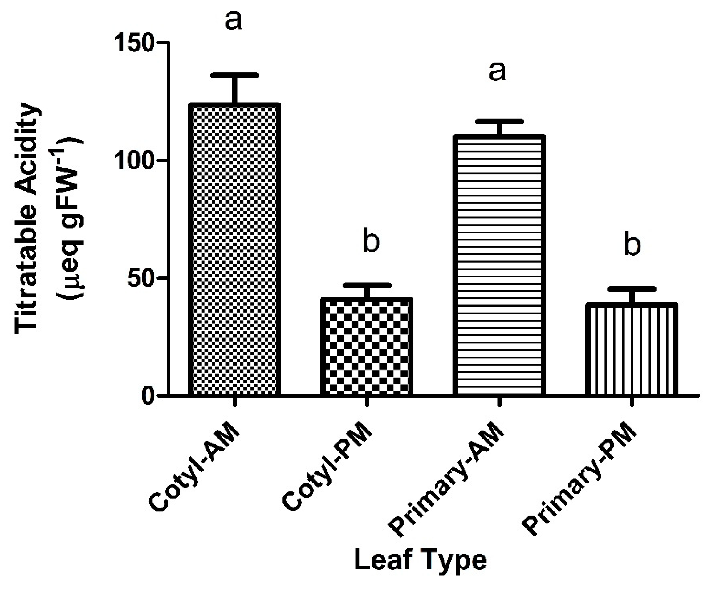
2.1. Titratable Acidity

Titratable acidity levels for 10 days old cotyledons were at approximately 50–60 µeq gFW−1 (FW = Fresh Weight; Figure 1). At 10 days, the control groups and water-stressed leaves showed a slight acid fluctuation of 10–20 µeq gFW−1 from a.m. to p.m. (Figure 1). There was no significant difference between a.m. and p.m. acid levels. At 20–25 days old, cotyledons, under control conditions, there was no acid fluctuation observed from a.m. to p.m. levels. Under water stress, cotyledons showed a small significant titratable acid fluctuation from the morning to the evening (Figure 1). Continued water stress to 7 days of the 20–25 days old cotyledons induced a large and significant acid fluctuation of 83 µeq gFW−1 in both the cotyledons and primary leaves of P. grandiflora (Figure 2). The a.m. acid levels had increased to more than double the control cotyledons.

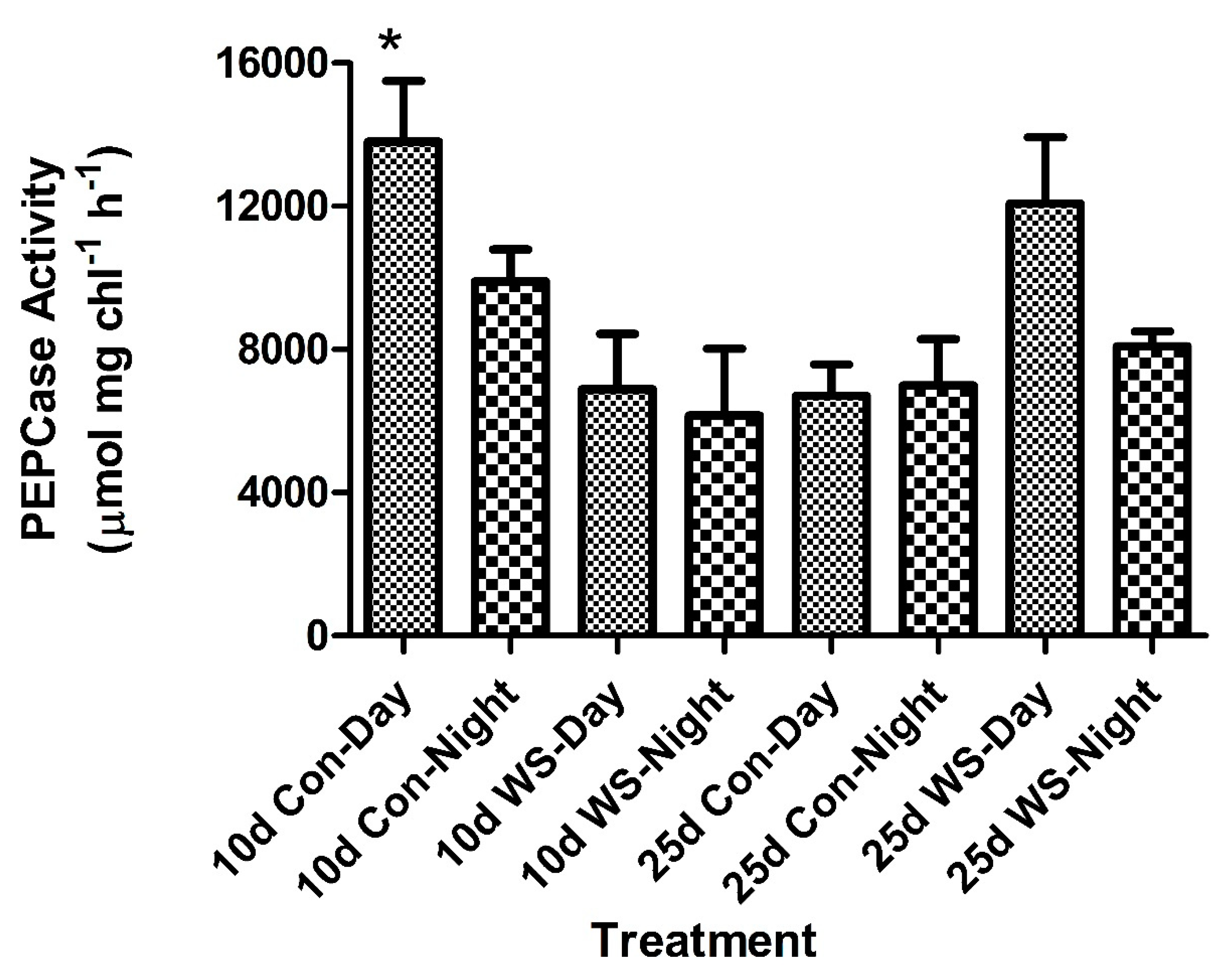
2.2. Enzyme Activity: PEP Carboxylase and NADP-ME

Ten day old cotyledons showed PEPCase activity higher during the day than at night (Figure 3). Water stress lowered the activity of PEPCase in the 10 days old cotyledons during the day and night. There was a significant difference in daytime activity between the control and water stress at 10 days. At 25 days old, the activity remained high during the day but lower at midnight (Figure 3). In the 25 days old cotyledons, water stress had less of an effect on the overall activity of PEPCase. The PEPCase activity during the day increased in the water-stress plants compared to the control plants (Figure 3). There was no significant differences in PEPCase activity in the 25 days old cotyledons (Figure 3).
The decarboxylase, NADP-Malic Enzyme (ME), showed high daytime activity in both the 10 days and 25 days old control cotyledons (Figure 4). Water stress significantly lowered the NADP-ME activity at 10 days, while at 25 days there was no significant change in the level of activity between the control and water-stressed plants.

2.3. Electron Transport Rate/JO2
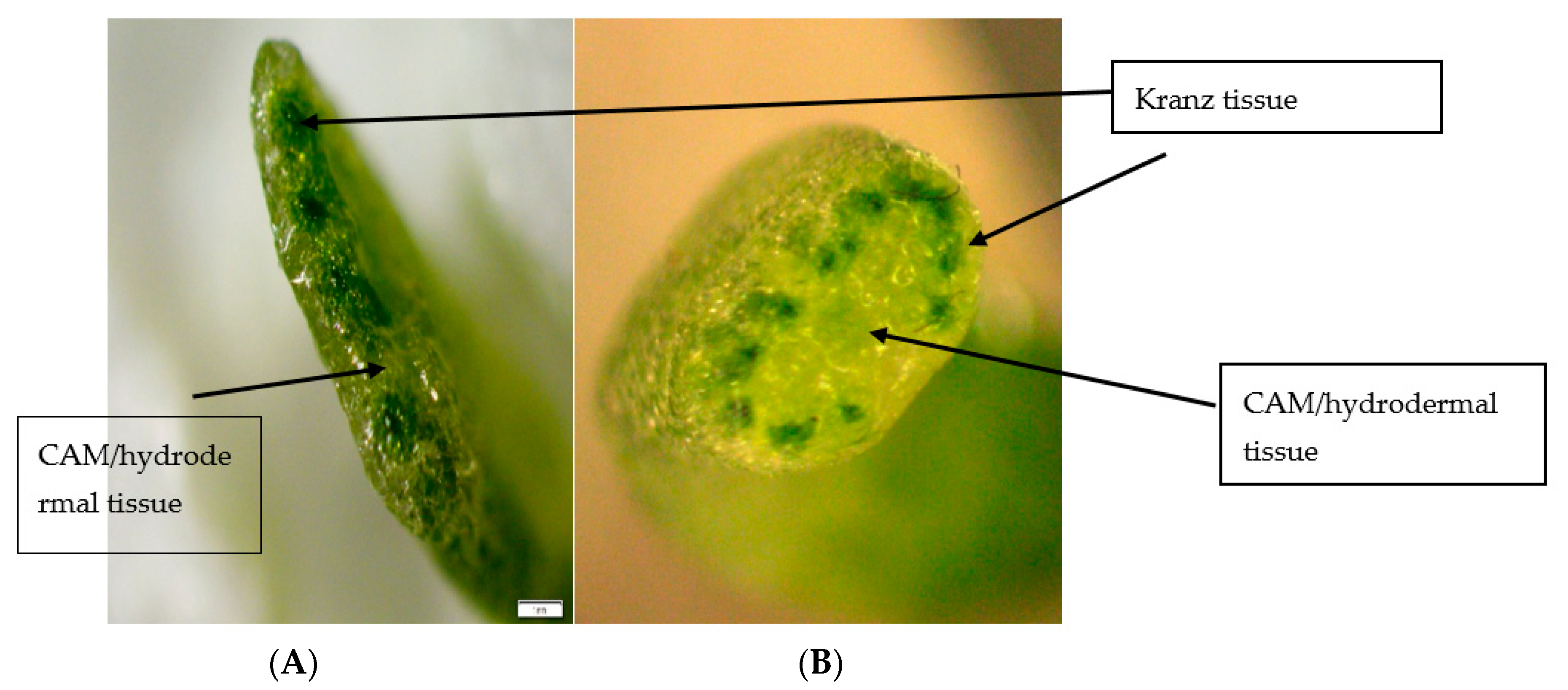
The JO2 rates showed no differences between control and water-stressed plants in 10 days old and 25 days old cotyledons (Figure 5). The JO2 rates were lower in 25 days old cotyledons than in the 10 days old cotyledons.
The gross rate of photosynthesis (JO2) showed a peak at midday for the well-watered 10 days old cotyledons while the stressed cotyledons showed a peak earlier in the morning (Figure 5). In the 25 days old cotyledons, the JO2 calculated rates were relatively constant over the course of the light period. There was no difference between the control and water-stressed plants in the JO2 rates (Figure 5).

2.4. Leaf Anatomy

Portulaca grandiflora cotyledons were dissected and photographed to display the internal gross anatomy of the tissue (Figure 6). The sections illustrate the Atriplicoid Kranz anatomy in the cotyledon (Figure 6A), and its linear arrangement compared to a mature leaf where the Kranz anatomy shows a radial arrangement (Figure 6B). A low-magnification view of a cotyledon leaf tissue showed a well-developed Atriplicoid Kranz anatomy arranged in a linear fashion (Figure 7). Mesophyll cells were well developed with chloroplast along the cell wall (Figure 7A). The bundle sheath cells were also well developed with chloroplasts located along the periphery of the inner cell wall close to the vascular bundles (Figure 7A). The CAM/hydrodermal tissue showed fewer chloroplasts than the C4 tissue (Figure 7A). At 25 days, the Kranz anatomy was very well developed and the vascular tissue was more prominent. Under water-stress conditions, the CAM/hypodermal tissue at 10 days showed signs of water loss compared to the control cotyledon tissue. The cells appeared to have some shriveling and a more irregular shape. At 25 days, there were less noticeable changes under water-stress conditions in the succulent mesophyll and hypodermal tissue (Figure 7).
The sections illustrate the Atriplicoid Kranz anatomy in its linear arrangement compared to a mature leaf where the Kranz anatomy shows a radial arrangement (Figure 6B). We performed tissue prints of cotyledons to determine the presence of glycine decarboxylase (GDC, a mitochondrial marker for the photorespiratory pathway). GDC was found to be present in the CAM/hydrodermal tissue of P. grandiflora cotyledons (Figure 8B). In mature leaves of P. grandiflora, GDC was also located in the inner CAM/hydrodermal tissue (data not shown). For comparison, Portulacaria afra, a facultative CAM species, showed the presence of GDC throughout the spongy parenchyma mesophyll tissue (8b).

3. Discussion

Guralnick and Jackson [10] previously suggested the CAM pathway appeared evolutionarily first in the leaf and the C4 photosynthetic pathway overlaid the CAM tissue. These conclusions were supported by research which showed the CAM PEPCase gene was more primitive to the C4 derived PEPCase gene [22]. Sage [21] described the leaf anatomy found in the Portulaca as being unique because of the apparent incompatibility of the photosynthetic pathways. The genus Portulaca shows nearly all members display C4 photosynthesis with attributes of the CAM pathway in the same leaf tissue and are considered facultative CAM species [18]. Previous research showed the CAM pathway aids in the retention of water for maintenance of the C4 pathway and the recycling of carbon to the mesophyll and bundle sheath cells [20]. This cooperation between the tissues may be the result of the close proximity of the tissues to each other because previous work has proposed that the tissues are functioning independently of each other [20].

3.1. C4 Development

Research has been performed on the physiological and structural development of C4 photosynthesis in cotyledons of the Portulaca but little of CAM development in these C4 species [23]. The research presented here indicated it was apparent that the C4 pathway was well developed in 10 days old cotyledons based on the anatomy and physiology. The leaf anatomy of the cotyledons showed a well-developed Kranz C4 anatomy as found previously in the genus Portulaca [23]. P. grandiflora cotyledons displayed an Atriplicoid-type anatomy where the vascular bundles are in one plane of the leaf. The Atriplicoid Kranz anatomy showed CAM tissues around the periphery of the leaf surrounding the Kranz bundles. This differs from the Pilosoid Kranz anatomy found in mature leaves of P. grandiflora with the C4 tissue in a ring surrounding the CAM water storage tissue [2,20].
The enzymes of the C4 pathway were fully functional at 10 days, as shown by a high activity of PEPCase and NADP-ME. This was comparable to the conclusions reached by Dengler et al. [24] for Atirplex rosea, which showed accumulation of PEPCase was detected 2–4 days after leaf development and expansion. The expression was limited to mesophyll tissue adjacent to the bundle sheath tissue. The JO2 studies in this study showed high rates of photosynthesis indicative of C4 activity and were comparable to the rates previously measured in mature leaf tissue [20]. The C4 pathway is the primary CO2 acquiring pathway in this genus and it was predictable the C4 pathway developed quickly for seedling establishment.

3.2. CAM Development

The development of CAM, which can be measured by titratable acidity levels and diurnal acid fluctuations, showed a slower development in cotyledons from that of the C4 pathway. The study of CAM development showed comparable titratable acidity levels but slightly lower and comparable to mature leaf tissues under control conditions [20]. One can measure significant total titratable acidity levels in the cotyledons at 10 and 25 days. The acid levels found in the 10 days old cotyledons of P. grandiflora are similar but larger than those found in well-watered cotyledons of Mesembryanthemum crystallinum, a facultative CAM species [25], and higher than the acid levels in the CAM-cycling species, Lewisia cotyledon [26]. The acid levels of P. grandiflora were lower than those found in a number of columnar cactus seedlings at 1 day and 7 days old [27]. The acid levels were lower than those for Opuntia elatior at 23 days old cotyledons [28]. However, these cactus species primarily utilize the CAM pathway for CO2 uptake, even in cotyledons. The cotyledon anatomy in cactus has succulent anatomy with a spongy parenchyma tissue with little airspace, which is more conducive to CAM photosynthesis [29,30]. The results in the present study indicated P. grandiflora cotyledons showed similar total titratable acid levels comparable to other CAM species.
We investigated the potential for CAM activity by water stressing the cotyledons for three and seven days. Our results differed with the age of the cotyledons and duration of the drought. Ten days old cotyledons showed small acid fluctuations indicative of CAM activity after three days of water stress. The 25 days old water-stressed cotyledons showed a small acid fluctuation compared to control cotyledons which showed no acid fluctuation. Continuation of water stress for seven days induced a very large and significant CAM acid fluctuation in both cotyledons and primary leaf tissue. The acid fluctuation measured in the cotyledons was comparable to acid levels in mature leaf tissue. This indicated an induction of CAM similar to previous research on P. grandiflora [20] and was indicative of CAM activity.
In addition, enzyme activity, as measured by nocturnal PEPCase activity, was quite high and comparable to C4 rates of PEPCase measured during the day. Water stress only had a slight effect on the overall activity of PEPCase. NADP-ME was affected more by water stressed conditions and this result had been observed previously in P. grandiflora [20]. Water stress in other facultative CAM plants, such as Portulacaria afra, show the decarboxylase enzymes are more affected than PEPCase [31]. The enzyme activities of PEPCase and NADP-ME reported here in cotyledons of P. grandiflora were much higher than those reported for cotyledons of Salsola spp. [32] and for mature leaves of P. grandiflora [20] and may be related to the lower chlorophyll concentrations.
The physiological and anatomical changes differed with the age. At 10 days, water stressing the cotyledons induced a small acid fluctuation and it lowered both the day and night activity of PEPCase. NADP-ME activity also decreased in 10 days old cotyledons. This indicated a decrease in C4 and CAM activity. It appears that NADP-ME is more sensitive to water stress than Rubisco and PEPCase. Anatomically, the mesophyll CAM tissue showed some shrinkage, which may be due to a redistribution of water from the CAM cells to the C4 metabolic cells. This has been observed in mature leaves of P. grandiflora during a 10 days drought [20]. The sensitivity of the enzymes due to drought at 10 days may be due more in part to structural development of the CAM tissue and water storage at this stage. The enzymatic activity appeared earlier prior to full anatomical development of the CAM tissue. This is similar to the development of C4 photosynthesis in A. rosea, which showed differential enzyme expression of the C4 photosynthetic enzymes by 4 days [24].
By 20–25 days old, the response to water stress and CAM activity was similar to the 10 days old cotyledons. There was a similar induction of an acid fluctuation in water-stressed plants. Seven days of water stress induced a much larger acid fluctuation indicative of CAM activity and greater than observed after three days of water stress. This acid fluctuation was similar to acid fluctuations in other CAM-cycling species [10,20]. The results indicated an induction of CAM activity similar to control plants of Lewisia cotyledon [26]. Induction of water stress did not lower PEPCase activity in 25 days old cotyledons compared to 10 days old cotyledons. NADP-ME did not show a decrease in activity due to water stress and showed a slight increase in activity, which may be due to increased CAM activity of the leaf. The enzyme activity indicated a maintenance of the C4 pathway in the 25 days old cotyledons. This was supported by the JO2 data, which showed no changes in activity during three days of drought. Structurally, the CAM/hydrodermal tissue was more mature in the 25 days old cotyledons and did not appear to show as much water loss as the 10 days old cotyledons. This may explain the ability of older cotyledons to maintain C4 activity when compared to 10 days old cotyledons. The continuation of drought showed a much stronger induction of CAM and was very similar to the response of mature leaves [20]. The CAM tissue in mature leaves recycles CO2, redistributes water to the C4 tissue to aid in the survival of the plant, and may play a similar role in cotyledons [20].
Mature leaves of P. grandiflora have a Pilosoid-type anatomy with a ring of bundles surrounding the water storage tissue [2,20]. P. grandiflora cotyledons have an Atriplicoid-type anatomy where the vascular bundles are in one plane of the leaf. This arrangement is considered ancestral to the Pilosoid arrangement with the water storage tissue found on both the abaxial and adaxial sides of the leaf [33]. Voznesenskaya et al. [2] reported on the diversity of structure in the Portulaca and found the arrangement of an inner water storage, the derived condition found in P. grandiflora, P. pilosa, and P. amilis [2], to be due to the ecological constraints of being found in more semi-arid regions than other Portulaca spp. However, they did not report any CAM activity in the cotyledons of the Portulaca spp. in their study. Further, we report the presence of glycine decarboxylase, a mitochondrial marker for the C2 photorespiratory pathway in the CAM tissue of cotyledons. Previous research has shown the GDC to be localized in the mitochondria of bundle sheath cells in cotyledons [2]. The presence of GDC adds another dimension to another function of CAM in P. grandiflora. Previous work indicated CAM tissue might transfer water from the tissue to the Kranz anatomy to help maintain C4 photosynthetic activity under water stress. Additionally, decarboxylation of the acid may produce CO2 for the adjacent mesophyll and bundle sheath tissue. We now suggest the C2 pathway may provide an additional source of CO2 for the C4 pathway. It was shown the photorespiratory pathway may elevate CO2 levels three-fold in the leaf in Flaveria pubescens, (a C3-C4 intermediate) [34]. The photorespiratory pathway in the CAM tissue of P. grandiflora may provide an additional source of CO2 for the C4 pathway. Since CAM tissue has reduced airspace, the Atriplicoid arrangement may reduce diffusion of water out of the leaf and maintain CO2 levels around the Kranz anatomy. This may aid in survival of the seedlings. Further work will be needed to investigate the role of photorespiration in the different Kranz types CAM tissue in P. grandiflora.
This is the first report of CAM activity in the cotyledons and the results indicated CAM can be induced in cotyledons of P. grandiflora. The development of CAM occurred after the development of the C4 photosynthetic pathway and may be due to more of a constraint of leaf anatomy of the CAM tissue in cotyledons. The retention of CAM in the cotyledons of P. grandiflora further supports the idea that CAM was an ancient pathway in the genus Portulaca including the retention of the C2 pathway. Since the cotyledons have a different type of Kranz anatomy and water storage tissue than the mature leaves in P. grandiflora, this leads to questions on the evolution of C4 and CAM within the genus Portulaca. Research on the origins of CAM in Portulaca using PEPCase have indicated the CAM-specific gene was similar in sequence in other species utilizing CAM [22]. The question that arises: Could C4 photosynthesis have evolved from the CAM cells? Christin et al. [22] have shown the C4 specific PEPCase gene appears to have evolved from a non-photosynthetic form rather than the CAM form. They also suggest the other enzymes required for C4 photosynthesis may not have required new genes but utilization of genes already present in the cell. Since CAM cells in the Portulaca retain all features of C3 and CAM photosynthesis including the C2 pathway, it maybe that novel cells types of the mesophyll and bundle sheath may have differentiated from CAM-like cells. During the slow evolution of C4 photosynthesis as suggested by Christin et al. [22], a change in the regulatory sequences of PEPCase and GDC would be required as the cells evolved. Current research does not support the idea but questions remain on the change from a CAM-type leaf structure evolved into a C4 photosynthetic leaf in Portulaca.

4. Materials and Methods

4.1. Plant Material

Seeds of Portulaca grandiflora were germinated in flats (Figure 9A,B). All plants grown were irrigated with half-strength Hoagland’s solution prior to sampling. The plants were grown under natural light conditions, supplemented with artificial light for a light intensity of 300–400 µmol m−2 s−1.
The day/night temperature in the glasshouse was 27 °C/17 °C. Seedlings were irrigated daily to maintain high water potential, and water stressed for three days prior to sampling. Experiments were performed when the leaf tissues were 10–14 days old and 22–26 days old post-germination. Leaf samples were taken during the course of the day/night cycle and frozen until assayed for titratable acidity and enzyme activity.

4.2. Titratable Acidity

Cotyledons were harvested in the morning and evening, and frozen (−80 °C) until assayed. Leaf samples were weighed, ground in glass-distilled water, and titrated with 0.01 N KOH to a pH 7 endpoint as described by Guralnick et al. [20]. Results were expressed as µeq gFW−1.

4.3. Enzyme Activity

Frozen leaf tissue collected midday and during the middle of the night period was utilized for phosphoenolpyruvate carboxylase (PEPCase) and NADP-malic enzyme (ME) activity. Assays of crude extracts were done in triplicate under well-watered and water-stress conditions. The samples were assayed spectrophotometrically by following the oxidation of NADH (for PEPCase) or the reduction of NADP+ at 340 nm as previously described by Guralnick and Ting [31]. Results were expressed as µmol mg chl−1 h−1.

4.4. Leaf Anatomy

Cotyledons were harvested and cut into sections. They were fixed in 1.5% glutaraldehyde in sodium cacadylate buffer followed by fixation in osmium. Sections were dehydrated in a series to 80% ethanol. The leaf material for transverse cross-section was fixed with fluoroacetic acid and then embedded in JB4 plastic resin (2-hydroxyethyl methacrylate). Sections were cut with a microtome and stained with 1% aniline B.

4.5. Electron Transport and JO2

The electron transport rate was determined using the pulse amplitude modulated fluorometer (OSP1 Fluorometer), according to Guralnick et al. [20]. Samples were taken over the course of the light period beginning at 6:00 in the greenhouse. Intact leaf tissue of well-watered and water-stressed cotyledons were measured [20]. The true rate of O2 evolution (JO2) from Photosystem II activity was calculated, according to the method of Lal and Edwards [35].

4.6. Tissue Printing

Cotyledons were cut with a razor and then photographed. The sections were pressed onto nitroceullusoe membranes. Protein localization of Glycine decarboxylase (GDC-H) antibodies was performed according to the method of Guralnick et al. [20]. Rabbit anti-GDC-H antibodies were purchased from Agrisera and diluted to a 1:5000 concentration. The presence of GDC was visualized using a secondary antibody linked to alkaline phosphatase and then photographed. GDC was a mitochondrial marker for the C2 photorespiratory pathway.

5. Conclusions

Research has shown the cotyledons of Portulaca grandiflora could be induced to perform CAM similar to mature leaves [20]. The CAM pathway appears to develop later due to anatomical but not physiological aspects of CAM. In the species, P. grandiflora, both pathways are present and appear to be operating independently in the cotyledons but CAM still aids the C4 pathway under water-stress conditions. Additionally, the C2 pathway may raise CO2 concentrations inside the cotyledons, aiding in raising the internal CO2 concentrations. More research will be needed to understand the evolution of CAM and C4 photosynthesis in the genus Portulaca.

Author Contributions

The authors on this manuscript contributed to many portions as undergraduate researchers. Conceptualization, L.J.G.; methodology, L.J.G., K.E.G., D.D., and N.A.; data collection, L.J.G., K.E.G., D.D., and N.A.; data analysis, L.J.G., K.E.G., D.D., and N.A.; writing, original draft preparation, L.J.G. and D.D.; writing, review, L.J.G., K.E.G., and N.A.; supervision and project administration, L.J.G.; funding acquisition, L.J.G., D.D., and N.A. All authors have read and agreed to the published version of the manuscript.

Funding

The research received no external funding.

Acknowledgments

The research was supported in part from an Internal RI-INBRE fellowship (IDeA Networks of Biomedical Research Excellence) and a Center for Economic and Environmental Development (CEED) fund grant awarded to Diana Denio and Nicolas Antico. Part of this research was presented at the CAM 2010 conference held at the Smithsonian Tropical Research Institute in Panama City, Panama, by Diana Denio. The authors would like to thank Tammy Sage and Rowan Sage for their help with the light microscopy. The authors would like to thank Tim Pelletier and Colleen McKnight-Torres for their support with equipment and supplies for this study.

Conflicts of Interest

The authors declare no conflict of interest.

References

  1. Sage, R.F.; Monson, R.K. Preface. In C4 Plant Biology; Academic: Philip Drive Norwell, MA, USA, 1999; pp. xiii–xv. [Google Scholar]
  2. Voznesenskaya, E.V.; Koteyeva, N.K.; Edwards, G.E.; Ocampo, G. Revealing diversity in structural and biochemical forms of C4 photosynthesis and a C3-C4 intermediate in genus Portulaca L. (Portulacaceae). J. Exp. Bot. 2010, 61, 3647–3662. [Google Scholar] [CrossRef] [PubMed]
  3. Voznesenskaya, E.V.; Franceschi, V.R.; Kiirats, O.; Freitag, H.; Edwards, G.E. Kranz anatomy is not essential for terrestial C4 plant photosynthesis. Nature 2001, 414, 543–546. [Google Scholar] [CrossRef] [PubMed]
  4. Voznesenskaya, E.V.; Franceschi, V.R.; Kiirats, O.; Artyusheva, E.G.; Freitag, H.; Edwards, G.E. Proof of C4 photosynthesis without Kranz anatomy in Bienertia cycloptera (Chenopodiaceae). Plant J. 2002, 31, 649–662. [Google Scholar] [CrossRef] [PubMed] [Green Version]
  5. Osmond, C.B. Crassulacean acid metabolism: A curiosity in context. Ann. Rev. Plant Physiol. 1978, 29, 379–414. [Google Scholar] [CrossRef]
  6. Ting, I.P. Crassulacean acid metabolism. Ann. Rev. Plant Physiol. 1985, 36, 595–622. [Google Scholar] [CrossRef]
  7. Gibson, A. Anatomy of Succulence. In Crassulacean Acid Metabolism: Proceedings of the Fifth Symposium in Botany; Ting, I.P., Gibbs, M., Eds.; Waverly Press: Baltimore, MD, USA, 1982; pp. 1–17. [Google Scholar]
  8. Winter, K.; Smith, J.A.C. An Introduction to Crassulacean acid metabolism. In Crassulacean Acid Metabolism: Biochemical Principles and Ecological Diversity; Springer: Berlin/Heidelberg, Germany, 1996; pp. 1–13. [Google Scholar]
  9. Eggli, U.; Ford-Werntz, D. Portulacaceae. In Illustrated Handbook of Succulent Plants: Dicotyledons; Springer: Berlin/Heidelberg, Germany, 2002; p. 370. [Google Scholar]
  10. Guralnick, L.J.; Jackson, M.D. The occurrence and phylogenetics of Crassulacean acid metabolism activity in the Portulacaceae. Int. J. Plant Sci. 2001, 162, 257–262. [Google Scholar] [CrossRef]
  11. Guralnick, L.J.; Cline, A.; Smith, M.; Sage, R. Evolutionary Physiology: The extent of C4 and CAM photosynthesis in the Genera Anacampseros and Grahamia of the Portulacaceae. J. Exp. Bot. 2008, 59, 1735–1742. [Google Scholar] [CrossRef] [Green Version]
  12. Hershokivitz, M.A.; Zimmer, E.A. On the evolutionary origins of the cacti. Taxon 1997, 46, 217–232. [Google Scholar] [CrossRef]
  13. Lara, M.V.; Andreo, C.S. Photosynthesis in nontypical C4 species. In Handbook of Photosyntheis, 2nd ed.; Pessarakli, M., Ed.; Taylor & Francis: London, UK, 2005; pp. 392–421. [Google Scholar]
  14. Koch, K.E.; Kennedy, R.A. Characteristics of Crassulacean acid metabolism in the succulent C4 dicot, Portulaca oleracea L. Plant Physiol. 1980, 65, 193–197. [Google Scholar] [CrossRef] [Green Version]
  15. Koch, K.E.; Kennedy, R.A. Crassulacean acid metabolism in the succulent C4 dicot, Portulaca oleracea L. under natural environmental conditions. Plant Physiol. 1982, 69, 757–761. [Google Scholar] [CrossRef] [Green Version]
  16. Kraybill, A.A.; Martin, C.E. Crassulacean acid metabolism in three of the C4 genus Portulaca. Int. J. Plant Sci. 1996, 57, 103–109. [Google Scholar] [CrossRef] [Green Version]
  17. Mazen, A.M.A. Changes in the levels of phosphoenolpyruvate carboxylase with induction of Crassulacean acid metabolism (CAM)-like behaviour in the C4 plant Portulaca oleracea. Physiol. Plant. 1996, 98, 111–116. [Google Scholar] [CrossRef]
  18. Winter, K.; Sage, R.F.; Edwards, E.J.; Virgo, A.; Holtum, J.A.M. Facultative Crassulacean acid metabolism in a C3-C4 intermediate. J. Exp. Bot. 2019. [Google Scholar] [CrossRef] [PubMed]
  19. Ku, S.B.; Shieh, Y.J.; Reger, B.J.; Black, C.C. Photosynthetic characteristics of Portulaca grandiflora, a succulent C4 dicot. Plant Physiol. 1981, 68, 1073–1080. [Google Scholar] [CrossRef] [Green Version]
  20. Guralnick, L.J.; Edwards, G.E.; Ku, M.S.B.; Hockema, B.; Franceschi, V.R. Photosynthetic and anatomical characteristics in the C4-crassulacean acid metabolism-cycling plant, Portulaca grandiflora. Funct. Plant Biol. 2002, 29, 763–773. [Google Scholar] [CrossRef]
  21. Sage, R.F. Are CAM and C4 photosynthesis incompatible? Funct. Plant Biol. 2002, 29, 775–785. [Google Scholar] [CrossRef]
  22. Chrisitin, P.A.; Arakaki, M.; Osborne, C.P.; Brautigam, A.; Sage, R.F.; Hibberd, J.M.; Kelly, S.; Covshoff, S.; Wong, G.K.S.; Hancock, L.; et al. Shared origins of key enzyme during the evolution of C4 photosynthesis and CAM metabolism. J. Exp. Bot. 2014, 65, 3609–3621. [Google Scholar] [CrossRef] [Green Version]
  23. Voznesenskaya, E.V.; Koteyeva, N.K.; Edwards, G.E.; Ocampo, G. Unique photsynthetic phenotypes in Portulaca (Portulacceae): C3-C4 intermediates and NAD-ME C4 species with Pilosoid-type Kranz anatomy. J. Exp. Bot. 2017, 68, 225–239. [Google Scholar] [CrossRef] [Green Version]
  24. Dengler, N.G.; Dengler, R.E.; Donnelly, P.M.; Filosa, M.F. Expression of the C4 pattern of photosynthetic enzyme accumulation during leaf development in Atriplex rosea (Chenopodiaceae). Am. J. Bot. 1995, 82, 319–327. [Google Scholar] [CrossRef]
  25. Von Willert, D.J.; EUer, B.M.; Werger, M.J.A.; Brinckmann, E.; Ihlenfeldt, H.-D. Life Strategies of Succulents in Deserts. With Special Reference to the Namib Desert. Cambridge Studies in Ecology; von Willert, D.J., Matyssek, R., Herppich, W.B., Eds.; Cambridge University Press: Cambridge, UK, 1992. [Google Scholar]
  26. Guralnick, L.J.; Marsh, C.; Asp, R.; Karjala, A. Physiological and anatomical aspects of CAM-cycling in Lewisia cotyledon var cotyledon (Portulacaceae). Madrono 2001, 48, 131–137. [Google Scholar]
  27. Hernandez-Gonzales, O.; Villarreal, O.B. Crassulacean acid metabolism photosynthesis in columnar cactus seedling during ontogeny: The effect of light on nocturnal acidity accumulation and chlorophyll fluorescence. Am. J. Bot. 2007, 94, 1344–1351. [Google Scholar] [CrossRef] [PubMed]
  28. Winter, K.; Garcia, M.; Holtum, J.A.M. Drought-stress-induced up-regulation of CAM in seedlings of a tropical cactus, Opuntia elatior, operating predominantly in the C3 mode. J. Exp. Bot. 2011, 62, 4037–4042. [Google Scholar] [CrossRef] [PubMed] [Green Version]
  29. Ayala-Cordero, G.; Terrazas, T.; Lopez-Mata, L.; Trejo, C. Morpho-anatomical changes and photosynthetic metabolism of Stenocereus beneckei seedlings under soil water deficit. J. Exp. Bot. 2006, 57, 3165–3174. [Google Scholar] [CrossRef] [PubMed] [Green Version]
  30. Secorum, A.C.; de Souza, L.A. Morphology and anatomy of Rhipsalis cereuscula, Rhipsalis floccosa subsp. Hohenauensis and Lepismium cruciforme (Cactaceae) seedlings. Rev. Mex. Biodivers. 2011, 82, 131–143. [Google Scholar]
  31. Guralnick, L.J.; Ting, I.P. Seasonal response to drought and rewatering in Portulacaria afra (L.) Jacq. Oecologia 1987, 70, 85–91. [Google Scholar] [CrossRef] [PubMed]
  32. Pyankov, V.I.; Voznesenskaya, E.V.; Kuz’min, A.N.; Ku, M.S.B.; Ganko, E.; Franceschi, V.R.; Black, C.C.; Edwards, G.E. Occurrence of C3 and C4 photosynthesis in coltyledons of leaves of Salsola species (Chenopodiaceae). Photosynth. Res. 2000, 63, 69–84. [Google Scholar] [CrossRef]
  33. Artyushera, E.G.; Edwards, G.E.; P’yankov, V.I. Photosynthesizing tissue development in C4 cotyledons of two Salsola species (Chenopodiaceae). Russ. J. Plant Physiol. 2003, 50, 4–18. [Google Scholar] [CrossRef]
  34. Keerberg, O.; Parnik, T.; Ivanova, H.; Bassuner, B.; Bauwe, H. C2 photosynthesis generates about 3-fold elevated leaf CO2 levels in the C3–C4 intermediate species Flaveria pubescens. J. Exp. Bot. 2014, 65, 3649–3656. [Google Scholar] [CrossRef] [Green Version]
  35. Lal, A.; Edwards, G.E. Analysis of inhibition of photosynthesis wunder waer stress in the C4 species Amaranthus cruentus and Zea mays: Electron transport, CO2 fixation, and carboxylation capacity. Australian J. Exp. Bot. 1996, 23, 403–412. [Google Scholar]
Figure 1. Titratable acidity of Portulaca grandiflora in 10 days and 25 days old cotyledons under control and 3 days water-stress conditions. Bars represent the means (SEM). For 10 days old, N = 9–11 leaves per treatment; 25 days old, N = 8–13 leaves per treatment; and * indicates a significant difference between a.m. and p.m. acid levels for 25 days old treatment. Con = Control; WS = Water Stress for all figures.
Plants 09 00055 g001
Figure 2. Diurnal titratable acidity levels in cotyledons and primary leaves of P. grandiflora after 7 days of water stress. Bars represent the means (SEM). Bars with different letters are significantly different (p < 0.05, N = 4).
Plants 09 00055 g002
Figure 3. PEP carboxylase activity of P. grandiflora in 10 days and 25 days old cotyledons under control and 3 days water-stress conditions. Bars represent the means (SEM); * = 10 days Con-Day significantly different from 10 days WS-Day (p < 0.05, N = 8–12); 25 days old cotyledons N = 21–25 control plants, N = 8–11 water-stress plants.
Plants 09 00055 g003
Figure 4. NADP-ME activity of P. grandiflora in 10 days and 25 days old cotyledons under control and 3 days water-stress conditions. Bars represent the means (SEM). N = 6–13. Bars with different letters are significantly different (p < 0.05).
Plants 09 00055 g004
Figure 5. JO2 rates of leaf samples for 10 days and 25 days control and water-stress treatments. Error bars represent one standard error of the mean. N = 12–14 leaves per time point.
Plants 09 00055 g005
Figure 6. Cross section of 1.7× magnification of (A) ~20 days old cotyledon and (B) mature leaf of P. grandiflora. The dark green bundles within the leaf tissue are the C4 Kranz anatomy with high levels of chlorophyll. The lighter areas are the CAM/hydrodermal tissue.
Plants 09 00055 g006
Figure 7. Light microscope images of cotyledons of P. grandiflora at low magnification, (A) 10 days control, (B) 10 days water stress, (C) 25 days control, (D) 25 days water stress. Bars = 50 µm, VB = vascular bundle, BS = bundle sheath, M = mesophyll, CAM = hypodermal tissue.
Plants 09 00055 g007
Figure 8. Glycine decarboxylase (GDC) protein presence by tissue printing. (A) Cotyledon leaf samples were sectioned and photographed, then printed onto nitrocellulose, incubated with GDC antibody, and visulaized. The arrow points to the bundle sheath tissue which showed more GDC protein. The GDC protein was also found present in the surroounding succulent CAM tissue. (B) Portulacaria afra, a facultative CAM species, leaf tissue print with the arrow indicating GDC antibody found throughout the spongy parenchyma tissue.
Plants 09 00055 g008
Figure 9. (A) Ten-day-old cotyledons growing flat. (B) Approximately 20–25 days old P. grandiflora seedling with cotyledons and primary leaves.
Plants 09 00055 g009

