Phylogenetic Analysis and Development of Molecular Tool for Detection of Diaporthe citri Causing Melanose Disease of Citrus

 ,
, 

Abstract
:1. Introduction
2. Results
2.1. Isolation of Diaporthe Species
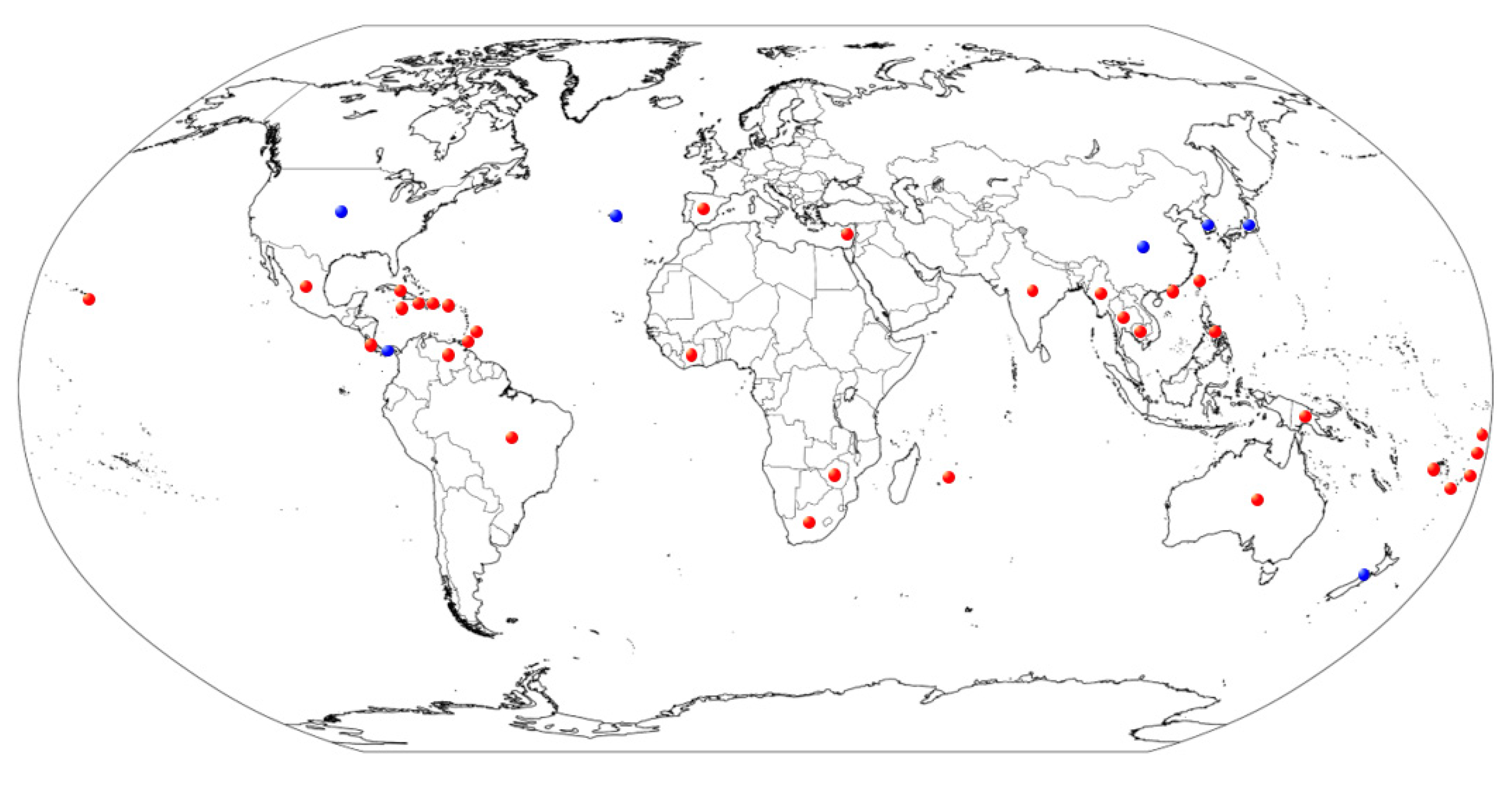
2.2. Geographic Distribution of D. citri
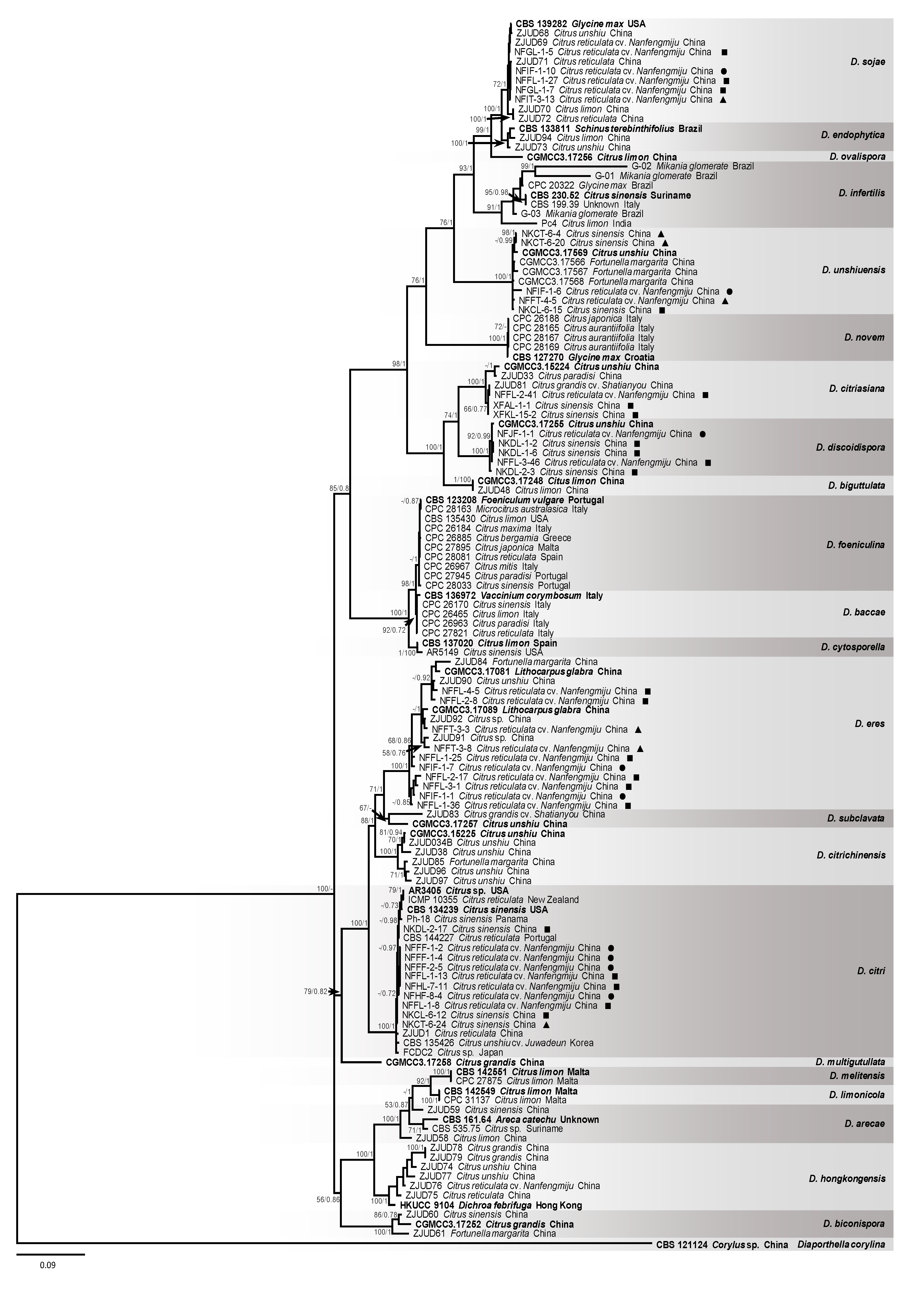
2.3. Phylogenetic Analysis of Diaporthe Species
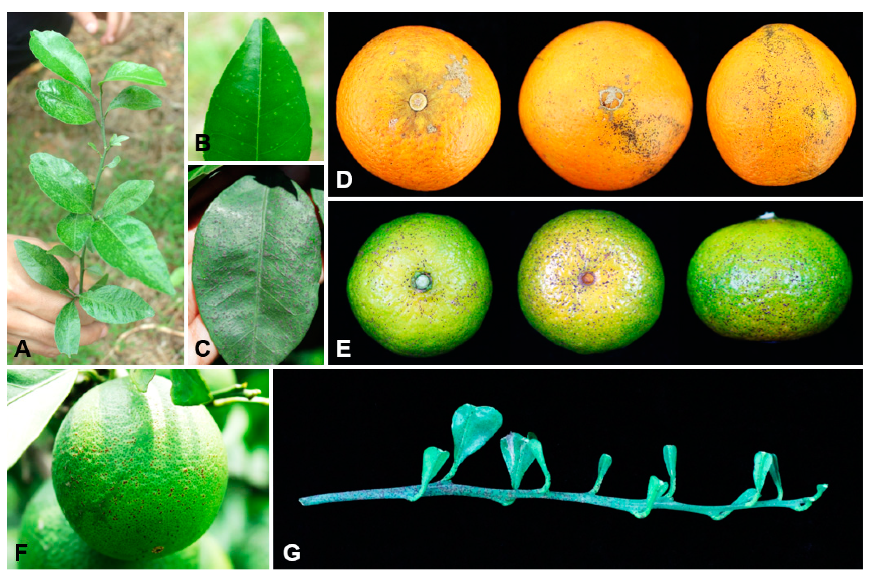
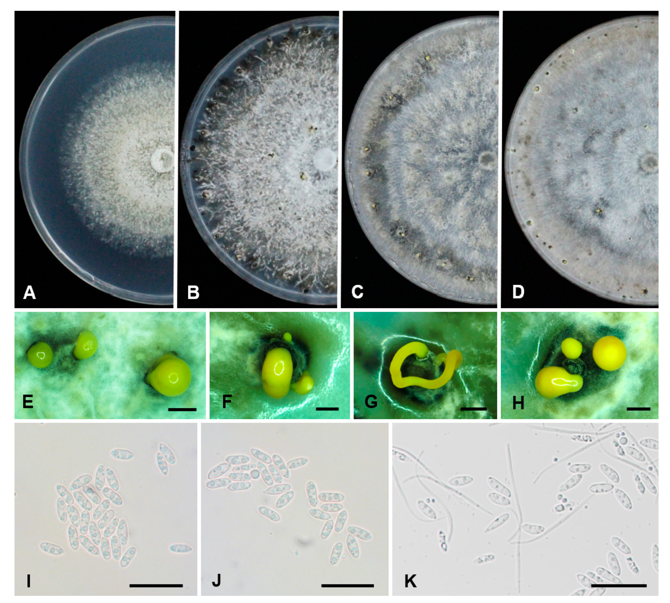
2.4. Morphological Characterization of D. citri
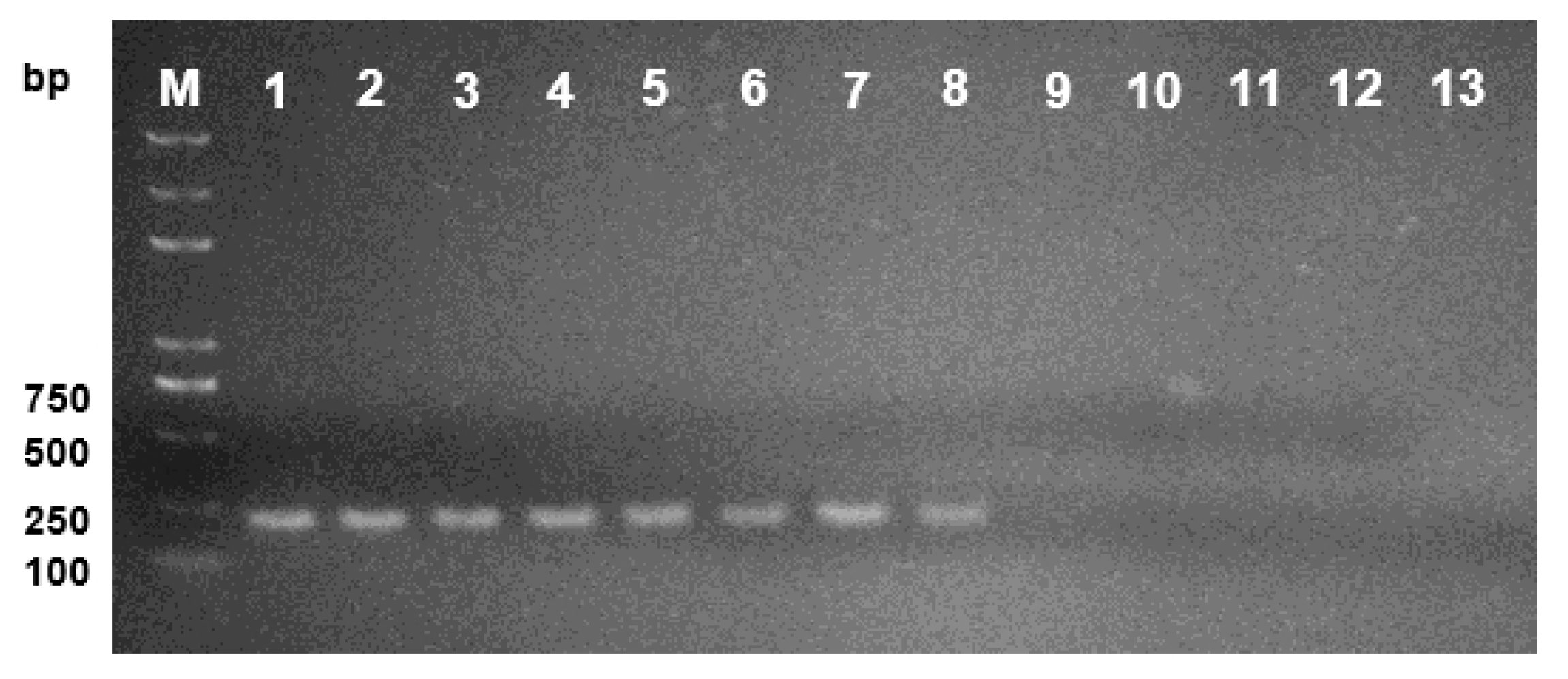
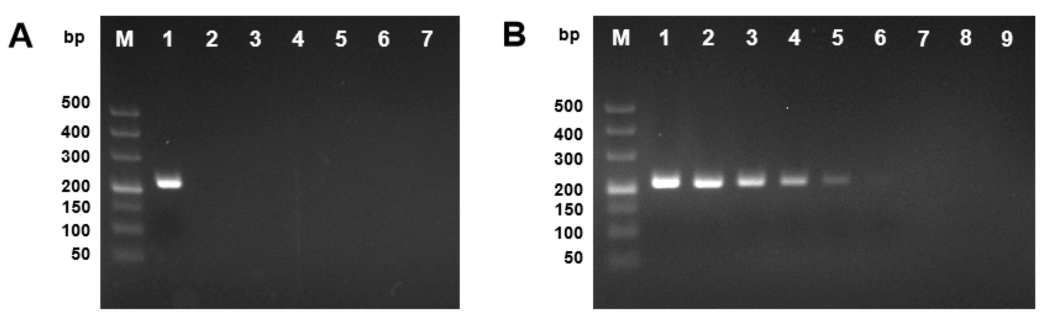
2.5. Specificity and Sensitivity of PCR Method for Detection of D. citri
3. Discussion
4. Materials and Methods
4.1. Sample Collection and Fungal Isolation
4.2. Geographic Distribution of D. citri
4.3. DNA Extraction from Fungal Mycelia
4.4. Sequencing of PCR Products
4.5. Phylogenetic Analyses of Diaporthe Species
4.6. Morphology and Culture Characteristics of D. citri
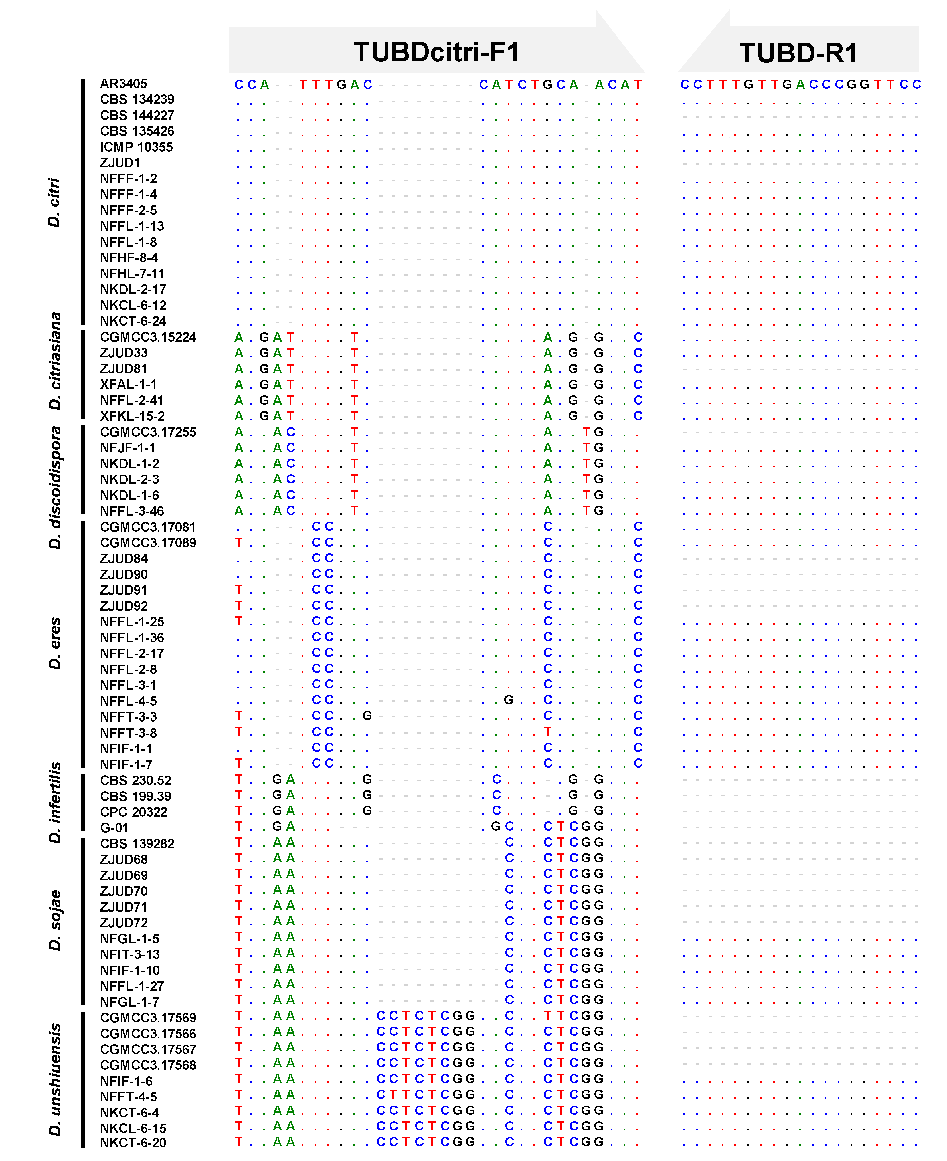
4.7. Primer Design and Development of the Molecular Tool to Detect D. citri
5. Conclusions
Supplementary Materials
Author Contributions
Funding
Acknowledgments
Conflicts of Interest
References
- Wu, G.A.; Terol, J.; Ibanez, V.; López-García, A.; Pérez-Román, E.; Borredá, C.; Domingo, C.; Tadeo, F.R.; Carbonell-Caballero, J.; Alonso, R.; et al. Genomics of the origin and evolution of Citrus. Nature 2018, 554, 311–330. [Google Scholar] [CrossRef] [Green Version]
- FAO. Citrus fruit—Fresh and processed statistical bulletin 2016; Food and Agriculture Organization of the United Nations: Rome, Italy, 2017. [Google Scholar]
- Deng, X.X.; Peng, C.J.; Chen, Z.S.; Deng, Z.N.; Xu, J.G.; Li, J. Citrus Varieties in China; China Agriculture Press: Beijing, China, 2008. [Google Scholar]
- Boddy, L.; Griffth, G.S. Role of endophytes and latent invasion in the development of decay communities in sapwood of angiospermous trees. Sydowia 1989, 41, 41–73. [Google Scholar]
- Carroll, G.C. The Biology of Endophytism in Plants with Particular Reference to Woody Perennials; Cambridge University Press: Cambridge, UK, 1986. [Google Scholar]
- Gomes, R.R.; Glienke, C.; Videira, S.I.R.; Lombard, L.; Groenewald, J.Z.; Crous, P.W. Diaporthe: A genus of endophytic, saprobic and plant pathogenic fungi. Persoonia 2013, 31, 1–41. [Google Scholar] [CrossRef] [PubMed] [Green Version]
- Marin-Felix, Y.; Hernández-Restrepo, M.; Wingfield, M.J.; Akulov, A.; Carnegie, A.J.; Cheewangkoon, R.; Gramaje, D.; Groenewald, J.Z.; Guarnaccia, V.; Halleen, F.; et al. Genera of phytopathogenic fungi: GOPHY 2. Stud. Mycol. 2019, 92, 47–133. [Google Scholar] [CrossRef] [PubMed]
- Suryanarayanan, T.S.; Devarajan, P.T.; Girivasan, K.P.; Govindarajulu, M.B.; Kumaresan, V.; Murali, T.S.; Rajamani, T.; Thirunavukkarasu, N.; Venkatesan, G. The host range of multi-host endophytic fungi. Curr. Sci. 2018, 115, 1963–1969. [Google Scholar] [CrossRef]
- Guarnaccia, V.; Vitale, A.; Cirvilleri, G.; Aiello, D.; Susca, A.; Epifani, F.; Perrone, G.; Polizzi, G. Characterisation and pathogenicity of fungal species associated with branch cankers and stem-end rot of avocado in Italy. Eur. J. Plant Pathol. 2016, 146, 963–976. [Google Scholar] [CrossRef]
- Mostert, L.; Crous, P.W.; Kang, J.C.; Phillips, A.J.L. Species of Phomopsis and a Libertella sp. occurring on grapevines with specific reference to South Africa: Morphological, cultural, molecular and pathological characterization. Mycologia 2001, 93, 146–167. [Google Scholar] [CrossRef]
- Rehner, S.A.; Uecker, F.A. Nuclear ribosomal internal transcribed spacer phylogeny and host diversity in the coelomycete Phomopsis. Can. J. Bot. 1994, 72, 1666–1674. [Google Scholar] [CrossRef]
- Santos, J.M.; Vrandečić, K.; Ćosić, J.; Duvnjak, T.; Phillips, A.J.L. Resolving the Diaporthe species occurring on soybean in Croatia. Persoonia 2011, 27, 9–19. [Google Scholar] [CrossRef] [Green Version]
- Thompson, S.M.; Tan, Y.P.; Young, A.J.; Neate, S.M.; Aitken, E.A.B.; Shivas, R.G. Stem cankers on sunflower (Helianthus annuus) in Australia reveal a complex of pathogenic Diaporthe (Phomopsis) species. Persoonia 2011, 27, 80–89. [Google Scholar] [CrossRef] [Green Version]
- Cai, L.; Giraud, T.; Zhang, N.; Begerow, D.; Cai, G.H.; Shivas, R.G. The evolution of species concepts and species recognition criteria in plant pathogenic fungi. Fungal Divers. 2011, 50, 121–133. [Google Scholar] [CrossRef]
- Duan, W.J.; Yan, J.; Liu, F.; Cai, L.; Zhu, S.F. The list of Chinese quarantine fungi is in need of revision and renewal (in Chinese). Mycosystema 2015, 34, 942–960. [Google Scholar]
- Rossman, A.Y.; Palm-Hernández, M.E. Systematics of plant pathogenic fungi: Why it matters. Plant Dis. 2008, 92, 1376–1386. [Google Scholar] [CrossRef] [PubMed] [Green Version]
- Shivas, R.G.; Cai, L. Cryptic fungal species unmasked. Microbiol. Aust. 2012, 33, 36–37. [Google Scholar]
- Timmer, L.W.; Garnsey, S.M.; Graham, J.H. Scab Diseases; revised edition: 31–32 ed.; American Phytopathological Society Press: St. Paul, MN, USA, 2000; p. 92. [Google Scholar]
- Whiteside, J.O.; Timmer, L.W. Citrus Diseases: General Concepts; revised edition: 3–4 ed.; American Phytopathological Society: St. Paul, MN, USA, 2000. [Google Scholar]
- Guarnaccia, V.; Crous, P.W. Emerging citrus diseases in Europe caused by species of Diaporthe. IMA Fungus 2017, 8, 317–334. [Google Scholar] [CrossRef] [Green Version]
- Huang, F.; Hou, X.; Dewdney, M.M.; Fu, Y.S.; Chen, G.Q.; Hyde, K.D.; Li, H.Y. Diaporthe species occurring on citrus in China. Fungal Divers. 2013, 61, 237–250. [Google Scholar] [CrossRef]
- Kucharek, T.; Whiteside, J.; Brown, E. Melanose and Stem End Rot of Citrus; Florida Cooperative Extension Service, Institute of Food and Agricultural Sciences, University of Florida: Gainesville, FL, USA, 1983. [Google Scholar]
- Mondal, S.N.; Vicent, A.; Reis, R.F.; Timmer, L.W. Saprophytic colonization of citrus twigs by Diaporthe citri and factors affecting pycnidial production and conidial survival. Plant Dis. 2007, 91, 387–392. [Google Scholar] [CrossRef] [Green Version]
- Udayanga, D.; Castlebury, L.A.; Rossman, A.Y.; Hyde, K.D. Species limits in Diaporthe: Molecular re-assessment of D. citri, D. cytosporella, D. foeniculina and D. rudis. Persoonia 2014, 32, 83–101. [Google Scholar] [CrossRef] [Green Version]
- Swingle, W.T.; Webber, H.J. The principal disease of citrus fruits in Florida. USDA Div. Veg. Physiol. Pathol. Bull. 1896, 8, 9–14. [Google Scholar]
- Fawcett, H.S. The cause of stem-end rot of citrus fruits (Phomopsis citri n. sp.). Phytopathology 1912, 2, 109–113. [Google Scholar]
- Floyd, B.F.; Stevens, H.E. Melanose and stem-end rot. Fla. Agr. Expt. Sta. Bull. 1912, 111, 1–16. [Google Scholar]
- Rehm, H. Ascomycetes philippinenses VI. Leafl. Philipp. Bot. 1914, 6, 2258–2281. [Google Scholar]
- Horne, W.T. A Phomopsis in grape fruit from the isle of Pines W. I., with notes on Diplodia natalensis. Phytopathology 1922, 12, 414–418. [Google Scholar]
- Nitschke, T.R.J. Pyrenomycetes germanici. In Die kernpilze Deutschlands Bearbeitet Von Dr. Th. Nitschke; Eduard Trewendt: Breslau, Germany, 1870; Volume 2, pp. 161–320. [Google Scholar]
- Fawcett, H.S. A Phomopsis of citrus in California. Phytopathology 1922, 12, 107. [Google Scholar]
- Bach, W.J.; Wolf, F.A. The isolation of the fungus that causes citrus melanose and the pathological anatomy of the host. J. Agric. Res. 1928, 37, 243–252. [Google Scholar]
- Ruehle, G.D.; Kuntz, W.A. Melanose of Citrus and Its Commercial Control; Florida Agricultural Experiment Station Bulletin, University of Florida: Gainesville, FL, USA, 1940. [Google Scholar]
- Castlebury, L. The Diaporthe vaccinii complex of fruit pathogens. Inoculum 2005, 56, 12. [Google Scholar]
- Santos, J.M.; Correia, V.G.; Phillips, A.J.L.; Spatafora, J.W. Primers for mating-type diagnosis in Diaporthe and Phomopsis: Their use in teleomorph induction in vitro and biological species definition. Fungal Biol. 2010, 114, 255–270. [Google Scholar] [CrossRef]
- Santos, J.M.; Phillips, A.J.L. Resolving the complex of Diaporthe (Phomopsis) species occurring on Foeniculum vulgare in Portugal. Fungal Divers. 2009, 34, 111–125. [Google Scholar]
- Guarnaccia, V.; Groenewald, J.Z.; Woodhall, J.; Armengol, J.; Cinelli, T.; Eichmeier, A.; Ezra, D.; Fontaine, F.; Gramaje, D.; Gutierrez-Aguirregabiria, A.; et al. Diaporthe diversity and pathogenicity revealed from a broad survey of grapevine diseases in Europe. Persoonia 2018, 40, 135–153. [Google Scholar] [CrossRef] [Green Version]
- Santos, L.; Alves, A.; Alves, R. Evaluating multi-locus phylogenies for species boundaries determination in the genus Diaporthe. PeerJ 2017, 5, 1–26. [Google Scholar] [CrossRef] [Green Version]
- Udayanga, D.; Castlebury, L.A.; Rossman, A.Y.; Chukeatirote, E.; Hyde, K.D. Insights into the genus diaporthe: Phylogenetic species delimitation in the D. eres species complex. Fungal Divers. 2014, 67, 203–229. [Google Scholar] [CrossRef] [Green Version]
- Yang, Q.; Fan, X.L.; Guarnaccia, V.; Tian, C.M. High diversity of Diaporthe species associated with dieback diseases in China, with twelve new species described. MycoKeys 2018, 39, 97–149. [Google Scholar] [CrossRef] [PubMed] [Green Version]
- Hyde, K.D.; Nilsson, R.H.; Alias, S.A.; Ariyawansa, H.A.; Blair, J.E.; Cai, L.; de Cock, A.W.A.M.; Dissanayake, A.J.; Glockling, S.L.; Goonasekara, I.D.; et al. One stop shop: Backbones trees for important phytopathogenic genera: I (2014). Fungal Divers. 2014, 67, 21–125. [Google Scholar] [CrossRef] [Green Version]
- Zhang, A.W.; Hartman, G.L.; Riccioni, L.; Chen, W.D.; Ma, R.Z.; Pedersen, W.L. Using PCR to distinguish Diaporthe phaseolorum and Phomopsis longicolla from other soybean fungal pathogens and to detect them in soybean tissues. Plant Dis. 1997, 81, 1143–1149. [Google Scholar] [CrossRef] [PubMed] [Green Version]
- Prasad, M.N.N.; Bhat, S.S.; Raj, A.P.C.; Janardhana, G.R. Molecular detection of Phomopsis azadirachtae, the causative agent of dieback disease of neem by polymerase chain reaction. Curr. Sci. 2006, 91, 158–159. [Google Scholar]
- Vedashree, S.; Sateesh, M.K.; Chowdappa, P.; Nirmalkumar, B.J. Species-specific PCR-based assay for identification and detection of Phomopsis (Diaporthe) azadirachtae causing die-back disease in Azadirachta indica. J. Phytopathol. 2015, 163, 818–828. [Google Scholar] [CrossRef]
- Shishido, M.; Sato, K.; Yoshida, N.; Tsukui, R.; Usami, T. PCR-based assays to detect and quantify Phomopsis sclerotioides in plants and soil. J. Gen. Plant Pathol. 2010, 76, 21–30. [Google Scholar] [CrossRef]
- Shirahatti, P.; Ramu, R.; Purushothama, C.R.A.; Prasad, M.N.N. Development of a simple and reliable species-species detection of Phomopsis azadirachtae, using the translation elongation factor 1-alpha gene. Eur. J. Plant Pathol. 2015, 141, 769–778. [Google Scholar] [CrossRef]
- Anonymous. List of Plant Diseases in Taiwan; Plant Protection Soc: Taichung, China, 1979; p. 404. [Google Scholar]
- Huang, F.; Udayanga, D.; Wang, X.H.; Hou, X.; Mei, X.F.; Fu, Y.S.; Hyde, K.D.; Li, H.Y. Endophytic Diaporthe associated with citrus: A phylogenetic reassessment with seven new species from China. Fungal Biol. 2015, 119, 331–347. [Google Scholar] [CrossRef]
- Lu, B.S.; Hyde, K.D.; Ho, W.H.; Tsui, K.M.; Taylor, J.E.; Wong, K.M.; Yanna, Z.D.; Zhou, D.Q. Checklist of Hong Kong Fungi; Fungal Diversity Press: Hong Kong, China, 2000; p. 207. [Google Scholar]
- Zhuang, W.Y. Higher Fungi of Tropical China; Mycotaxon, Ltd.: Ithaca, NY, USA, 2001; p. 485. [Google Scholar]
- Udayanga, D.; Castlebury, L.A.; Rossman, A.Y.; Chukeatirote, E.; Hyde, K.D. The Diaporthe sojae species complex: Phylogenetic re-assessment of pathogens associated with soybean, cucurbits and other field crops. Fungal Biol. 2015, 119, 383–407. [Google Scholar] [CrossRef]
- Bonants, P.J.M.; Carroll, G.C.; de Weerdt, M.; van Brouwershaven, I.R.; Baayen, R.P. Development and validation of a PCR-based detection method for pathogenic isolates of the citrus black spot fungus, Guignardia citricarpa. Eur. J. Plant Pathol. 2003, 109, 503–513. [Google Scholar] [CrossRef]
- Wang, X.H.; Chen, G.Q.; Huang, F.; Zhang, J.Z.; Hyde, K.D.; Li, H.Y. Phyllosticta species associated with citrus diseases in China. Fungal Divers. 2012, 52, 209–224. [Google Scholar] [CrossRef]
- Meyer, L.; Sanders, G.M.; Jacobs, R.; Korsten, L. A one-day sensitive method to detect and distinguish between the citrus black spot pathogen Guignardia citricarpa and the endophyte Guignardia mangiferae. Plant Dis. 2006, 90, 97–101. [Google Scholar] [CrossRef] [PubMed] [Green Version]
- Peres, N.A.; Harakava, R.; Carroll, G.C.; Adaskaveg, J.E.; Timmer, L.W. Comparison of molecular procedures for detection and identification of Guignardia citricarpa and G. mangiferae. Plant Dis. 2007, 91, 525–531. [Google Scholar] [CrossRef]
- van Gent-Peizer, M.P.E.; van Brouwershaven, I.R.; Kox, L.F.F.; Bonants, P.J.M. A Taqman PCR method for routine diagnosis of the quarantine fungus Guignardia citricarpa on citrus fruit. J. Phytopathol. 2007, 155, 357–363. [Google Scholar] [CrossRef]
- Schirmacher, A.M.; Tomlinson, J.A.; Barnes, A.V.; Barton, V.C. Species-specific real-timle PCR for diagnosis of Phyllosticta citricarpa on citrus species. Bull. OEPP/EPPO Bull. 2019, 49, 306–313. [Google Scholar] [CrossRef]
- Yang, Y.H.; Hu, J.H.; Chen, F.J.; Ding, D.K.; Zhou, C.Y. Development of a SCAR marker-based diagnostic method for the detection of the citrus target spot pathogen Pseudofabraea citricarpa. Biomed. Res. Int. 2018, 2018, 7128903. [Google Scholar] [CrossRef] [Green Version]
- Das, A.K.; Nerkar, S.; Gawande, N.; Thakre, N.; Kumar, A. Scar marker for phytophthora nicotianae and a multiplex PCR assay for simultaneous detection of P. nicotianae and Candidatus liberibacter asiaticus in citrus. J. Appl. Microbiol. 2019, 127, 1172–1183. [Google Scholar] [CrossRef]
- Pereira, W.V.; Bertolini, E.; Cambra, M.; Junior, N.S.M. Multiplex real-time PCR for detection and quantification of Colletotrichum abscissum and C. gloeosporioides on Citrus leaves. Eur. J. Plant Pathol. 2019, 155, 1–13. [Google Scholar] [CrossRef]
- Goh, T.K. Single-spore isolation using a hand-made glass needle. Fungal Divers. 1999, 2, 47–63. [Google Scholar]
- Yin, L.F.; Chen, S.N.; Chen, G.K.; Schnabel, G.; Du, S.F.; Chen, C.; Li, G.Q.; Luo, C.X. Identification and characterization of three Monilinia species from plum in China. Plant Dis. 2015, 99, 1775–1783. [Google Scholar] [CrossRef] [PubMed] [Green Version]
- Farr, D.F.; Rossman, A.Y. Fungal Databases; U.S. National Fungus Collections, ARS, USDA: Washington, DC, USA, 2018.
- Chi, M.H.; Park, S.Y.; Lee, Y.H. A quick and safe method for fungal DNA extraction. Plant Pathol. J. 2009, 25, 108–111. [Google Scholar] [CrossRef]
- White, T.J.; Bruns, T.; Lee, S.; Taylor, J. Amplification and direct sequencing of fungal ribosomal RNA genes for phylogenetics. In PCR Protocols: A Guide to Methods and Applications; Innis, M.A., Gelfand, D.H., Sninsky, J.J., White, T.J., Eds.; Academic Press: San Diego, CA, USA, 1990; pp. 315–322. [Google Scholar]
- Carbone, I.; Kohn, L.M. A method for desianing primer sets for speciation studies in filamentous ascomycetes. Mycologia 1999, 91, 553–556. [Google Scholar] [CrossRef]
- Glass, N.L.; Donaldson, G.C. Development of primer sets designed for use with the PCR to amplify conserved genes from filamentous ascomycetes. Appl. Environ. Microb. 1995, 61, 1323–1330. [Google Scholar] [CrossRef] [Green Version]
- Crous, P.W.; Groenewald, J.Z.; Risède, J.M.; Simoneau, P.; Hywel-Jones, N.L. Calonectria species and their Cylindrocladium anamorphs: Species with clavate vesicles. Stud. Mycol. 2004, 50, 415–430. [Google Scholar] [CrossRef] [Green Version]
- Hall, A.T. Bioedit: A user-friendly biological sequence alignment editor and analysis program for windows 95/98/nt. Nucleic Acids Res. 1999, 41, 95–98. [Google Scholar]
- Swofford, D.L. PAUP* Phylogenetic Analysis Using Parsimony, (*and Other Methods); Version 4.0 b10; Sinauer Associates: Sunderland, MA, USA, 2003. [Google Scholar]
- Hillis, D.M.; Bull, J.J. An empirical test of bootstrapping as a method for assessing confidence in phylogenetic analysis. Syst. Biol. 1993, 42, 182–192. [Google Scholar] [CrossRef]
- Ronquist, F.; Teslenko, M.; Van der Mark, P.; Ayres, D.L.; Darling, A.; Hӧhna, S.; Larget, B.; Liu, L.; Suchard, M.A.; Huelsenbeck, J.P. Mrbayes 3.2: Efficient bayesian phylogenetic tnference and model choice across a large model space. Syst. Biol. 2012, 61, 539–542. [Google Scholar] [CrossRef] [Green Version]
- Nylander, J.A.A. Mrmodeltest v.2. Program Distributed by the Author; Evolitionary Biology Centre, Uppsala Univeristy: Uppsala, Sweden, 2004. [Google Scholar]
- Rambaut, A. Figtree v.1.4.2; Institute of Evolutionary Biology, Ashworth Laboratories, University of Edinburgh: Edinburgh, UK, 2014. [Google Scholar]
- Lombard, L.; van Leeuwen, G.C.M.; Guarnaccia, V.; Polozzi, G.; van Rijswick, P.C.J.; Rosendahl, C.H.M.; Gabler, J.; Crous, P.W. Diaporthe species associated with Vaccinium, with specific reference to Europe. Phytopathol. Mediterr. 2014, 53, 287–299. [Google Scholar]
- Aguilera-Cogley, V.; Vicent, A. Etiology and distribution of foliar fungal diseases of citrus in Panama. Trop. Plant Pathol. 2019, 44, 519–532. [Google Scholar] [CrossRef]
- Kanematsu, S.; Kobayashi, T.; Kudo, A.; Ohtsu, Y. Conidial morphology, pathogenicity and culture characteristics of Phomopsis isolates from peach, Japanese pear and apple in Japan. Jpn. J. Phytopathol. 1999, 65, 264–273. [Google Scholar] [CrossRef]
- Kanematsu, S. Phylogeny of phomopsis species from fruit trees. In Direct Submission Sequence of Diaporthe citri Strian FCDC2; National Institute of Fruit Tree Science, Apple Research Station: Morioka, Japan, 2007. [Google Scholar]
- Gao, Y.H.; Su, Y.Y.; Sun, W.; Cai, L. Diaporthe species occurring on Lithocarpus glabra in China, with descriptions of five new species. Fungal Biol. 2015, 119, 295–309. [Google Scholar] [CrossRef] [PubMed]
- Mahadevakumar, S.; Yadav, V.; Tejaswini, G.S.; Sandeep, S.N.; Janardhana, G.R. First report of Phomopsis citri associated with dieback of Citrus lemon in India. Plant Dis. 2014, 98, 1281. [Google Scholar] [CrossRef] [PubMed]
- Polonio, J.C.; Almeida, T.T.; Garcia, D.; Mariucci, G.E.G.; Azevedo, J.L.; Rhoden, S.A.; Pamphile, J.A. Biotechnological prospecting of foliar endophytic fungi of guaco (Mikania glomerata Spreng.) with antibacterial and antagonistic activity against phytopathogens. Genet. Mol. Res. 2015, 14, 7297–7309. [Google Scholar] [CrossRef]
- Polonio, J.C.; Ribeiro, M.A.S.; Rhoden, S.A.; Sarragiotto, M.H.; Azevedo, J.L.; Pamphile, J.A. 3-nitropropionic acid production by the endophytic Diaporthe citri: Molecular taxonomy, chemical characterization, and quantification under ph variation. Fungal Biol. 2016, 120, 1600–1608. [Google Scholar] [CrossRef]
- Vasilyeva, L.N.; Rossman, A.Y.; Farr, D.F. New species of the Diaporthales from Eastern Asia and Eastern North America. Mycologia 2007, 99, 916–923. [Google Scholar] [CrossRef]
- Rayner, R.W. A Mycological Colour Chart; Commonwealth Mycological Institute and British Mycological Society: Kew, Surrey, UK, 1970. [Google Scholar]


| Diaporthe Species | Isolate Number | Plant Host | Tissue | Locality | GenBank Accession Numbers 1 | ||||
|---|---|---|---|---|---|---|---|---|---|
| ITS | TUB | TEF | CAL | HIS | |||||
| D. citri | NFFF-1-2 | Citrus reticulata cv. Nanfengmiju | fruit | China: Jiangxi: Nanfeng | MN816394 | MN894454 | MN894415 | MN894355 | MN894380 |
| NFFF-1-4 | Citrus reticulata cv. Nanfengmiju | fruit | China: Jiangxi: Nanfeng | MN816395 | MN894455 | MN894416 | MN894356 | MN894381 | |
| NFFF-2-5 | Citrus reticulata cv. Nanfengmiju | fruit | China: Jiangxi: Nanfeng | MN816396 | MN894456 | MN894417 | MN894357 | MN894382 | |
| NFFL-1-13 | Citrus reticulata cv. Nanfengmiju | leaf | China: Jiangxi: Nanfeng | MN816397 | MN894457 | MN894418 | MN894358 | – | |
| NFFL-1-8 | Citrus reticulata cv. Nanfengmiju | leaf | China: Jiangxi: Nanfeng | MN816398 | MN894458 | MN894419 | MN894359 | – | |
| NFHF-8-4 | Citrus reticulata cv. Nanfengmiju | fruit | China: Jiangxi: Nanfeng | MN816399 | MN894459 | MN894420 | MN894360 | – | |
| NFHL-7-11 | Citrus reticulata cv. Nanfengmiju | leaf | China: Jiangxi: Nanfeng | MN816400 | MN894460 | MN894421 | MN894361 | MN894383 | |
| NKDL-2-17 | Citrus sinensis | leaf | China: Jiangxi: Nankang | MN816401 | MN894461 | MN894422 | MN894362 | MN894384 | |
| NKCL-6-12 | Citrus sinensis | leaf | China: Jiangxi: Nankang | MN816402 | MN894462 | MN894423 | MN894363 | MN894385 | |
| NKCT-6-24 | Citrus sinensis | twig | China: Jiangxi: Nankang | MN816403 | MN894463 | MN894424 | MN894364 | MN894386 | |
| D. citriasiana | XFAL-1-1 | Citrus sinensis | leaf | China: Jiangxi: Xinfeng | MN816404 | MN894464 | MN894425 | – | MN894387 |
| NFFL-2-41 | Citrus reticulata cv. Nanfengmiju | leaf | China: Jiangxi: Nanfeng | MN816405 | MN894465 | MN894426 | – | MN894388 | |
| XFKL-15-2 | Citrus sinensis | leaf | China: Jiangxi: Xinfeng | MN816406 | MN894466 | MN894427 | – | MN894389 | |
| D. discoidispora | NFJF-1-1 | Citrus reticulata cv. Nanfengmiju | fruit | China: Jiangxi: Nanfeng | MN816407 | MN894467 | MN894428 | – | MN894390 |
| NKDL-1-2 | Citrus sinensis | leaf | China: Jiangxi: Nankang | MN816408 | MN894468 | MN894429 | – | MN894391 | |
| NKDL-2-3 | Citrus sinensis | leaf | China: Jiangxi: Nankang | MN816409 | MN894469 | MN894430 | – | MN894392 | |
| NKDL-1-6 | Citrus sinensis | leaf | China: Jiangxi: Nankang | MN816410 | MN894470 | MN894431 | – | MN894393 | |
| NFFL-3-46 | Citrus reticulata cv. Nanfengmiju | leaf | China: Jiangxi: Nanfeng | MN816411 | MN894471 | MN894432 | – | MN894394 | |
| D. eres | NFFL-1-25 | Citrus reticulata cv. Nanfengmiju | leaf | China: Jiangxi: Nanfeng | MN816412 | MN894472 | MN894433 | – | MN894395 |
| NFFL-1-36 | Citrus reticulata cv. Nanfengmiju | leaf | China: Jiangxi: Nanfeng | MN816413 | MN894473 | MN894434 | MN894365 | MN894396 | |
| NFFL-2-17 | Citrus reticulata cv. Nanfengmiju | leaf | China: Jiangxi: Nanfeng | MN816414 | MN894474 | MN894435 | – | MN894397 | |
| NFFL-2-8 | Citrus reticulata cv. Nanfengmiju | leaf | China: Jiangxi: Nanfeng | MN816415 | MN894475 | MN894436 | MN894366 | MN894398 | |
| NFFL-3-1 | Citrus reticulata cv. Nanfengmiju | leaf | China: Jiangxi: Nanfeng | MN816416 | MN894476 | MN894437 | MN894367 | MN894399 | |
| NFFL-4-5 | Citrus reticulata cv. Nanfengmiju | leaf | China: Jiangxi: Nanfeng | MN816417 | MN894477 | MN894438 | MN894368 | MN894400 | |
| NFFT-3-3 | Citrus reticulata cv. Nanfengmiju | twig | China: Jiangxi: Nanfeng | MN816418 | MN894478 | MN894439 | – | MN894401 | |
| NFFT-3-8 | Citrus reticulata cv. Nanfengmiju | twig | China: Jiangxi: Nanfeng | MN816419 | MN894479 | MN894440 | MN894369 | MN894402 | |
| NFIF-1-1 | Citrus reticulata cv. Nanfengmiju | fruit | China: Jiangxi: Nanfeng | MN816420 | MN894480 | MN894441 | MN894370 | MN894403 | |
| NFIF-1-7 | Citrus reticulata cv. Nanfengmiju | fruit | China: Jiangxi: Nanfeng | MN816421 | MN894481 | MN894442 | – | MN894404 | |
| D. sojae | NFGL-1-5 | Citrus reticulata cv. Nanfengmiju | leaf | China: Jiangxi: Nanfeng | MN816422 | MN894482 | MN894443 | MN894371 | MN894405 |
| NFIT-3-13 | Citrus reticulata cv. Nanfengmiju | twig | China: Jiangxi: Nanfeng | MN816423 | MN894483 | MN894444 | MN894372 | MN894406 | |
| NFIF-1-10 | Citrus reticulata cv. Nanfengmiju | fruit | China: Jiangxi: Nanfeng | MN816424 | MN894484 | MN894445 | MN894373 | MN894407 | |
| NFFL-1-27 | Citrus reticulata cv. Nanfengmiju | leaf | China: Jiangxi: Nanfeng | MN816425 | MN894485 | MN894446 | MN894374 | MN894408 | |
| NFGL-1-7 | Citrus reticulata cv. Nanfengmiju | leaf | China: Jiangxi: Nanfeng | MN816426 | MN894486 | MN894447 | MN894375 | MN894409 | |
| D. unshiuensis | NFIF-1-6 | Citrus reticulata cv. Nanfengmiju | fruit | China: Jiangxi: Nanfeng | MN816427 | MN894487 | MN894448 | MN894376 | – |
| NFFT-4-5 | Citrus reticulata cv. Nanfengmiju | twig | China: Jiangxi: Nanfeng | MN816428 | MN894488 | MN894449 | – | MN894410 | |
| NKCT-6-4 | Citrus sinensis | twig | China: Jiangxi: Nankang | MN816429 | MN894489 | MN894450 | – | MN894411 | |
| NKCL-6-15 | Citrus sinensis | leaf | China: Jiangxi: Nankang | MN816430 | MN894490 | MN894451 | MN894377 | MN894412 | |
| NKCT-6-20 | Citrus sinensis | twig | China: Jiangxi: Nankang | MN816431 | MN894491 | MN894452 | MN894378 | MN894413 | |
| Gene/Locus | ITS | TEF | TUB | CAL | HIS | Combined |
|---|---|---|---|---|---|---|
| No. of taxa | 129 | 124 | 124 | 68 | 112 | 129 |
| Aligned length (with gaps) | 645 | 472 | 893 | 617 | 540 | 3183 |
| Invariable characters (%) | 414 (64.19) | 186 (39.41) | 523 (58.57) | 310 (50.24) | 352 (65.19) | 1801 (56.58) |
| Phylogenetically informative characters (%) | 123 (19.07) | 230 (48.73) | 263 (29.45) | 238 (38.57) | 143 (26.48) | 997 (31.32) |
| Uninformative variable characters (%) | 108 (16.74) | 56 (11.86) | 107 (11.98) | 69 (11.18) | 45 (8.33) | 385 (12.10) |
| Tree length (TL) | 670 | 856 | 745 | 554 | 538 | 3,654 |
| Consistency index (CI) | 0.506 | 0.575 | 0.686 | 0.773 | 0.55 | 0.565 |
| Retention index (RI) | 0.901 | 0.948 | 0.94 | 0.952 | 0.926 | 0.921 |
| Rescaled consistency index (RC) | 0.456 | 0.545 | 0.645 | 0.735 | 0.509 | 0.521 |
| Homoplasy index (ID) | 0.494 | 0.425 | 0.314 | 0.227 | 0.45 | 0.435 |
| Nucleotide substitution model | GTR + I + G | GTR + I + G | HKY + G | GTR + G | GTR + I + G | GTR + I + G |
| Primer Name | Primer Sequences (5´ to 3´) | Length (nt) 1 | Ta (°C) 2 | %GC | Reference |
|---|---|---|---|---|---|
| ITS1 | TCCGTAGGTGAACCTGCGG | 19 | 55.0 | 63.2 | White, et al. [65] |
| ITS4 | TCCTCCGCTTATTGATATGC | 20 | 45.0 | White, et al. [65] | |
| EF1-728F | CATCGAGAAGTTCGAGAAGG | 20 | 58.0 | 50.0 | Carbone and Kohn [66] |
| EF1-986R | TACTTGAAGGAACCCTTACC | 20 | 45.0 | Carbone and Kohn [66] | |
| Bt2a | GGTAACCAAATCGGTGCTGCTTTC | 24 | 58.0 | 50.0 | Glass and Donaldson [67] |
| Bt2b | ACCCTCAGTGTAGTGACCCTTGGC | 24 | 58.0 | Glass and Donaldson [67] | |
| TUBDcitri-F1 | CCATTTGACCATCTGCAACAT | 21 | 55.0 | 42.9 | This study |
| TUBD-R1 | CCTTGGCCCAGTTGTTTCC | 19 | 57.9 | This study | |
| CAL-228F | GAGTTCAAGGAGGCCTTCTCCC | 22 | 55.0 | 59.0 | Carbone and Kohn [66] |
| CAL-737R | CATCTTCTGGCCATCATGG | 19 | 52.6 | Carbone and Kohn [66] | |
| CYLH3F | AGGTCCACTGGTGGCAAG | 18 | 58.0 | 61.1 | Crous, et al. [68] |
| H3-1b | GCGGGCGAGCTGGATGTCCTT | 21 | 66.6 | Glass and Donaldson [67] |
| Species | Isolate Number 1,2 | Plant Host | Locality | GenBank Accession Numbers 3 | Reference(s) | ||||
|---|---|---|---|---|---|---|---|---|---|
| ITS | TUB | TEF | CAL | HIS | |||||
| Diaporthe arecae | CBS 161.64 IT | Areca catechu | Unknown | KC343032 | KC344000 | KC343758 | KC343274 | KC343516 | Gomes, et al. [6] |
| CBS 535.75 | Citrus sp. | Suriname | KC343033 | KC344001 | KC343759 | KC343275 | KC343517 | Gomes, et al. [6] | |
| ZJUD58 | Citrus limon | China: Yunnan | KJ490593 | KJ490414 | KJ490472 | – | KJ490535 | Huang, et al. [48] | |
| ZJUD59 | Citrus sinensis | China: Jiangxi | KJ490594 | KJ490415 | KJ490473 | – | KJ490536 | Huang, et al. [48] | |
| D. baccae | CBS 136,972 T | Vaccinium corymbosum | Italy: Sicily, Catania | KJ160565 | MF418509 | KJ160597 | – | MF418264 | Guarnaccia and Crous [20], Lombard, et al. [75] |
| CPC 26170 | Citrus sinensis | Italy: Catania | MF418351 | MF418510 | MF418430 | MF418185 | MF418265 | Guarnaccia and Crous [20] | |
| CPC 26465 | Citrus limon | Italy: Catania | MF418352 | MF418511 | MF418431 | MF418186 | MF418266 | Guarnaccia and Crous [20] | |
| CPC 26963 | Citrus paradisi | Italy: Vibo Valentia | MF418353 | MF418512 | MF418432 | MF418187 | MF418267 | Guarnaccia and Crous [20] | |
| CPC 27821 | Citrus reticulata | Italy: Cosenza | MF418357 | MF418516 | MF418436 | MF418191 | MF418271 | Guarnaccia and Crous [20] | |
| D. biconispora | CGMCC3.17252 T | Citrus grandis | China: Fujian | KJ490597 | KJ490418 | KJ490476 | – | KJ490539 | Huang, et al. [48] |
| ZJUD60 | Citrus sinensis | China: Jiangxi | KJ490595 | KJ490416 | KJ490474 | – | KJ490537 | Huang, et al. [48] | |
| ZJUD61 | Fortunella margarita | China: Guangxi | KJ490596 | KJ490417 | KJ490475 | – | KJ490538 | Huang, et al. [48] | |
| D. biguttulata | CGMCC3.17248 T | Citus limon | China: Yunnan | KJ490582 | KJ490403 | KJ490461 | – | KJ490524 | Huang, et al. [48] |
| ZJUD48 | Citrus limon | China: Yunnan | KJ490583 | KJ490404 | KJ490462 | – | KJ490525 | Huang, et al. [48] | |
| D. citri | AR3405 T | Citrus sp. | USA: Florida | KC843311 | KC843187 | KC843071 | KC843157 | MF418281 | Guarnaccia and Crous [20], Udayanga, et al. [24] |
| CBS 134,239 T | Citrus sinensis | USA: Florida | KC357553 | KC357456 | KC357522 | KC357488 | MF418280 | Guarnaccia and Crous [20], Huang, et al. [21] | |
| ZJUD1 | Citrus reticulata | China: Zhejiang | JQ954654 | KJ490395 | JQ954671 | – | KJ490514 | Huang, et al. [21], Huang, et al. [48] | |
| CBS 144227 | Citrus reticulata | Portugal: Azores | MH063904 | MH063916 | MH063910 | MH063892 | MH063898 | Guarnaccia and Crous [20] | |
| CBS 135426 | Citrus unshiu cv. Juwadeun | Korea: Odeung-dong | KC843324 | KC843200 | KC843084 | KC843170 | – | Udayanga, et al. [24] | |
| ICMP 10355 | Citrus reticulata | New Zealand: Kerikeri | KC843314 | KC843190 | KC843074 | KC843160 | – | Udayanga, et al. [24] | |
| Ph-18 | Citrus sinensis | Panama: Coclé | MK214464 | – | MK283703 | – | – | Aguilera-Cogley and Vicent [76] | |
| FCDC2 | Citrus sp. | Japan: Fukuoka | AB302249 | – | – | – | – | Kanematsu, et al. [77], Kanematsu [78] | |
| D. citriasiana | CGMCC3.15224 T | Citrus unshiu | China: Shaanxi | JQ954645 | KC357459 | JQ954663 | KC357491 | MF418282 | Guarnaccia and Crous [20], Huang, et al. [21] |
| ZJUD33 | Citrus paradisi | China: Jiangxi | JQ954658 | KC357460 | JQ972716 | KC357493 | – | Huang, et al. [21] | |
| ZJUD81 | Citrus grandis cv. Shatianyou | China: Zhejiang | KJ490616 | KJ490437 | KJ490495 | – | KJ490558 | Huang, et al. [48] | |
| D. citrichinensis | CGMCC3.15225 T | Citrus unshiu | China: Shaanxi | JQ954648 | MF418524 | JQ954666 | KC357494 | KJ490516 | Guarnaccia and Crous [20], Huang, et al. [21,48] |
| ZJUD034B | Citrus unshiu | China: Shaanxi | KJ210539 | KJ420829 | KJ210562 | KJ435042 | KJ420879 | Udayanga, et al. [24], Udayanga, et al. [39] | |
| ZJUD38 | Citrus unshiu | China: Shaanxi | KC357558 | KC357463 | KC357527 | KC357498 | – | Huang, et al. [21] | |
| ZJUD85 | Fortunella margarita | China: Guangxi | KJ490620 | KJ490441 | KJ490499 | – | KJ490562 | Huang, et al. [48] | |
| ZJUD96 | Citrus unshiu | China: Fujian | KJ490631 | KJ490452 | KJ490510 | – | KJ490573 | Huang, et al. [48] | |
| ZJUD97 | Citrus grandis | China: Fujian | KJ490632 | KJ490453 | KJ490511 | – | KJ490574 | Huang, et al. [48] | |
| D. cytosporella | CBS 137,020 T | Citrus limon | Spain | KC843307 | KC843221 | KC843116 | KC843141 | MF418283 | Guarnaccia and Crous [20], Udayanga, et al. [24] |
| AR5149 | Citrus sinensis | USA: California | KC843309 | KC843222 | KC843118 | KC843143 | – | Udayanga, et al. [24] | |
| D. discoidispora | CGMCC3.17255 T | Citrus unshiu | China: Jiangxi | KJ490624 | KJ490445 | KJ490503 | – | KJ490566 | Huang, et al. [48] |
| D. endophytica | CBS 133,811 T | Schinus terebinthifolius | Brazil | KC343065 | KC344033 | KC343791 | KC343307 | KC343549 | Gomes, et al. [6] |
| ZJUD73 | Citrus unshiu | China: Fujian | KJ490608 | KJ490429 | KJ490487 | – | KJ490550 | Huang, et al. [48] | |
| ZJUD94 | Citrus limon | China: Yunnan | KJ490629 | KJ490450 | KJ490508 | – | KJ490571 | Huang, et al. [48] | |
| D. eres | CGMCC3.17081 T | Lithocarpus glabra | China: Zhejiang | KF576282 | KF576306 | KF576257 | – | – | Gao, et al. [79] |
| CGMCC3.17089 T | Lithocarpus glabra | China: Zhejiang | KF576267 | KF576291 | KF576242 | – | – | Gao, et al. [79] | |
| ZJUD84 | Fortunella margarita | China: Guangxi | KJ490619 | KJ490440 | KJ490498 | – | KJ490561 | Huang, et al. [48] | |
| ZJUD90 | Citrus unshiu | China: Jiangxi | KJ490625 | KJ490446 | KJ490504 | – | KJ490567 | Huang, et al. [48] | |
| ZJUD91 | Citrus sp. | China: Jiangxi | KJ490626 | KJ490447 | KJ490505 | – | KJ490568 | Huang, et al. [48] | |
| ZJUD92 | Citrus sp. | China: Zhejiang | KJ490627 | KJ490448 | KJ490506 | – | KJ490569 | Huang, et al. [48] | |
| D. foeniculina | CBS 123,208 T | Foeniculum vulgare | Portugal: Évora | KC343104 | KC344072 | KC343830 | KC343346 | KC343588 | Gomes, et al. [6] |
| CBS 135430 | Citrus limon | USA: California | KC843301 | KC843215 | KC843110 | KC843135 | MF418284 | Guarnaccia and Crous [20], Udayanga, et al. [24] | |
| CPC 26184 | Citrus maxima | Italy: Messina | MF418365 | MF418525 | MF418444 | MF418199 | MF418285 | Guarnaccia and Crous [20] | |
| CPC 26885 | Citrus bergamia | Greece: Missolonghi | MF418374 | MF418534 | MF418453 | MF418208 | MF418294 | Guarnaccia and Crous [20] | |
| CPC 26967 | Citrus mitis | Italy: Messina | MF418379 | MF418539 | MF418458 | MF418213 | MF418299 | Guarnaccia and Crous [20] | |
| CPC 27895 | Citrus japonica | Malta: Gozo | MF418391 | MF418551 | MF418470 | MF418225 | MF418311 | Guarnaccia and Crous [20] | |
| CPC 27945 | Citrus paradisi | Portugal: Faro | MF418397 | MF418557 | MF418476 | MF418231 | MF418317 | Guarnaccia and Crous [20] | |
| CPC 28033 | Citrus sinensis | Portugal: Mesquita | MF418402 | MF418562 | MF418481 | MF418236 | MF418322 | Guarnaccia and Crous [20] | |
| CPC 28081 | Citrus reticulata | Spain: Algemesi | MF418415 | MF418575 | MF418494 | MF418249 | MF418335 | Guarnaccia and Crous [20] | |
| CPC 28163 | Microcitrus australasica | Italy: Catania | MF418416 | MF418576 | MF418495 | MF418250 | MF418336 | Guarnaccia and Crous [20] | |
| D. hongkongensis | HKUCC 9104 T | Dichroa febrifuga | Hong Kong: China | KC343119 | KC344087 | KC343845 | KC343361 | KC343603 | Gomes, et al. [6] |
| ZJUD74 | Citrus unshiu | China: Fujian | KJ490609 | KJ490430 | KJ490488 | – | KJ490551 | Huang, et al. [48] | |
| ZJUD75 | Citrus reticulata | China: Fujian | KJ490610 | KJ490431 | KJ490489 | – | KJ490552 | Huang, et al. [48] | |
| ZJUD76 | Citrus reticulata cv. Nanfengmiju | China: Jiangxi | KJ490611 | KJ490432 | KJ490490 | – | KJ490553 | Huang, et al. [48] | |
| ZJUD77 | Citrus unshiu | China: Zhejiang | KJ490612 | KJ490433 | KJ490491 | – | KJ490554 | Huang, et al. [48] | |
| ZJUD78 | Citrus grandis | China: Fujian | KJ490613 | KJ490434 | KJ490492 | – | KJ490555 | Huang, et al. [48] | |
| ZJUD79 | Citrus grandis | China: Fujian | KJ490614 | KJ490435 | KJ490493 | – | KJ490556 | Huang, et al. [48] | |
| D. infertilis | CBS 230.52 T | Citrus sinensis | Suriname: Paramaribo | KC343052 | KC344020 | KC343778 | KC343294 | KC343536 | Gomes, et al. [6] |
| CBS 199.39 | Unknown | Italy | KC343051 | KC344019 | KC343777 | KC343293 | KC343535 | Gomes, et al. [6] | |
| CPC 20322 | Glycine max | Brazil | KC343053 | KC344021 | KC343779 | KC343295 | KC343537 | Gomes, et al. [6] | |
| Pc4 | Citrus limon | India | KJ477016 | – | – | – | – | Mahadevakumar, et al. [80] | |
| G-01 | Mikania glomerata | Brazil | KJ934221 | KT962837 | KT962838 | – | – | Polonio, et al. [81], Polonio, et al. [82] | |
| G-02 | Mikania glomerata | Brazil | KJ934219 | – | – | – | – | Polonio, et al. [81] | |
| G-03 | Mikania glomerata | Brazil | KJ934220 | – | – | – | – | Polonio, et al. [81] | |
| D. limonicola | CBS 142,549 T | Citrus limon | Malta: Gozo | MF418422 | MF418582 | MF418501 | MF418256 | MF418342 | Guarnaccia and Crous [20] |
| CPC 31137 | Citrus limon | Malta: Zurrieq | MF418423 | MF418583 | MF418502 | MF418257 | MF418343 | Guarnaccia and Crous [20] | |
| D. melitensis | CBS 142,551 T | Citrus limon | Malta: Gozo | MF418424 | MF418584 | MF418503 | MF418258 | MF418344 | Guarnaccia and Crous [20] |
| CPC 27875 | Citrus limon | Malta: Gozo | MF418425 | MF418585 | MF418504 | MF418259 | MF418345 | Guarnaccia and Crous [20] | |
| D. multigutullata | CGMCC3.17258 T | Citrus grandis | China: Fujian | KJ490633 | KJ490454 | KJ490512 | – | KJ490575 | Huang, et al. [48] |
| D. novem | CBS 127,270 T | Glycine max | Croatia | KC343156 | KC344124 | KC343882 | KC343398 | KC343640 | Gomes, et al. [6] |
| CPC 26188 | Citrus japonica | Italy: Messina | MF418426 | MF418586 | MF418505 | MF418260 | MF418346 | Guarnaccia and Crous [20] | |
| CPC 28165 | Citrus aurantiifolia | Italy: Catania | MF418427 | MF418587 | MF418506 | MF418261 | MF418347 | Guarnaccia and Crous [20] | |
| CPC 28167 | Citrus aurantiifolia | Italy: Catania | MF418428 | MF418588 | MF418507 | MF418262 | MF418348 | Guarnaccia and Crous [20] | |
| CPC 28169 | Citrus aurantiifolia | Italy: Catania | MF418429 | MF418589 | MF418508 | MF418263 | MF418349 | Guarnaccia and Crous [20] | |
| D. ovalispora | CGMCC3.17256 T | Citrus limon | China: Yunnan | KJ490628 | KJ490449 | KJ490507 | – | KJ490570 | Huang, et al. [48] |
| D. sojae | CBS 139,282 ET | Glycine max | USA: Ohio | KJ590719 | KJ610875 | KJ590762 | KJ612116 | KJ659208 | Udayanga, et al. [51] |
| ZJUD68 | Citrus unshiu | China: Zhejiang | KJ490603 | KJ490424 | KJ490482 | – | KJ490545 | Huang, et al. [48] | |
| ZJUD69 | Citrus reticulata cv. Nanfengmiju | China: Jiangxi | KJ490604 | KJ490425 | KJ490483 | – | KJ490546 | Huang, et al. [48] | |
| ZJUD70 | Citrus limon | China: Yunnan | KJ490605 | KJ490426 | KJ490484 | – | KJ490547 | Huang, et al. [48] | |
| ZJUD71 | Citrus reticulata | China: Zhejiang | KJ490606 | KJ490427 | KJ490485 | – | KJ490548 | Huang, et al. [48] | |
| ZJUD72 | Citrus reticulata | China: Yunnan | KJ490607 | KJ490428 | KJ490486 | – | KJ490549 | Huang, et al. [48] | |
| D. subclavata | CGMCC3.17257 T | Citrus unshiu | China: Fujian | KJ490630 | KJ490451 | KJ490509 | – | KJ490572 | Huang, et al. [48] |
| ZJUD83 | Citrus grandis cv. Shatianyou | China: Guangdong | KJ490618 | KJ490439 | KJ490497 | – | KJ490560 | Huang, et al. [48] | |
| D. unshiuensis | CGMCC3.17569 T | Citrus unshiu | China: Zhejiang | KJ490587 | KJ490408 | KJ490466 | – | KJ490529 | Huang, et al. [48] |
| CGMCC3.17566 | Fortunella margarita | China: Guilin | KJ490584 | KJ490405 | KJ490463 | – | KJ490526 | Huang, et al. [48] | |
| CGMCC3.17567 | Fortunella margarita | China: Guilin | KJ490585 | KJ490406 | KJ490464 | – | KJ490527 | Huang, et al. [48] | |
| CGMCC3.17568 | Fortunella margarita | China: Guilin | KJ490586 | KJ490407 | KJ490465 | – | KJ490528 | Huang, et al. [48] | |
| Diaporthella corylina | CBS 121,124 T | Corylus sp. | China: Heilongjiang | KC343004 | KC343972 | KC343730 | KC343246 | KC343488 | Gomes, et al. [6], Vasilyeva, et al. [83] |
© 2020 by the authors. Licensee MDPI, Basel, Switzerland. This article is an open access article distributed under the terms and conditions of the Creative Commons Attribution (CC BY) license (http://creativecommons.org/licenses/by/4.0/).
Share and Cite
Chaisiri, C.; Liu, X.-Y.; Lin, Y.; Li, J.-B.; Xiong, B.; Luo, C.-X. Phylogenetic Analysis and Development of Molecular Tool for Detection of Diaporthe citri Causing Melanose Disease of Citrus. Plants 2020, 9, 329. https://doi.org/10.3390/plants9030329
Chaisiri C, Liu X-Y, Lin Y, Li J-B, Xiong B, Luo C-X. Phylogenetic Analysis and Development of Molecular Tool for Detection of Diaporthe citri Causing Melanose Disease of Citrus. Plants. 2020; 9(3):329. https://doi.org/10.3390/plants9030329
Chicago/Turabian StyleChaisiri, Chingchai, Xiang-Yu Liu, Yang Lin, Jiang-Bo Li, Bin Xiong, and Chao-Xi Luo. 2020. "Phylogenetic Analysis and Development of Molecular Tool for Detection of Diaporthe citri Causing Melanose Disease of Citrus" Plants 9, no. 3: 329. https://doi.org/10.3390/plants9030329