Article

Production of Non-Volatile Metabolites from Sooty Molds and Their Bio-Functionalities

by
Sukanya Haituk
1,
Patchareeya Withee
1,
Jiraporn Sangta
2,
Chanokned Senwanna
1,
Pattarapol Khamsaw
1,
Anuruddha Karunarathna
1,
Surat Hongsibsong
3,4,
Korawan Sringarm
5,
Shashanka K. Prasad
6,
Sarana Rose Sommano
2,7,* and
Ratchadawan Cheewangkoon
1,8,9,*
1
Department of Entomology and Plant Pathology, Faculty of Agriculture, Chiang Mai University, Chiang Mai 50200, Thailand
2
Plant Bioactive Compound Laboratory, Faculty of Agriculture, Chiang Mai University, Chiang Mai 50200, Thailand
3
School of Health Science Research, Chiang Mai University, Chiang Mai 50200, Thailand
4
Research Institute for Health Science, Chiang Mai University, Chiang Mai 50200, Thailand
5
Department of Animal and Aquatic Science, Faculty of Agriculture, Chiang Mai University, Chiang Mai 50200, Thailand
6
Department of Biotechnology and Bioinformatics, Faculty of Life Sciences, JSS Academy of Higher Education and Research (JSSAHER), Mysuru 570015, Karnataka, India
7
Department of Plant and Soil Sciences, Faculty of Agriculture, Chiang Mai University, Chiang Mai 50200, Thailand
8
Innovative Agriculture Research Centre, Faculty of Agriculture, Chiang Mai University, Chiang Mai 50200, Thailand
9
Research Center of Microbial Diversity and Sustainable Utilization, Chiang Mai University, Chiang Mai 50200, Thailand
*
Authors to whom correspondence should be addressed.
Processes 2022, 10(2), 329; https://doi.org/10.3390/pr10020329
Submission received: 14 December 2021 / Revised: 26 January 2022 / Accepted: 28 January 2022 / Published: 8 February 2022

Abstract

:
In the current study, eleven sooty mold isolates were collected from different tropical host plants. The isolates were identified under Capnodium, Leptoxyphium and Trichomerium, based on morphology and phylogeny. For the secondary metabolite analysis, the isolates were grown on Potato Dextrose Broth (PDB). The well-grown mycelia were filtered and extracted over methanol (MeOH). The metabolites in the growth medium (or filtrate) were extracted over ethyl acetate (EtOAc). The antifungal activities of each crude extract were tested over Alternaria sp., Colletotrichum sp., Curvularia sp., Fusarium sp. and Pestalotiopsis sp. The metabolites were further tested for their total phenolic, flavonoid and protein content prior to their antioxidant and anti-fungal potential evaluation. The MeOH extracts of sooty molds were enriched with proteins and specifically inhibited Curvularia sp. The total phenolic content and 2,2-azino-bis-(3-ethylbenzothiazoline-6-sulfonic acid) (ABTS) activity was largely recovered from the filtrate corresponding to the inhibition of Alternaria sp.; while the flavonoid and free radical reduction suggested a relative induction of growth of the Fusarium sp., Colletotrichum sp. and Pestalotiopsis sp. Hence, this study reveals the diversity of sooty molds in Thailand by a modern phylogenetic approach. Furthermore, the preliminary screening of the isolates reveals the potential of finding novel compounds and providing insights for the future research on secondary metabolites of bio-trophic fungi and their potential usage on sustainable agriculture.

1. Introduction

Sooty molds are saprophytic fungi that induce surface discoloration and sooty-like structures on the vegetative surfaces of the plants. The honeydew exudate from plants and insect sap serve as the nutrient source for sooty molds and they are reported in all climatic regions while being highly abundant in the tropical rainforests [1,2]. Over 200 species of sooty molds have been isolated and taxonomically identified from various hosts [2,3,4,5]. Sooty molds belong to several different families of the Capnodiales and Chaetothyriales, including Antennulariellaceae, Capnodiaceae, Chaetothyriaceae, Coccodiniaceae, Euantennariaceae, Metacapnodiaceae and Trichomeriaceae [2,6,7,8]. Sooty molds reduce the photosynthetic ability of plants through extensive mycelial networks and resultant chlorosis occurring under the mycelia. Hence sooty molds negatively affect crop yield and also cause quality reduction [2,9]. Global warming causes climate change and erratic rain falls [10]. Such phenomena affect the species richness and abundance of sooty molds particularly in more humid climates [11,12]. In addition, the adaptation of plants and insects to the changing climate could also play a vital role in fungal communities and diversity [5].
Fungi play an important role in agro-industry as pathogens, nutrient decomposers, biocontrol agents, plant growth regulators, disease resistance boosters and biofertilizers [13]. Fungi produce a wide spectrum of secondary metabolites as a response to the environmental stimuli to withstand unfavorable environments [14,15]. These metabolites are either volatile or non-volatile organic compounds with a variety of biological activities [15]. Hence, the fungal secondary metabolites can beutilized as antimicrobial agents, enzyme inhibitors, anti-tumor agents, immunological suppressants, antiparasitic agents, plant growth stimulators, herbicides, insecticides and anthelmintics [16]. Phenolic compounds show a vast range of biological activities towards the stress resistance of the plant [16,17,18,19]. Furthermore, the phenolic secondary metabolites protect the plants against UV radiation and the oxidative stress scavenging of free radicals in the plant cells [16,17,18,19]. Hence, the phenolic secondary metabolites produced by the sooty molds may support the plant host for stress tolerance. However, there are limited studies on secondary metabolites of sooty molds and their biological potentials. With the current global warming issues, the tropical ecosystem is highly threatened. Therefore, it is important to understand the diversity of sooty molds and their secondary metabolites. Taxonomy and phylogeny provide insight into the diversity of sooty molds. Furthermore, taxonomy and phylogeny is used to identify the taxonomic placement of each strain. Hence, secondary metabolite studies together with taxonomy and phylogeny provide more accurate data. Currently, the tropical and subtropical ecosystems are highly threatened. Hence, the economic value of secondary metabolites of threatened fungi in such ecosystems will draw much attention to protect those fungi and their ecosystems. This study was aimed at isolating several sooty molds from their natural habitats in Thailand and identifying them using taxonomy and phylogeny as well as identifying the bio-functionalities of their non-volatile secondary metabolites. Specifically, the non- volatile secondary metabolites of each isolate were extracted, and their antioxidant and antifungal potentials were evaluated.

2. Materials and Methods

2.1. Sample Collection and Isolation

Infected plant materials with sooty mold-like taxa were collected from various regions of Chiang Mai, Chiang Rai, Chanthaburi and Songkhla provinces in Thailand, during the rainy season of 2017 to 2019. Specimens were kept in plastic bags and transported to the laboratory within 24 h. Single spore isolation was done as described by Senwanna et al. [20]. The fungal specimens with desired structures were mounted on lactic acid and microphotographs were taken using the Axiovision Zeiss Scope-A1 microscope (Zeiss, Jena, Germany) fitted with a Canon EOS 6D digital camera (Canon, Tokyo, Japan). The morphological measurements were carried out using the Tarosoft (R) Image Frame Work program (Tarosoft, Bangkok, Thailand). The specimens were deposited in the fungal collection library at the Department of Entomology and Plant Pathology, Faculty of Agriculture, Chiang Mai University, Chiang Mai, Thailand with the assigned number as listed in the Table 1. Pure cultures were used for secondary metabolite production and molecular investigations.

2.2. DNA Extraction, Amplification and Sequencing

The genomic DNA from the fungal mycelia were extracted using the DNA Extraction Mini Kit (FAVORGEN, Ping Tung, Taiwan) following the manufacturer’s protocol. The extracted DNA was amplified through polymerase chain reaction (PCR). The desired gene regions, partial large subunit nuclear rDNA (LSU) and internal transcribed spacer (ITS), were amplified using the primer pairs ITS5/ITS4 and LROR/LR5, respectively [21,22]. PCR was performed following the protocol described in Haituk et al. [23]. The sequences were obtained from commercial sequence provider 1st BASE Company (Kembangan, Malaysia). The acquired sequence data were deposited in the GenBank (Table 1).

2.3. Phylogenetic Analysis

The novel sequencing data generated in this work were analyzed using a nucleotide BLAST (BLASTn) search of the NCBI database (http://blast.ncbi.nlm.nih.gov/, accessed on 13 November 2021). The highly similar sequences were downloaded according to the BLASTn results and from recent publications [3,23,24]. The sequences were aligned using MAFFT v7 (RIMD, Osaka, Japan) [25]. Alignment was manually improved, where necessary, using MEGA 7 (MEGA, Tokyo, Japan) [26]. Phylogenetic trees were reconstructed using the MEGA 7 (MEGA, Tokyo, Japan) neighbor-joining and bootstrap resampling approach with 1000 replications to assess the confidence values of nodes [27]. Single gene trees were prepared in order to find incongruences and the combined LSU and ITS trees were obtained afterwards. Two phylogenetic trees were generated to illustrate the phylogenetic relationships of fungi used in this study (Figure 1 and Figure 2).

2.4. Preparation of Fungal Extract

The fungal metabolite extraction was performed based on the protocol published by Kumla et al. [28] and several modifications were done where necessary. The fungal colonies on Potato Dextrose Agar (PDA) were transferred to 70 mL of Potato Dextrose Broth (PDB) and cultured at room temperature for 14 days on a shaker-incubator (OS-300 Shaker, Seoul, Korea), with continuous agitation set at 120 rpm. The fungal mycelium and the fungal filtrate were extracted with methanol (MeOH) and ethyl acetate (EtOAc), respectively. The crude extracts were prepared by evaporating the excess solvent using a rotary evaporator (Buchi R-215 Rotavapor, New Castle, DE, USA) and the yield was noted.

2.5. Protein Analysis

Using a protein assay kit (Bio-Rad, Gladesville, NSW 2111, Australia) and bovine serum albumin (BSA) solution (0.1–0.5 mg/mL) as the standard, the total protein content of each extract was determined according to the manufacturer’s protocol.

2.6. Quantitative Polyphenol Analysis

2.6.1. Total Phenolic Content

The estimation of total polyphenol content was performed according to Sangta et al. [29]. Dried extract was dissolved in MeOH to achieve the final concentration of 30 mg/mL prior to the analysis. Each fungal extract was combined with 60 µL of Folin–Ciocalteu reagent, then neutralized with 210 µL of 6.0% w/v saturated NaHCO3 and incubated for 2 h in the dark at room temperature. Analysis of the total polyphenol content was carried out by measuring the absorbance at 725 nm using a UV-VIS spectrophotometer (SPECTROstar, BMG LABTECH, Offenburg, Germany). A Gallic acid standard solutions range (10–200 mg/mL) was used to make the calibration curve. The total phenolic content of the dried extract was measured in mg gallic acid equivalents per g.
Total phenolic content (%w/w) = GAE × V × D × 10−6 × 100/W
where GAE = Gallic acid equivalent (μg/mL), V = Total volume of sample (mL), D = Dilution factor, W = Sample weight (g).

2.6.2. Total Flavonoid Content

Total flavonoid content was determined according to Sangta et al. [29]. The diluted extract (30 mg/mL) (25 µL) was mixed with 125 µL of distilled water followed by 7.5 µL of a 5% NaNO2 solution. After allowing for a 5 min incubation at room temperature, 15 µL of 10%AlCl3·6H2O was added and incubated for 6 min. After that, 50 µL of 1 M NaOH and 27.5 µL of distilled water were added to the mixture. A UV-Vis spectrophotometer was used to detect absorbance at 510 nm. The total flavonoid concentration in the samples (30–300 mg/mL) was then calculated using a catechin standard calibration curve. The total flavonoid concentration of the dried extract was measured in mg catechin equivalents per g.
Total flavonoids content (%w/w) = CE × V × D × 10−6 × 100/W
where CE = Catechin equivalent (μg/mL), V = Total volume of sample (mL), D = Dilution factor, W = Sample weight (g).

2.7. Antioxidant Activities

2.7.1. DPPH• Radical Scavenging Activity

The free radical-scavenging activity was measured according to Sangta et al. [29]. Initially, 25 µL of the crude extract was mixed with 250 µL of 0.20 mM DPPH (2,2-diphenyl-1-picrylhydrazyl) and incubated at room temperature for 30 min in the dark. The UV-Vis spectrophotometer was used to measure the absorbance at 510 nm wavelength. The following formula was used to calculate the DPPH radical scavenging:
DPPH radical scavenging activity (%) = [(Abscontrol − Abssample)]/(Abscontrol)] × 100
where Abscontrol is the absorbance of DPPH radical mixed with methanol; Abssample is the absorbance of DPPH radical reacted with sample extract/standard.

2.7.2. ABTS + Radical Cation Decolorization Assay

For the ABTS [2,2-azino-bis-(3-ethylbenzothiazoline-6-sulfonic acid)] assay, the method described by Adedapo et al. [30] was used and modified accordingly. The working solution was made by combining equal volumes of 7.00 mM ABTS solution and 2.45 mM K2S2O8 solution. The working solution was incubated for 12–16 h at room temperature in the dark. The ABTS solution was made by diluting 1 mL ABTS with 60 mL of 80% methanol until the absorbance at 734 nm was 0.7 ± 0.02 units. Fungal extract (10 µL) and 200 µL of ABTS working solution were pipetted into microtiter wells, agitated and allowed to stand for 30 min at room temperature.
The extract’s ABTS scavenging capacity was estimated using the equation below:
ABTS radical scavenging activity (%) = [(Abscontrol − Abssample)]/(Abscontrol)] × 100
where Abscontrol is the absorbance of ABTS radical mixed with 80% methanol; Abssample is the absorbance of ABTS radical reacted with sample extract/standard.

2.8. Antifungal Activities

2.8.1. Microorganisms

The microorganisms used in this study consisted of five strains: Alternaria sp. (CRC 152), Colletotrichum sp. (CRC 149), Curvularia sp. (CRC 162), Fusarium sp. (CRC 161) and Pestalotiopsis sp. (CRC 151). All of the microorganisms originate from the Department of Entomology and Plant Pathology, Faculty of Agriculture, Chiang Mai University fungal collections. They were originally isolated from lettuce (Brassica rapa subsp. pekinensis), mango (Mangifera indica Linn.), rice (Oryza sativa L.) and palm oil (Elaeis guineensis L.). The stock cultures were stored in 10% glycerol and maintained at 4 °C.

2.8.2. Determination of Antifungal Activity

The antifungal activity of the extract was evaluated in Potato Dextrose Agar (PDA) plates using the paper disc diffusion technique [31]. The Fungal spore suspension (on distilled water) of each fungus Alternaria sp. (CRC 152), Colletotrichum sp. (CRC 149), Curvularia sp. (CRC 162), Fusarium sp. (CRC 161) and Pestalotiopsis sp. (CRC 151) was adjusted to 1 Optical Density (OD) and swabbed onto the agar surface. The crude extracts were dissolved in methanol (MeOH) and the final concentration was adjusted to 30 mg/mL. The extract was impregnated onto sterile paper discs (6 mm in diameter). The discs were allowed to dry before being placed on the tested plate agar surface. As a negative control, discs impregnated in MeOH were used. The plates were incubated for 24–48 h at 28 °C. The inhibition zone surrounding the discs was measured.

2.8.3. Chemometric and Statistical Analyses

All the tests were triplicated. The differences of the inhibition zones and the metabolites of two different initial extracts (MeOH and EtOAc), one-way and two-way analysis of variance (ANOVA) together with Duncan’s Multiple Range test (MRT) from SPSS 23.0 (SPSS Inc.; Chicago, IL, USA) were used. Statistical significance was defined as a p-value of less than (<0.05). The relationships between the different extracting solvents, contents of polyphenol, antioxidants, protein and antifungal activities were analyzed using Principal Component Analysis (PCA) by the XLSTAT version 2020 (Addinsoft Inc, New York, NY, USA).

3. Results

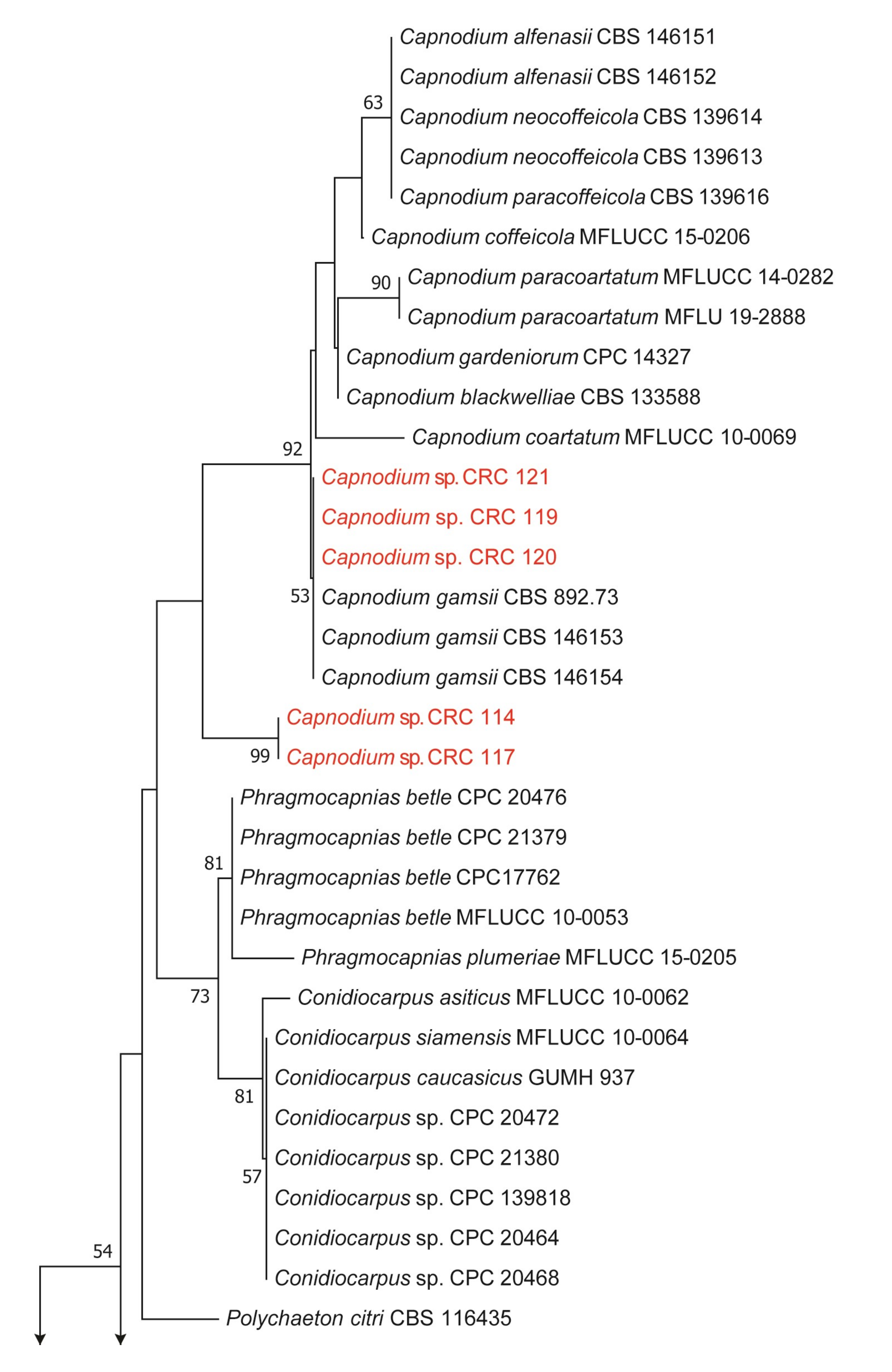
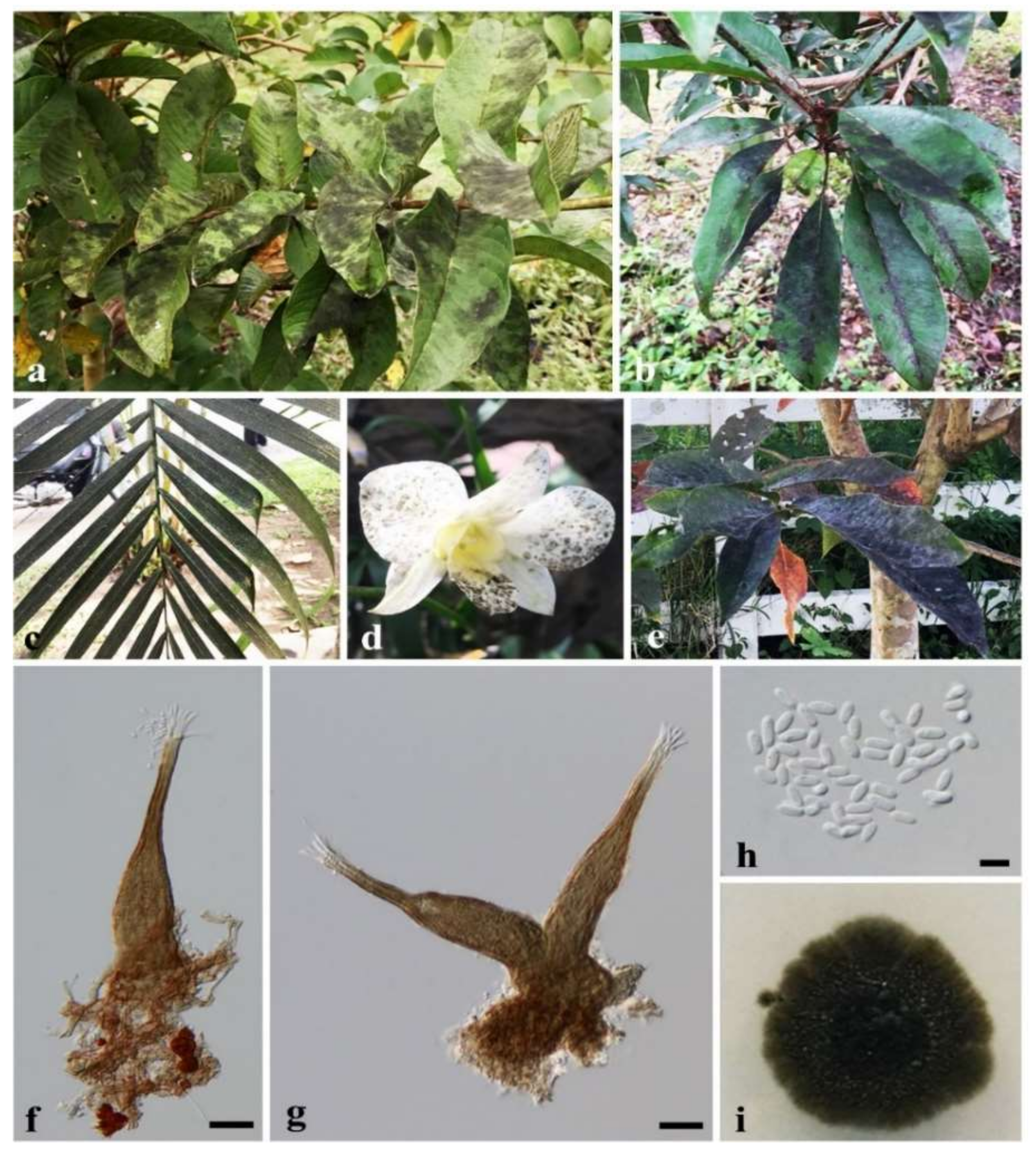
3.1. Characteristics and Phylogenetic Analyses

Eleven sooty mold strains from different plant hosts were isolated. The neighbor-joining phylogeny of each isolate is shown in Figure 1 and Figure 2. Based on the GenBank BLAST results, the phylogenetic analysis was performed separately for the two families Capnodiaceae (Figure 1) and Trichomeriaceae (Figure 2). The newly generated sequences of 10 isolates of sooty mold were grouped within Capnodiaceae. However, LSU and ITS phylogeny could not identify our isolates to the species level. Hence, those taxa were identified only to the generic level: Capnodium (Figure 3) and Leptoxyphium (Figure 4). The Phylogenetic tree of the Trichomeriaceae (Figure 2), based on the combined LSU and ITS genes, showed that our isolate was clustered with T. deniqulatum (Figure 5) with 99% bootstrap support. Therefore, our strain (CRC 141) was identified as T. deniqulatum and introduced as a new host record on Shorea roxburghii herein.

3.2. Quantitative Analysis of the Metabolites

The fungal mycelia were extracted in MeOH, and the growth medium/culture filtrate was extracted in EtOAc. Yields of the EtOAc and MeOH fractions are shown in Table 2. The yields of the EtOAc extract were in the range of 3.90–16.63 mg, with CRC 98 producing the highest amount, while the yields of the MeOH extracts were in the range of 11.70 (CRC 120)–74.13 (CRC 117) mg. The two-way ANOVA analysis denotes that the data were significantly independent within the isolates and solvent types. Both solvent fractions from the identified isolates were tested for total polyphenol and flavonoid content as presented in Table 2. The total polyphenol content of the EtOAc fraction (0.50–2.45 mg gallic/g sample) were significantly higher than the MeOH fraction (0.14–0.44 mg gallic/g sample). However, the total flavonoid content was not significantly different between MeOH and EtOAc extracts. Based on the data, the highest total flavonoid was obtained from CRC 141 (2.03 mg catechin/g sample) in the EtOAc extract and CRC 117 (0.71 mg catechin/g sample) in the MeOH extract. The total protein content in the EtOAc extracts were apparently lower (0.08–0.32 mg/mL) than those of the MeOH ones (1.18–6.22 mg/mL). The isolate CRC 121 provided the highest value of total protein in both extracts.

3.3. Biological Activities

3.3.1. Antioxidant Assay

The antioxidant activities of the extracts according to the DPPH and ABTS assays are also shown in Table 2. The DPPH activity of the EtOAc and MeOH extracts were in the range of 10.49–80.44 mg/mL and 36.86–77.77 mg/mL, respectively. The data were independent of the isolate as well as the solvent used. The ABTS assay of EtOAc and MeOH extracts were in the range 53.07–99.11 mg/mL and 0.58–51.81 mg/mL, respectively. Based on the results, the EtOAc fraction of CRC 119 had higher DPPH activity while the MeOH extract had lower ABTS activity. In contrast, the isolate CRC 141 showed higher ABTS activity for the MeOH fraction while the EtOAc fraction had the lowest DPPH score.

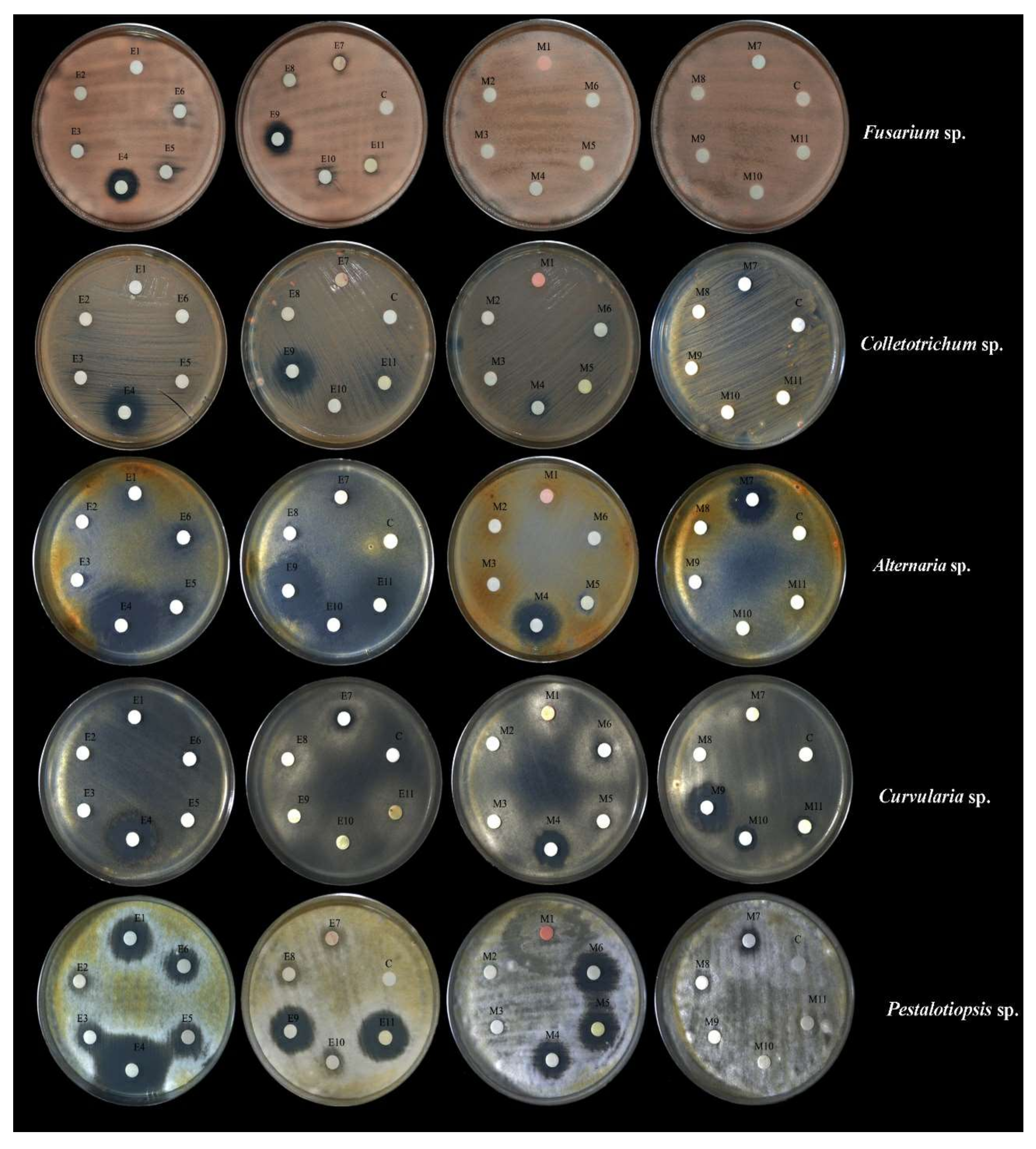
3.3.2. Antifungal Activities

The formation of inhibition zones by each extract after 3 days is shown in Table 3. The result indicates that the EtOAc extracts, especially from the CRC 108 and CRC 120, had higher inhibitory activity than MeOH ones against various plant pathogens (Figure 6). EtOAc extract of CRC 108 showed significant anti-fungal activity against Pestatotiopsis sp., Alternaria sp., Fusarium sp. and Colletotrichum sp. with mean diameter of inhibitory zones 28.93 ± 0.12, 26.13 ± 0.03, 16.5 ± 0.05 and 13.60 ± 0.06, respectively (Figure 6, Table 3). While EtOAc extract of CRC 120 illustrated significant antifungal activity against all plant pathogens except Curvularia sp., the strongest inhibition activity was against Alternaria sp. by CRC 120 with mean diameter of inhibition zone of 26.93 ± 0.05 (Figure 6, Table 3). On the other hand, the MeOH extract of CRC 102, CRC 121, and CRC 108 showed relatively high anti-fungal activity against Pestalotiopsis sp., Curvularia sp. and Alternaria sp. with inhibitory zones of 24.73 ± 0.33, 20.60 ± 0.57 and 19.50 ± 0.10, respectively (Table 3).

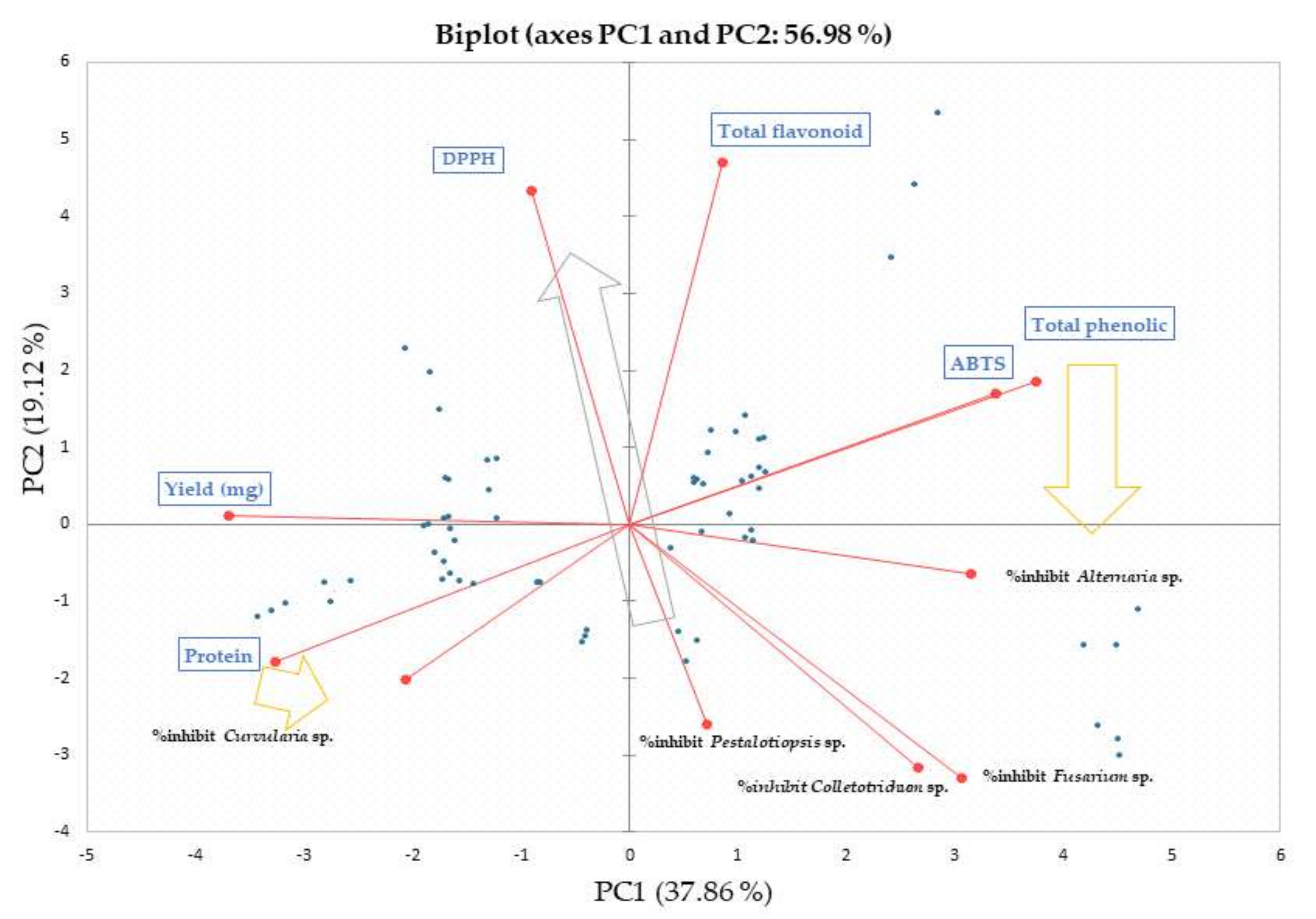
3.4. Chemometric Relations

The principle component analysis (PCA) between MeOH and EtOAc extracts together with their corresponding biological activities are shown in Figure 7 and Figure 8. The variables accounted for a total of 56.98% across the score plot (PC1: 37.86% and PC2: 19.12%). It was clear that the component in the extracts were highly varied upon the solvent. The EtOAc extracts of CRC 141 (E11), CRC 120 (E9) and CRC 108 (E4) were separated from the others. CRC 141 is Trichomerium deniqulatum, while CRC120 and CRC 108 are Capnodium spp. The MeOH extracts, on the other hand, were clustered closer together (Figure 7). A PCA-biplot was performed to elucidate the relationship among metabolites and biological activities (Figure 8). Based on the plot, ABTS antioxidant activity was related to the total phenolic compounds, while the DPPH scavenging activity was far more related to total flavonoid content, and EtOAc extracts contributed vastly to these trends.
The EtOAc fraction contributed to the inhibition of the fungal pathogens. It can be seen that total phenolic content and ABTS activity contributed to the inhibition of Alternaria sp. The total flavonoid content and DPPH activity, however, had reversely induced the antifungal activities on the Fusarium sp., Colletotrichum sp. and Pestalotiopsis sp. On the other hand, the protein content in MeOH fractions contributed to the inhibition of Curvularia sp.

4. Discussion

Capnodium, Leptoxyphium and Trichomerium are sooty molds found in tropical and subtropical climates on a variety of plant hosts [2]. Plant-stunting disease, low yield, and marketability issues have all been linked to Capnodium and Leptoxyphium species [2]. Sooty molds were previously known as host specific fungi. However, currently, they were found on a wide range of host families such as Apocynaceae, Lythraceae, Malvaceae, Myrtaceae, and Rutaceae [3,4]. Furthermore, Trichomerium were found as extremotolerant fungi on rock surfaces [32]. Currently, seven Trichomerium species have been reported in Thailand: T. camporesii on the leaf surface of guava (Psidium guajava) [33], T. chiangmaiensis from coffee leaves [24], T. gloeosporum on fig (Ficus sp.) leaves [6] and gardenia (Gardenia sp.) [34], T. bambusae on culm of bamboo [35], T. siamensis on yellow elder (Tecoma sp.) leaves [36] and T. foliicola on orange jasmine (Murraya paniculate) leaves, mango (Mangifera indica), guava (Psidium guajava) and date palm (Phoenix dactylifera) [6]. In our study we provide a novel host record and asexual morph for the T. deniqulatum on white meranti (Shorea roxburghii) in Thailand. Trichomerium was firstly introduced with its sexual morph [6]. Moreover, in this study, we provide new host record for the Capnodium: Areca palm (Dypsis lutescens), orchid (Dendrobium hybrid) and queen’s crepe myrtle (Lagerstroemia speciose). The genus Leptoxyphium on durian, bird of paradise (Strelitzia reginae), globe amaranth (Gomphrena globose) and queen’s crepe myrtle (L. speciose). Consequently, it can be assumed that the genus Capnodium, Leptoxyphium and Trichomerium are not host-specific.
The total yield of extracted metabolites depends upon the polarity of the solvents used [37]. It is more likely that these sooty mold strains produce high-polarity secondary metabolites in the cellular structure of the mycelium. Selim et al. [38] reported a higher yield of MeOH over EtOAc from endophytic fungi.
In our study, the total polyphenol and total flavonoid content was higher in the EtOAc fraction than in the MeOH fraction. Pinta et al. [39] claimed that EtOAc has a higher selectivity for less polar polyphenols and produces extracts with higher purity than MeOH. Therefore, it is possible that sooty molds produce polyphenols which are slightly polar. Gonçalves et al. [40] reported higher total polyphenols on EtOAc than in our study. The total flavonoid content from the EtOAc extract of Penicillium griseoroseum, an endophytic microorganism from coffee beans was also higher [41]. In addition, the total polyphenol and flavonoid contents of MeOH extracts of tropical black bolete mushroom were reported higher than that in this study [28]. During mycelial growth, proteins are secreted from the tips of growing hyphae in the form of enzymes, which are released to the mycelial wall and media [42]. The extraction with methanol is directly carried out from the mycelium. Therefore, the amount of protein found in mycelia is greater than in the filtrate. In our antioxidant potential determination study, DPPH and ABTS activities of the EtOAc extracts were higher than the DPPH and ABTS activities of pigment producing fungi [40]. Furthermore, both DPPH and ABTS activity assays of MeOH extract of tropical black bolete mushroom were lower than the current study [28].
Sangta et al. [29] reported a crude methanolic fraction of coffee pulp powder against the same group of pathogens. Among other things, fungal metabolites are currently used for managing fungal diseases [43]. Plant pathogens, Alternaria sp., Colletotrichum sp., Curvularia sp., Fusarium sp. and Pestalotiopsis sp. are commonly foliar pathogens causing leaf spot, leaf blight on various hosts worldwide, thereby decreasing their yield and deteriorating crop quality [44]. In addition, Curvularia sp. and Fusarium sp. are seed-borne fungi affecting seed germination and reducing the grain yield and quality through seed deterioration [45].
Chemometric multivariate analysis evaluated the correlations among the data set including strains, metabolite substances and biological activities [29,46,47]. Phenolic compounds and phenol-derived structures can be biosynthesized in microorganisms via the shikimate pathway; these chemicals have a variety of bioactivities, including antioxidant, cytotoxic and antibacterial properties [48]. The polyketide and isoprenoid synthesis routes, for example, can create additional metabolites such as terpenoids, alkaloids, phenylpropanoids, aliphatic chemicals, polyketides and peptides [49,50]. From the chemometric results, we noticed the correlation between the content of flavonoids and DPPH scavenging activity. This relationship was also observed previously [50].
The results also showed that total phenolic content and ABTS activity had contributed to the inhibitions fungal pathogen. Caffeic acid activates lipolytic enzymes, which break down cell membranes and reduce fungal sporulation and germination, according to a prior investigation [29,51]. In another study, flavonoids induced relative growth of the F. oxysporum f. sp. lycopersici due to its specific stimulating activity [52]. Protein was dominant in the methanol extracts and contributed to the inhibition of Curvularia sp. The fungal metabolites could be synthesized in the form of non-ribosomal peptides such as antibiotics and polyketides such as mycotoxins and secreted enzymes [53,54]. The latter contribute largely to fungicidal activity which is in association with the inhibition activity of cell structure compartmentation [55]. The so-called cell-wall lytic enzymes such as chitinase, β 1,3-glucanase and protease were proven to be antifungal agents with industrial applications [56].

5. Conclusions

We were able to isolate and identify three sooty mold genera namely Capnodium, Leptoxyphium and Trichomerium. The extracts from these fungi comprised phenolic compounds, flavonoids and proteins having antioxidant and antifungal activity against plant pathogens: Alternaria sp., Colletotrichum sp., Curvularia sp., Fusarium sp., and Pestalotiopsis sp. Based on the results, the extracts of mid-polar polyphenols have higher solubility towards EtOAc. However, other studies are needed to confirm the compounds with desired biological activity. Our study reveals the biological activities of secondary metabolites of the least studied sooty molds. These findings can be useful towards the development of fungicides from sustainable biological sources and are a step forward into resilience in agricultural production.

Author Contributions

Conceptualization, S.H. (Sukanya Haituk), R.C. and S.R.S.; methodology, S.H. (Sukanya Haituk), S.H. (Surat Hongsibsong), P.W., J.S. and P.K.; writing original draft preparation, S.H. (Sukanya Haituk), R.C., S.R.S., S.K.P. and C.S.; writing review and editing, S.H. (Sukanya Haituk), R.C., A.K., P.W., S.R.S. and S.K.P.; visualization, S.H. (Sukanya Haituk); supervision, R.C. and S.R.S.; funding acquisition, R.C. and K.S. All authors have read and agreed to the published version of the manuscript.

Funding

This research received no external funding.

Acknowledgments

This research project is supported by TSRI. We would like to acknowledge TA/RA Scholarship offered to S.H. from the Graduate School, Chiang Mai University. S.K.P. thanks to JSS Academy of Higher Education and Research (JSSAHER), Mysuru, India, for the provision of supportive infrastructure.

Conflicts of Interest

The authors declare no conflict of interest.

References

  1. Callan, B.; Carris, L.M. Fungi on living plant substrata, including fruits. In Biodiversity of Fungi; Mueller, G.M., Bills, G.F., Foster, M.S., Eds.; Academic Press: Burlington, CA, USA, 2004; pp. 105–126. [Google Scholar]
  2. Chomnunti, P.; Hongsanan, S.; Aguirre-Hudson, B.; Tian, Q.; Peršoh, D.; Dhami, M.K.; Alias, A.S.; Xu, J.; Liu, X.; Stadler, M.; et al. The sooty moulds. Fungal Divers. 2014, 66, 1–36. [Google Scholar] [CrossRef]
  3. Abdollahzadeh, J.; Groenewald, J.Z.; Coetzee, M.P.A.; Wingfield, M.J.; Crous, P.W. Evolution of lifestyles in Capnodiales. Stud Mycol. 2020, 95, 381–414. [Google Scholar] [CrossRef]
  4. Chomnunti, P.; Schoch, C.L.; Aguirre-Hudson, B.; Ko-Ko, T.W.; Hongsanan, S.; Jones, E.B.G.; Kodsueb, R.; Phookamsak, R.; Chukeatirote, E.; Bahkali, A.H.; et al. Capnodiaceae. Fungal Divers. 2011, 51, 103–134. [Google Scholar] [CrossRef]
  5. Flessa, F.; Harjes, J.; Cáceres, M.E.S.; Rambold, G. Comparative analyses of sooty mould communities from Brazil and Central Europe. Mycol. Prog. 2021, 20, 869–887. [Google Scholar] [CrossRef]
  6. Chomnunti, P.; Bhat, D.J.; Jones, E.B.G.; Chukeatirote, E.; Bahkali, A.H.; Hyde, K.D. Trichomeriaceae, a new sooty mould family of Chaetothyriales. Fungal Divers. 2012, 56, 63–76. [Google Scholar] [CrossRef]
  7. Reynolds, D.R. Capnodiaceous sooty mold phylogeny. Can. J. Bot. 1998, 76, 2125–2130. [Google Scholar] [CrossRef]
  8. Winka, K.; Eriksson, O.E.; Bång, Å. Molecular evidence for recognizing the Chaetothyriales. Mycologia 2018, 90, 822–830. [Google Scholar] [CrossRef]
  9. Hongsanan, S.; Tian, Q.; Hyde, K.; Chomnunti, P. Two new species of sooty moulds, Capnodium coffeicola and Conidiocarpus plumeriae in Capnodiaceae. Mycosp 2015, 6, 814–824. [Google Scholar] [CrossRef]
  10. Wisetkomolmat, J.; Inta, A.; Krongchai, C.; Kittiwachana, S.; Jantanasakulwong, K.; Rachtanapun, P.; Sommano, S.R. Ethnochemometric of plants traditionally utilised as local detergents in the forest dependent culture. Saudi J. Biol. Sci. 2021, 28, 2858–2866. [Google Scholar] [CrossRef] [PubMed]
  11. Castaño, C.; Alday, J.; Lindahl, B.; de Aragón, M.J.; Miguel, S.D.; Colinas, C.; Parladé, J.; Pera, J.; Bonet, J. Lack of thinning effects over inter-annual changes in soil fungal community and diversity in a Mediterranean pine forest. For. Ecol. Manag. 2018, 424, 420–427. [Google Scholar] [CrossRef]
  12. McGuire, K.L.; Bent, E.; Borneman, J.; Majumder, A.; Allison, S.D.; Tresederi, K.K. Functional diversity in resource use by fungi. Ecology 2010, 91, 2324–2332. [Google Scholar] [CrossRef]
  13. Mukherjee, P.K.; Horwitz, B.A.; Kenerley, C.M. Secondary metabolism in Trichoderma--a genomic perspective. Microbiology 2012, 158, 35–45. [Google Scholar] [CrossRef] [PubMed] [Green Version]
  14. Pusztahelyi, T.; Holb, I.; Pócsi, I. Secondary metabolites in fungus-plant interactions. Front. Plant Sci. 2015, 6, 573. [Google Scholar] [CrossRef] [Green Version]
  15. Renshaw, J.C.; Robson, G.D.; Trinci, A.P.J.; Wiebe, M.G.; Livens, F.R.; Collison, D.; Taylor, R.J. Fungal siderophores: Structures, functions and applications. Mycol. Res. 2002, 106, 1123–1142. [Google Scholar] [CrossRef]
  16. Caicedo, N.H.; Davalos, A.F.; Puente, P.A.; Rodríguez, A.Y.; Caicedo, P.A. Antioxidant activity of exo-metabolites produced by Fusarium oxysporum: An endophytic fungus isolated from leaves of Otoba gracilipes. Microbiologyopen 2019, 8, 903. [Google Scholar] [CrossRef] [PubMed] [Green Version]
  17. Kumar, S.; Kaushik, N.; Kumar, S.; Kaushik, N. Metabolites of endophytic fungi as novel source of biofungicide: A review. Phytochem Rev. 2013, 11, 507–522. [Google Scholar] [CrossRef]
  18. Ribeiro, B.; Valentão, P.; Baptista, P.; Seabra, R.M.; Andrade, P.B. Phenolic compounds, organic acids profiles and antioxidative properties of beefsteak fungus (Fistulina hepatica). Food Chem. Toxicol. 2007, 45, 1805–1813. [Google Scholar] [CrossRef] [Green Version]
  19. Wang, P.; Yu, J.-H.; Zhu, K.; Wang, Y.; Cheng, Z.-Q.; Jiang, C.-S.; Dai, J.-G.; Wu, J.; Zhang, H. Phenolic bisabolane sesquiterpenoids from a Thai mangrove endophytic fungus, Aspergillus sp. xy02. Fitoterapia 2018, 127, 322–327. [Google Scholar] [CrossRef] [PubMed]
  20. Senwanna, C.; Wanasinghe, D.; Bulgakov, T.; Wang, Y.; Bhat, D.J.; Tang, A.; Mortimer, P.; Xu, J.; Hyde, K.D.; Phookamsak, R. Towards a natural classification of Dothidotthia and Thyrostroma in Dothidotthiaceae (Pleosporineae, Pleosporales). Mycosphere 2019, 10, 701–738. [Google Scholar] [CrossRef]
  21. Vilgalys, R.; Hester, M. Rapid genetic identification and mapping of enzymatically amplified ribosomal DNA from several Cryptococcus species. J. Bacteriol. 1990, 172, 4238–4246. [Google Scholar] [CrossRef] [Green Version]
  22. White, T.J.; Bruns, T.; Lee, S.; Taylor, J. Amplification and direct sequencing of fungal ribosomal RNA genes for phylogenetics. In PCR Protocols: A Guide to Methods and Amplifications; Innis, M.A., Gelfand, D.H., Sninsky, J.J., White, T.J., Eds.; Academic Press: San Diego, CA, USA, 1990; pp. 315–322. [Google Scholar]
  23. Haituk, S.; Suwannarach, N.; Hongsanan, S.; Senwanna, C.; Cheewangkoon, R. New epiphytic sooty molds: Alloscorias syngonii (Readerielliopsidaceae) from Thailand. Phytotaxa 2021, 507, 271–282. [Google Scholar] [CrossRef]
  24. Maharachchikumbura, S.S.N.; Haituk, S.; Pakdeeniti, P.; Al-Sadi, A.M.; Hongsanan, S.; Chomnunti, P.; Cheewangkoon, R. Phaeosaccardinula coffeicola and Trichomerium chiangmaiensis, two new species of Chaetothyriales (Eurotiomycetes) from Thailand. Mycosphere 2018, 9, 769–778. [Google Scholar] [CrossRef]
  25. Katoh, K.; Rozewicki, J.; Yamada, K.D. MAFFT online service: Multiple sequence alignment, interactive sequence choice and visualization. Brief Bioinform. 2019, 20, 1160–1166. [Google Scholar] [CrossRef] [PubMed] [Green Version]
  26. Kumar, S.; Stecher, G.; Tamura, K. MEGA7: Molecular Evolutionary Genetics Analysis Version 7.0 for Bigger Datasets. Mol. Biol Evol. 2016, 33, 1870–1874. [Google Scholar] [CrossRef] [Green Version]
  27. Saitou, N.; Nei, M. The neighbor-joining method: A new method for reconstructing phylogenetic trees. Mol. Biol. Evol. 1987, 4, 406–425. [Google Scholar] [CrossRef]
  28. Kumla, J.; Suwannarach, N.; Tanruean, K.; Lumyong, S. Comparative Evaluation of Chemical Composition, Phenolic Compounds, and Antioxidant and Antimicrobial Activities of Tropical Black Bolete Mushroom Using Different Preservation Methods. Foods 2021, 10, 781. [Google Scholar] [CrossRef] [PubMed]
  29. Sangta, J.; Wongkaew, M.; Tangpao, T.; Withee, P.; Haituk, S.; Arjin, C.; Sringarm, K.; Hongsibsong, S.; Sutan, K.; Pusadee, T.; et al. Recovery of Polyphenolic Fraction from Arabica Coffee Pulp and Its Antifungal Applications. Plants 2021, 10, 1422. [Google Scholar] [CrossRef]
  30. Adedapo, A.A.; Jimoh, F.O.; Koduru, S.; Afolayan, A.J.; Masika, P.J. Antibacterial and antioxidant properties of the methanol extracts of the leaves and stems of Calpurnia aurea. BMC Complement. Altern. Med. 2008, 8, 53. [Google Scholar] [CrossRef] [Green Version]
  31. Bauer, A.W.; Kirby, W.M.; Sherris, J.C.; Turck, M. Antibiotic susceptibility testing by a standardized single disk method. Am. J. Clin. Pathol. 1966, 45, 493–496. [Google Scholar] [CrossRef]
  32. Isola, D.; Zucconi, L.; Onofri, S.; Caneva, G.; de Hoog, G.S.; Selbmann, L. Extremotolerant rock inhabiting black fungi from Italian monumental sites. Fungal Divers. 2015, 76, 75–96. [Google Scholar] [CrossRef]
  33. Hyde, K.; Silva, N.; Abeywickrama, P.; Phookamsak, R.; Jeewon, R.; Jayawardena, R.; Boonmee, S.; Manawasinghe, I.; Tennakoon, D.; Wanasinghe, D.; et al. AJOM new records and collections of fungi: 1–100. Asian J. Mycol. 2020, 3, 22–294. [Google Scholar] [CrossRef]
  34. Hongsanan, S.; Tian, Q.; Hyde, K.D.; Hu, D.M. The asexual morph of Trichomerium gloeosporum. Mycosphere 2016, 7, 1473–1479. [Google Scholar] [CrossRef]
  35. Hyde, K.D.; Hongsanan, S.; Jeewon, R.; Bhat, D.J.; McKenzie, E.H.C.; Jones, E.B.G.; Phookamsak, R.; Ariyawansa, H.A.; Boonmee, S.; Zhao, Q.; et al. Fungal diversity notes 367–490: Taxonomic and phylogenetic contributions to fungal taxa. Fungal Divers. 2016, 80, 1–270. [Google Scholar] [CrossRef]
  36. Liu, J.K.; Hyde, K.D.; Jones, E.B.G.; Ariyawansa, H.A.; Bhat, D.J.; Boonmee, S.; Maharachchikumbura, S.S.N.; McKenzie, E.H.C.; Phookamsak, R.; Phukhamsakda, C.; et al. Fungal diversity notes 1–110: Taxonomic and phylogenetic contributions to fungal species. Fungal Divers. 2015, 72, 1–197. [Google Scholar] [CrossRef]
  37. Spigno, G.; Tramelli, L.; De Faveri, D.M. Effects of extraction time, temperature and solvent on concentration and antioxidant activity of grape marc phenolics. J. Food Eng. 2007, 81, 200–208. [Google Scholar] [CrossRef]
  38. Selim, K.; Elkhateeb, W.; Tawila, A.; El-Beih, A.; Abdel-Rahman, T.; El Diwany, A.; Ahmed, E. Antiviral and Antioxidant Potential of Fungal Endophytes of Egyptian Medicinal Plants. Fermentation 2018, 4, 49. [Google Scholar] [CrossRef] [Green Version]
  39. Pintać, D.; Majkić, T.; Torović, L.; Orčić, D.; Beara, I.; Simin, N.; Mimica–Dukić, N.; Lesjak, M. Solvent selection for efficient extraction of bioactive compounds from grape pomace. Ind. Crops Prod. 2018, 111, 379–390. [Google Scholar] [CrossRef]
  40. Gonçalves, T.D.; Viana, L.B.B.; Lopes, F.R.; Ferreira Duarte, W.; Gomes Cardoso, P. Antioxidant activity and phenolic compounds of the extract from pigment-producing fungi isolated from Brazilian caves. Biocatal. Agric. 2018, 16, 148–154. [Google Scholar] [CrossRef]
  41. Silva, J.; Fill, T.; Ferreira-Silva, B.; Rodrigues-Fo, E. Diclavatol and tetronic acids from Penicillium griseoroseum. Nat. Prod. Res. 2012, 27, 9–16. [Google Scholar] [CrossRef]
  42. Wösten, H.A.; Moukha, S.M.; Sietsma, J.H.; Wessels, J.G. Localization of growth and secretion of proteins in Aspergillus niger. J. Gen. Microbiol. 1991, 137, 2017–2023. [Google Scholar] [CrossRef] [Green Version]
  43. Brauer, V.S.; Rezende, C.P.; Pessoni, A.M.; De Paula, R.G.; Rangappa, K.S.; Nayaka, S.C.; Gupta, V.K.; Almeida, F. Antifungal Agents in Agriculture: Friends and Foes of Public Health. Biomolecules 2019, 9, 521. [Google Scholar] [CrossRef] [Green Version]
  44. Verma, S.K.; Kingsley, K.L.; Bergen, M.S.; Kowalski, K.P.; White, J.F. Fungal Disease Prevention in Seedlings of Rice (Oryza sativa) and Other Grasses by Growth-Promoting Seed-Associated Endophytic Bacteria from Invasive Phragmites australis. Microorganisms 2018, 6, 21. [Google Scholar] [CrossRef] [Green Version]
  45. Prom, L. The Effects of Fusarium thapsinum, Curvularia lunata, and Their Combination on Sorghum Germination and Seed Mycoflora. J. New Seeds 2004, 6, 39–49. [Google Scholar] [CrossRef]
  46. Suksathan, R.; Rachkeeree, A.; Puangpradab, R.; Kantadoung, K.; Sommano, S.R. Phytochemical and nutritional compositions and antioxidants properties of wild edible flowers as sources of new tea formulations. NFS J. 2021, 24, 15–25. [Google Scholar] [CrossRef]
  47. Wongkaew, M.; Tinpovong, B.; Sringarm, K.; Leksawasdi, N.; Jantanasakulwong, K.; Rachtanapun, P.; Hanmoungjai, P.; Sommano, S.R. Crude Pectic Oligosaccharide Recovery from Thai Chok Anan Mango Peel Using Pectinolytic Enzyme Hydrolysis. Foods 2021, 10, 627. [Google Scholar] [CrossRef] [PubMed]
  48. Negreiros, C.P.L.; Silva, E.O.; Chagas-Paula, D.A.; Hortolan, L.J.H.; Ikegaki, M. Importance and Implications of the Production of Phenolic Secondary Metabolites by Endophytic Fungi: A Mini-Review. Mini Rev. Med. Chem. 2016, 16, 259–271. [Google Scholar] [CrossRef]
  49. Mousa, W.K.; Raizada, M.N. The diversity of anti-microbial secondary metabolites produced by fungal endophytes: An interdisciplinary perspective. Front. Microbiol. 2013, 4, 65. [Google Scholar] [CrossRef] [PubMed] [Green Version]
  50. Palanichamy, P.; Krishnamoorthy, G.; Kannan, S.; Marudhamuthu, M. Bioactive potential of secondary metabolites derived from medicinal plant endophytes. Egypt. J. Basic Appl. Sci. 2018, 5, 303–312. [Google Scholar] [CrossRef] [Green Version]
  51. Shull, T.E.; Kurepa, J.; Miller, R.D.; Martinez-Ochoa, N.; Smalle, J.A. Inhibition of Fusarium oxysporum f. sp. nicotianae Growth by Phenylpropanoid Pathway Intermediates. Plant Pathol. J. 2020, 36, 637–642. [Google Scholar] [CrossRef]
  52. Steinkellner, S.; Mammerler, R. Effect of flavonoids on the development of Fusarium oxysporum f. sp. lycopersici. J. Plant Interact. 2007, 2, 17–23. [Google Scholar] [CrossRef]
  53. Baker, S.E. Grand Challenges in Fungal Biotechnology. Front. Fungal Biol. 2020, 1, 626551. [Google Scholar] [CrossRef]
  54. Bayram, Ö.; Braus, G.H. Coordination of secondary metabolism and development in fungi: The velvet family of regulatory proteins. FEMS Microbiol. Rev. 2012, 36, 1–24. [Google Scholar] [CrossRef] [PubMed] [Green Version]
  55. Ganendren, R.; Widmer, F.; Singhal, V.; Wilson, C.; Sorrell, T.; Wright, L. In vitro antifungal activities of inhibitors of phospholipases from the fungal pathogen Cryptococcus neoformans. Antimicrob. Agents Chemother. 2004, 48, 1561–1569. [Google Scholar] [CrossRef] [PubMed] [Green Version]
  56. Choudhary, B.; Nagpure, A.; Gupta, R.K. Fungal cell-wall lytic enzymes, antifungal metabolite(s) production, and characterization from Streptomyces exfoliatus MT9 for controlling fruit-rotting fungi. J. Basic Microbiol. 2014, 54, 1295–1309. [Google Scholar] [CrossRef] [PubMed]
Figure 1. Neighbor-joining phylogenetic tree of Capnodiaceae generated based on the combined LSU and ITS. Bootstrap support values for neighbor-joining phylogeny equal to or higher than 50% are displayed on the nodes. The newly generated strains in this study are indicated in red. The tree is rooted to Fumagospora salicinum (CBS 131.34).
Processes 10 00329 g001aProcesses 10 00329 g001b
Figure 2. Neighbor-joining phylogenetic tree of Trichomeriaceae generated based on the combined LSU and ITS. Bootstrap support values for neighbor-joining phylogeny equal to or higher than 75% are displayed on the nodes. The newly generated strain in this study is indicated in red. The tree is rooted to Bradymyces pullus (CGMCC 3.17305) and B. yunnanensis (CGMCC 3.17350).
Processes 10 00329 g002
Figure 3. Capnodium spp. (ae) host plants: (a) Psidium guajava (CRC 114), (b) Manilkara zapota (CRC 117), (c) Dypsis lutescens (CRC 119), (d) Dendrobium hybrid (CRC 120), (e) Lagerstroemia speciose (CRC 121); (f,g) pycnidia, (h) conidia, (i) colony on PDA. Scale bars: (f,g) = 50 μm; (h) = 10 μm.
Processes 10 00329 g003
Figure 4. Leptoxyphium spp. (ae) host plants: (a) Durio ziberhinus (CRC 98), (b) Annona squamosal (CRC 101), (c) Strelitzia reginae (CRC 102), (d) Gomphrena globose (CRC 108), (e) Lagerstroemia speciose (CRC 110); (fi) conidiomata, (j) conidia, (k) colony on PDA. Scale bars: (fi) = 50 μm; (j) = 10 μm.
Processes 10 00329 g004
Figure 5. Trichomerium deniqulatum (a) sooty mold on Shorea roxburghii, (b) colony with conidia on PDA, (c) conidia and conidiophore, (di) immature conidia at various stages of development attached to the prostrate mycelium, (j) conidia. Scale bars: (di) = 10 μm; (c,j) = 20 μm.
Processes 10 00329 g005
Figure 6. Inhibitory effect (inhibition zone, mm) after 3 days of fungal extract at a concentration of 30 mg/mL. Five plant pathogenic strains were tested: Fusarium sp. (CRC 161), Colletotrichum sp. (CRC 149), Alternaria sp. (CRC 152), Curvularia sp. (CRC 162), and Pestalotiopsis sp. (CRC 151). Abbreviations; E = filtrate (EtOAc) extract, M = mycelium (MeOH) extract, C = Negative control (MeOH), E1, M1 = CRC 98; E2, M2 = CRC 101; E3, M3 = CRC 102; E4, M4 = CRC 108; E5, M5 = CRC 110; E6, M6 = CRC 114; E7, M7 = CRC 117; E8, M8 = CRC 119; E9, M9 = CRC 120; E10, M10 = CRC 121; E11, M11 = CRC 141.
Processes 10 00329 g006
Figure 7. Principal component relationships of fungal filtrate and mycelium extract from eleven sooty molds isolates, with M and E indicating methanol and ethyl acetate. Abbreviations; E = filtrate (EtOAc) extract, M = mycelium (MeOH) extract, E1, M1 = CRC 98; E2, M2 = CRC 101; E3, M3 = CRC 102; E4, M4 = CRC 108; E5, M5 = CRC 110; E6, M6 = CRC 114; E7, M7 = CRC 117; E8, M8 = CRC 119; E9, M9 = CRC 120; E10, M10 = CRC 121; E11, M11 = CRC 141.
Processes 10 00329 g007
Figure 8. Chemometric score plot of the antifungal activities described as a percentage of inhibition. Alternaria sp. (CRC 152), Colletotrichum sp. (CRC 149), Curvularia sp. (CRC 162), Fusarium sp. (CRC 161), and Pestalotiopsis sp. (CRC 151).
Processes 10 00329 g008
Table 1. Morphology, host information, locality and accession number of sooty molds generated in this study.
StrainHostCollected LocationGenBank Accession NumberMorphological Description
LSUITS
Leptoxyphium sp. CRC 98Durian
(Durio ziberhinus)
Chanthaburi, ThailandMZ725032MZ725022Conidiomata synnematous, simple, 27–43 × 28–45 μm. Conidia ellipsoidal, hyaline, aseptate and guttulate, 4.1–6.7 × 2.1–2.7 μm
Leptoxyphium sp. CRC 101Sugar Apple
(Annona squamosal)
Chiang Rai, ThailandMZ725033MZ725023Conidiomata synnematous, simple, 27.5–43 × 28.5–46 μm. Conidia ellipsoidal, hyaline, aseptate and guttulate, 4.1–6.8 × 2–2.6 μm
Leptoxyphium sp. CRC 102Bird of paradise (Strelitzia reginae)Chiang Mai, ThailandMZ725034MZ725024Conidiomata synnematous, simple, 27–43 × 28–45 μm. Conidia ellipsoidal, hyaline, aseptate and guttulate, 4–6.7 × 2.2–2.8 μm
Leptoxyphium sp. CRC 114Globe amaranth (Gomphrena globosa)Chiang Mai, ThailandMZ725035MZ725025Conidiomata synnematous, simple, 28–44 × 28.5–46 μm. Conidia ellipsoidal, hyaline, aseptate and guttulate, 4.1–6.7 × 2.1–2.7 μm
Leptoxyphium sp. CRC 110Queen’s crepe-myrtle
(Lagerstroemia speciosa)
Chiang Mai, ThailandMZ725036MZ725026Conidiomata synnematous, simple, 27–43 × 28–45 μm. Conidia ellipsoidal, hyaline, aseptate and guttulate, 4.1–6.7 × 2.1–2.7 μm
Capnodium sp. CRC 108Guava
(Psidium guajava)
Songkhla, ThailandMZ725037-Conidiomata synnematous, simple or branched, 25–45 × 16.5–30 μm. Conidia oblong to ellipsoid, hyaline, aseptate, 4.5–6.5 × 1.8–2.7 μm
Capnodium sp. CRC 117Sapodilla
(Manilkara zapota)
Songkhla, ThailandMZ725038-Conidiomata synnematous, simple or branched, 24–43 × 17–29 μm. Conidia oblong to ellipsoid, hyaline, aseptate, 4.5–7 × 2–2.9 μm
Capnodium sp. CRC 119Areca palm
(Dypsis lutescens)
Chiang Mai, ThailandMZ725039MZ725027Conidiomata synnematous, simple or branched, 23–44 × 16–29 μm. Conidia oblong to ellipsoid, hyaline, aseptate, 4.5–6.5 × 1.7–2.5 μm
Capnodium sp. CRC 120Orchid
(Dendrobium hybrid)
Chiang Mai, ThailandMZ725040MZ725028Conidiomata synnematous, simple or branched, 23–44 × 16–29 μm. Conidia oblong to ellipsoid, hyaline, aseptate, 4–5.5 × 1.9–2.4 μm
Capnodium sp. CRC 121Queen’s crepe-myrtle (Lagerstroemia speciosa)Chiang Mai, ThailandMZ725041MZ725029Conidiomata synnematous, simple or branched, 23–42 × 16–28 μm. Conidia oblong to ellipsoid, hyaline, aseptate, 4–6 × 1.8–2.5 μm
Trichomerium deniqulatum CRC 141White meranti (Shorea roxburghii)Chiang Mai, ThailandMZ373260MZ373260lateral arms from 1–2 globose to subglobose, Conidial arms 1–3, rarely 4–5 septate, hyaline to pale brown, 12–19 × 2.8–3.5 μm
Table 2. Yield, polyphenol compositions, antioxidant activity and protein of the fungal extracts from the filtrate (EtOAc) and mycelium (MeOH).
No.IsolateYield (mg)TPC
(mg GAE/g)
TFC
(mg CE/g)
DPPH Assay (%)ABTS Assay (%)Total Protein (mg/mL)
EtOAcMeOHEtOAcMeOHEtOAcMeOHEtOAcMeOHEtOAcMeOHEtOAcMeOH
1CRC 9816.63 ± 0.03 h34.30 ± 0.12 c0.92 ± 0.02 bc0.20 ± 0.004 a0.08 ± 0.009 a0.12 ± 0.002 abc55.31 ± 1.33 f69.26 ± 0.07 d98.33 ± 2.00 c5.45 ± 5.45 a0.13 ± 0.001 b3.13 ± 0.15 b
2CRC 1017.50 ± 0.00 e43.067 ± 0.03 d1.49 ± 0.09 d0.14 ± 0.01 a0.21 ± 0.04 ab0.08 ± 0.01 ab57.26 ± 4.80 f50.46 ± 7.05 bc98.21 ± 0.00 c34.43 ± 2.64 bc0.09 ± 0.006 a1.83 ± 0.08 a
3CRC 1025.83 ± 0.03 c52.13 ± 0.09 f1.40 ± 0.08 d0.18 ± 0.01 a0.10 ± 0.06 ab0.28 ± 0.04 c43.90 ± 2.39 e48.71 ± 1.93 abc97.42 ± 0.47 c51.81 ± 4.50 c0.14 ± 0.001 b6.22 ± 0.47 d
4CRC 1083.90 ± 0.06 a23.20 ± 0.06 b1.59 ± 0.05 de0.20 ± 0.008 an/a0.17 ± 0.03 abc29.41 ± 4.88 bc52.09 ± 0.86 c92.50 ± 2.20 cn/a0.08 ± 0.0002 a1.71 ± 0.13 a
5CRC 1106.30 ± 0.12 d62.13 ± 0.07 h1.03 ± 0.01 c0.19 ± 0.003 a0.10 ± 0.06 ab0.16 ± 0.08 abc39.14 ± 3.69 de43.16 ± 0.24 abc99.11 ± 0.52 c6.18 ± 3.88 a0.10 ± 0.002 a5.14 ± 0.24 c
6CRC 1148.13 ± 0.09 f58.73 ± 0.03 g0.73 ± 0.04 ab0.19 ± 0.001 a0.16 ± 0.02 ab0.23 ± 0.02 bc28.63 ± 0.51 bc52.11 ± 1.07 c92.22 ± 1.37 c18.64 ± 18.64 ab0.21 ± 0.006 c1.18 ± 0.008 a
7CRC 1174.03 ± 0.03 a74.13 ± 0.03 i1.36 ± 0.08 d0.44 ± 0.05 b0.001 ± 0.002 a0.71 ± 0.03 d10.49 ± 4.03 a77.77 ± 10.04 dn/an/a0.16 ± 0.008 c1.40 ± 0.03 a
8CRC 1199.13 ± 0.09 g44.13 ± 0.03 e0.68 ± 0.02 ab0.18 ± 0.002 a0.61 ± 0.17 b0.17 ± 0.01 abc36.08 ± 0.32 cde36.86 ± 2.74 a92.17 ± 0.68 cn/a0.14 ± 0.049 b2.99 ± 0.10 b
9CRC 1204.53 ± 0.03 b11.7 ± 0.12 a1.83 ± 0.22 e0.15 ± 0.004 a0.31 ± 0.10 ab0.05 ± 0.002 a34.72 ± 0.14 cd38.03 ± 1.41 ab92.05 ± 1.49 cn/a0.14 ± 0.006 b3.39 ± 0.11 b
10CRC 1217.53 ± 0.09 e62.13 ± 0.09 h0.50 ± 0.02 a0.17 ± 0.01 a0.14 ± 0.08 ab0.14 ± 0.001 abc20.89 ± 1.25 b38.19 ± 0.46 ab53.07 ± 16.76 b0.58 ± 0.58 a0.3 2± 0.001 e5.92 ± 0.95 d
11CRC 1414.53 ± 0.09 b43.20 ± 0.06 d2.45 ± 0.01 f0.14 ± 0.002 a2.03 ± 0.49 c0.22 ± 0.02 bc80.44 ± 1.13 g38.94 ± 1.39 ab87.95 ± 0.88 c10.63 ± 1.08 a0.28 ± 0.001 d5.94 ± 0.38 d
Isolate (I)******
solvent type (S)**ns***
I*S******
Data are expressed as mean ± standard error, n = 3; values followed by different letter(s) in the same column are significantly different (p < 0.05). n/a = Not available, * = Significantly different, ns = Not significant different.
Table 3. Diameter of inhibition zone of the fungal extracts from the filtrate (EtOAc) and mycelium (MeOH).
No.IsolateInhibition of Fusarium sp. (CRC 161)Inhibition of Colletotrichum sp. (CRC 149)Inhibition of Pestalotiopsis sp. (CRC 151)Inhibition of Alternaria sp.
(CRC 152)
Inhibition of Curvularia sp. (CRC 162)
EtOAcMeOHEtOAcMeOHEtOAcMeOHEtOAcMeOHEtOAcMeOH
1CRC 98----18.33 ± 0.09 g14.73 ± 0.33 d---7.20 ± 0.05 b
2CRC 101----11.03 ± 0.03 d-----
3CRC 102----14.20 ± 0.058 e24.73 ± 0.33 f----
4CRC 10816.5 ± 0.05 d-13.60 ± 0.06 b-28.93 ± 0.12 g16.33 ± 0.33 e26.13 ± 0.03 f19.50 ± 0.10 c-13.90 ± 0.11 d
5CRC 110-----9.20 ± 0.05 a9.90 ± 0.05 c--12.06 ± 0.88 c
6CRC 114----17.60 ± 0.10 f-9.20 ± 0.057 b---
7CRC 1176.73 ± 0.03 b---7.93 ± 0.08 b---9.73 ± 0.08 b-
8CRC 119----8.80 ± 0.17 c-----
9CRC 12014.63 ± 0.03 c-17.37 ± 0.07 c11.80 ± 0.58 b-11.93 ± 0.33 b26.93 ± 0.05 g---
10CRC 121------13.3 ± 0.06 d--20.60 ± 0.57 e
11CRC 141-----14.43 ± 0.33 c22.26 ± 0.06 e9.90 ± 0.10 b--
Isolate (I)*****
solvent type (S)*****
I*S*****
Data are expressed as mean ± standard error, n = 3; values followed by different letter(s) in the same column are significantly different (p < 0.05). (-) = No inhibition zone, * = Significantly different.
Publisher’s Note: MDPI stays neutral with regard to jurisdictional claims in published maps and institutional affiliations.

