
Morphology, Phylogeny, and Pathogenicity of Pestalotioid Species on Camellia oleifera in China

1
Key Laboratory for Non-Wood Forest Cultivation and Conservation of the Ministry of Education, Central South University of Forestry and Technology, Changsha 410004, China
2
Key Laboratory of National Forestry and Grassland Administration for Control of Diseases and Pests of South Plantation, Central South University of Forestry and Technology, Changsha 410004, China
3
Hunan Provincial Key Laboratory for Control of Forest Diseases and Pests, Central South University of Forestry and Technology, Changsha 410004, China
4
Hunan Provincial Key Laboratory for Forestry Biotechnology, Central South University of Forestry and Technology, Changsha 410004, China
*
Authors to whom correspondence should be addressed.
J. Fungi 2021, 7(12), 1080; https://doi.org/10.3390/jof7121080
Submission received: 21 November 2021
/
Revised: 14 December 2021
/
Accepted: 14 December 2021
/
Published: 15 December 2021
(This article belongs to the Special Issue Polyphasic Identification of Fungi)
Abstract
:Tea-oil tree (Camellia oleifera) is an important edible oil woody plant with a planting area of over 3,800,000 hectares in southern China. Pestalotioid fungi are associated with a wide variety of plants worldwide along with endophytes, pathogens, and saprobes. In this study, symptomatic leaves of C. oleifera were collected from Guangdong, Guangxi, Hainan, Hunan, and Jiangsu Provinces and pestalotioid fungi are characterized based on combined sequence data analyses of internal transcribed spacer (ITS), beta tubulin (tub2), and translation elongation factor 1-alpha (tef-1α) coupled with morphological characteristics. As a result, seven species were confirmed, of which five species are described as new viz. N. camelliae-oleiferae, P. camelliae-oleiferae, P. hunanensis, P. nanjingensis, P. nanningensis, while the other two are reported as known species, viz., N. cubana and N. iberica. Pathogenicity assays showed that all species except for P. nanjingensis developed brown lesions on healthy leaves and P. camelliae-oleiferae showed stronger virulence.
1. Introduction
Tea-oil tree (Camellia oleifera Abel.) is a unique woody edible oil species in China, mainly distributed in the Qinling-Huaihe River area. It has a long history of cultivation and utilization for more than 2300 years since ancient China [1]. Statistical data for 2014 indicated that these plantations comprise over 3,800,000 hectares and produce 518,000 tons of edible oil (State-owned Forest Farms and Nurseries Station, State Forestry Administration of China, 2016). Camellia oil, obtained from C. oleifera seeds, is rich in unsaturated fatty acids and unique flavors, and has become a rising high-quality edible vegetable oil in China [2]. Thus, the development of the C. oleifera industry is of great significance for the national economy and poverty alleviation of local farmers in China.
The expanding cultivation of C. oleifera over the last several decades has also attracted increasing attention from plant pathologists to infectious diseases on this crop. Anthracnose disease caused by Colletotrichum species is one of the foremost diseases in southern China, which can infect leaves and fruits of C. oleifera, causing up to a 40% fruit drop and up to 40% camellia seeds loss [3]. Several studies have focused on the diversity and the pathogenicity of fungi in this special habitat [3,4,5]. However, relatively little is known about the taxonomy, genetic diversity, and pathogenicity of pestalotioid species on C. oleifera.
Pestalotioid species represent a cosmopolitan group of fungi occupying diverse ecological behavior as plant pathogens, endophytes, or saprobes, and are widely distributed throughout tropical and temperate regions [6,7,8]. However, species identification in this genus remains a major challenge because of overlapping conidial measurements [6,7,9,10]. Maharachchikumbura et al. [8] segregated Neopestalotiopsis and Pseudopestalotiopsis from Pestalotiopsis, based on conidial pigment color, conidiophores and multi-locus phylogenetic analyses. Neopestalotiopsis can be easily distinguished from Pseudopestalotiopsis and Pestalotiopsis by its versicolorous median cells [8]. Pseudopestalotiopsis differs from Pestalotiopsis by having three darker median cells and knobbed apical appendages [8]. Many novel species were introduced into this group during recent years through a polyphasic approaches together with morphology [11,12,13,14,15,16,17,18,19,20,21]. This study aimed to identify the pestalotioid fungi associated with Camellia oleifera in China based on both morphological characters and molecular phylogeny.
2. Materials and Methods
2.1. Sample Collection and Isolation
The isolates in this study were collected from Camellia oleifera with irregular, brownish-grey lesions on leaves, and accounted for 25% of the surveyed leaves. Samples were obtained from the main tea-oil camellia production fields in Guangdong, Guangxi, Hainan, Hunan, and Jiangsu Provinces in 2020. Small sections (3 × 3 mm) were cut from the margins of infected tissues, and surface-sterilized in 75% ethanol for 30 s, then sterilized in 5% (vol/vol) sodium hypochlorite for 1 min, followed by three rinses with sterilized water and finally dried on sterilized filter paper. The sections were then plated onto PDA plates and incubated at 25 °C. Fungal growth was examined daily for up to 7 d. Isolates were then transferred aseptically to fresh PDA and purified by single-spore culturing. All fungal isolates were placed on PDA slants and stored at 4 °C. Specimens and isolates of the new species have been deposited in the Central South University of Forestry and Technology Culture Collection (CSUFTCC).
2.2. Morphological and Cultural Characterization
Colony characteristics of cultures on potato dextrose agar (PDA) medium were recorded after 7 d incubation at 25 °C. Fungal morphology was recorded from colonies grown in the dark for 14 d at 25 °C on PDA. The morphological characteristics were examined by mounting fungal structures in clear lactic acid and 30 measurements at ×1000 magnification were determined for each isolate using a Leica compound microscope (DM 2500) with interference contrast (DIC) optics. Descriptions, nomenclature, and illustrations of taxonomic novelties are deposited in MycoBank [22].
2.3. DNA Extraction, PCR Amplification, and Sequencing
Genomic DNA was extracted from colonies grown on cellophane-covered PDA using a CTAB [cetyltrimethylammonium bromide] method [23]. For PCR amplifications of phylogenetic markers, three different primer pairs were used [19]. The PCR conditions were: an initial denaturation step of 5 min at 94 °C followed by 35 cycles of 30 s at 94 °C, 50 s at 48 °C (ITS), 54 °C (tef-1α), or 55 °C (tub2), and 1 min at 72 °C, and a final elongation step of 7 min at 72 °C. PCR amplification products were assayed via electrophoresis in 2% agarose gels. DNA sequencing was performed using an ABI PRISM® 3730XL DNA Analyzer with a BigDye Terminater Kit v.3.1 (Invitrogen, Waltham, MA, USA) at the Shanghai Invitrogen Biological Technology Company Limited (Beijing, China).
2.4. Phylogenetic Analyses
The quality of our amplified nucleotide sequences was checked and combined by SeqMan v.7.1.0 and reference sequences (Table 1) were retrieved from the National Center for Biotechnology Information (NCBI), according to recent publications of the genus [19,20,21]. Sequences were aligned using MAFFT v. 6 [24] and manually corrected using Bioedit 7.0.9.0 [25]. Phylogenetic analyses were carried out with maximum likelihood analysis (ML), which was performed at the CIPRES web portal [26], 1000 rapid bootstrap replicates were run with GTRGAMMA model of nucleotide evolution. Bayesian inference analysis (BI) was performed in MrBayes v. 3.2.0 [27,28]. The best-fit nucleotide substitution models for each gene were selected using jModelTest v. 2.1.7 [29] under the Akaike Information Criterion. GTR + I model was selected a best-fit model for the ITS (Neopestalotiopsis), HKY + I + G was selected as the best-fit model for the ITS (Pestalotiopsis), GTR + I + G model was selected as the best-fit model for the β-tubulin, HKY + G was selected as the best-fit model for the tef-1α. Phylogenetic trees were viewed in FigTree v1.4. The names of the isolates from the present study are marked in blue in the trees. Maximum likelihood bootstrap support values ≥50% (BT) and Bayesian posterior probabilities ≥0.90 (PP) are given at the nodes, respectively. Alignment and trees were deposited in TreeBASE (submission ID: S29114 and S29115).
2.5. Pathogenicity Testing
Young and healthy leaves of Camellia oleifera were collected from trees growing in the greenhouse. The leaves were washed with tap water, then submerged in 70% ethanol for 2 min, and finally rinsed in sterilized water twice. The petioles of leaves were wrapped with damp cotton wool and the leaves were placed into petri dishes, three leaves per dish. One piercing wounds of each leaf were made in the mid-region forming a tiny little dot using a sterilized needle. Three drops of 6 μL spore suspension (106 conidia/mL) were individually placed directly onto the leaf upper surfaces. For the control group, 6 μL of sterilized water was used. Each set of three leaves per petri dish was incubated with a different isolate. The petri dishes were placed inside a plastic box and the leaves incubated at 25 °C with humidity and 12/12 h fluorescent light/dark cycle. After 5 d, the leaves were examined for symptom development, and the diameter of diseased spot was measured.
3. Results
3.1. Phylogenetic Analyses
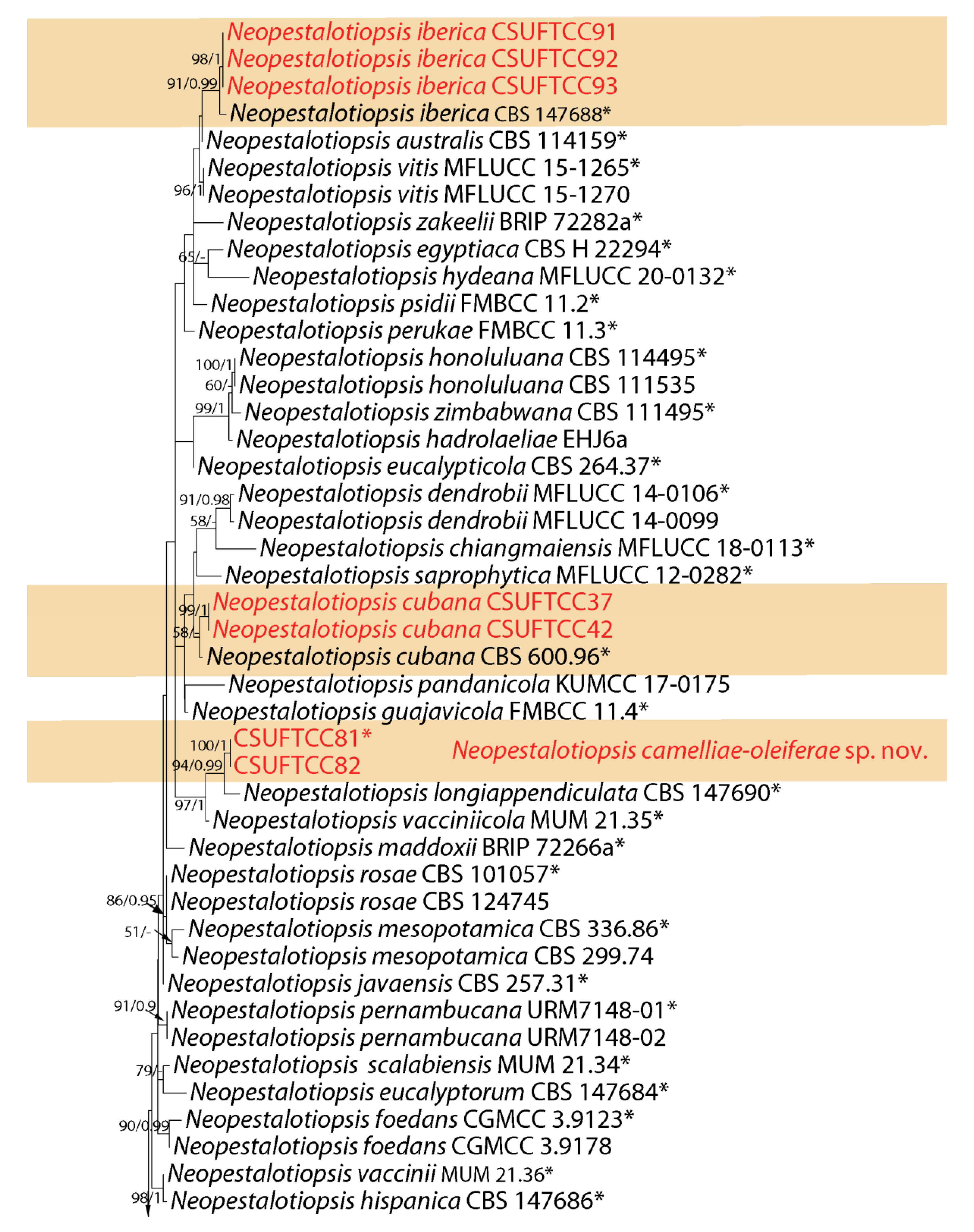
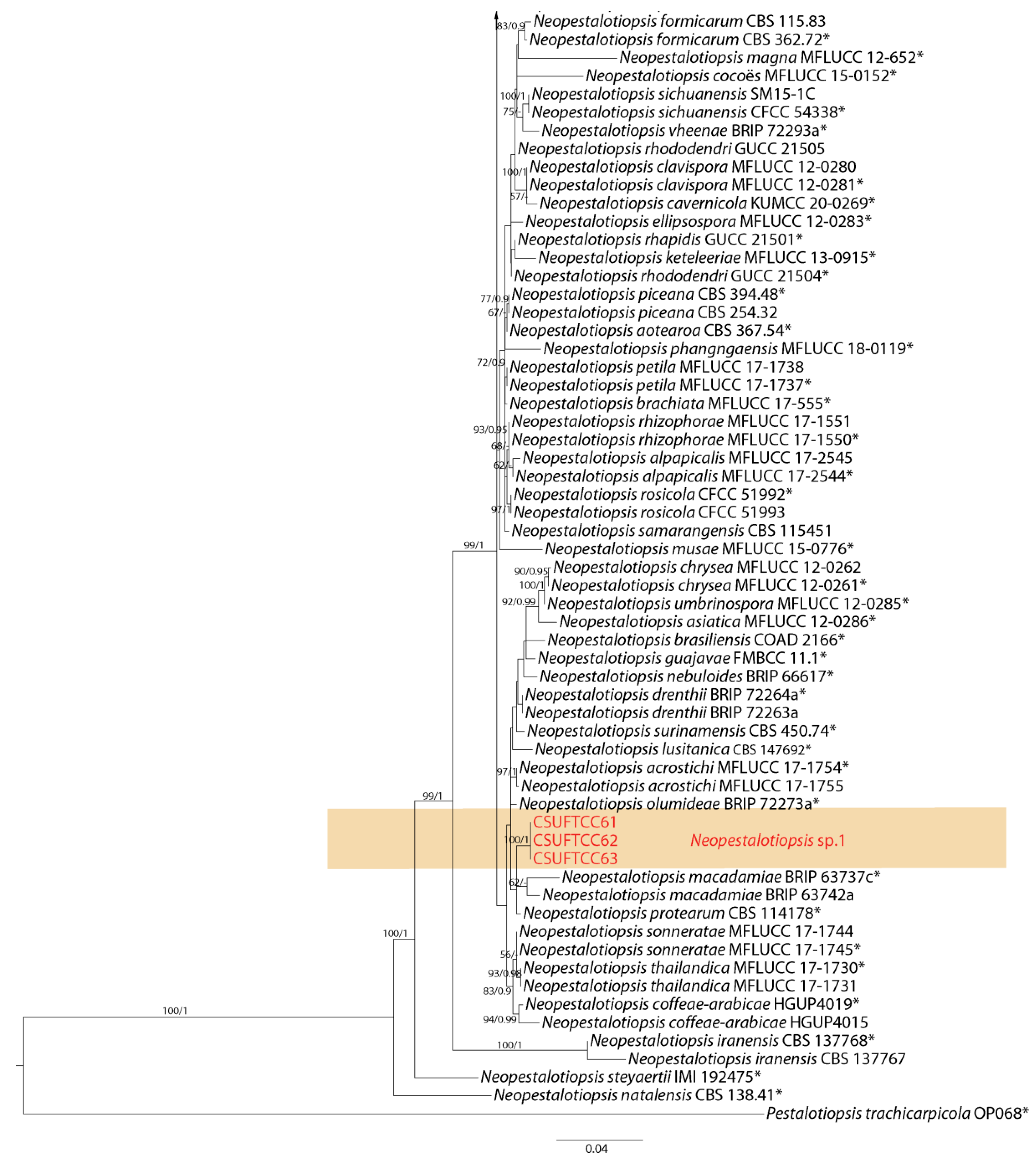
The first sequence datasets for the ITS, tef-1α and tub2, were analyzed in combination to infer the interspecific relationships within Neopestalotiopsis. The combined species phylogeny of the Neopestalotiopsis isolates consisted of 105 sequences, including the outgroup Pestalotiopsis trachicarpicola (culture OP068). A total of 1389 characters including gaps (479 for ITS, 498 for tef-1α, and 412 for tub2) were included in the phylogenetic analysis. Similar tree topologies were obtained by ML and BI methods, and the best scoring ML tree is shown in Figure 1. ML bootstrap values and BI posterior probabilities (MLBS/BIPP) are given at nodes of the phylogram (Figure 1). The phylogenetic tree inferred from the concatenated alignment resolved the ten Neopestalotiopsis isolates from symptomatic leaves of Camellia oleifera into four well-supported monophyletic clades that represent one novel species, one undetermined species and two known species of Neopestalotiopsis (Figure 1).
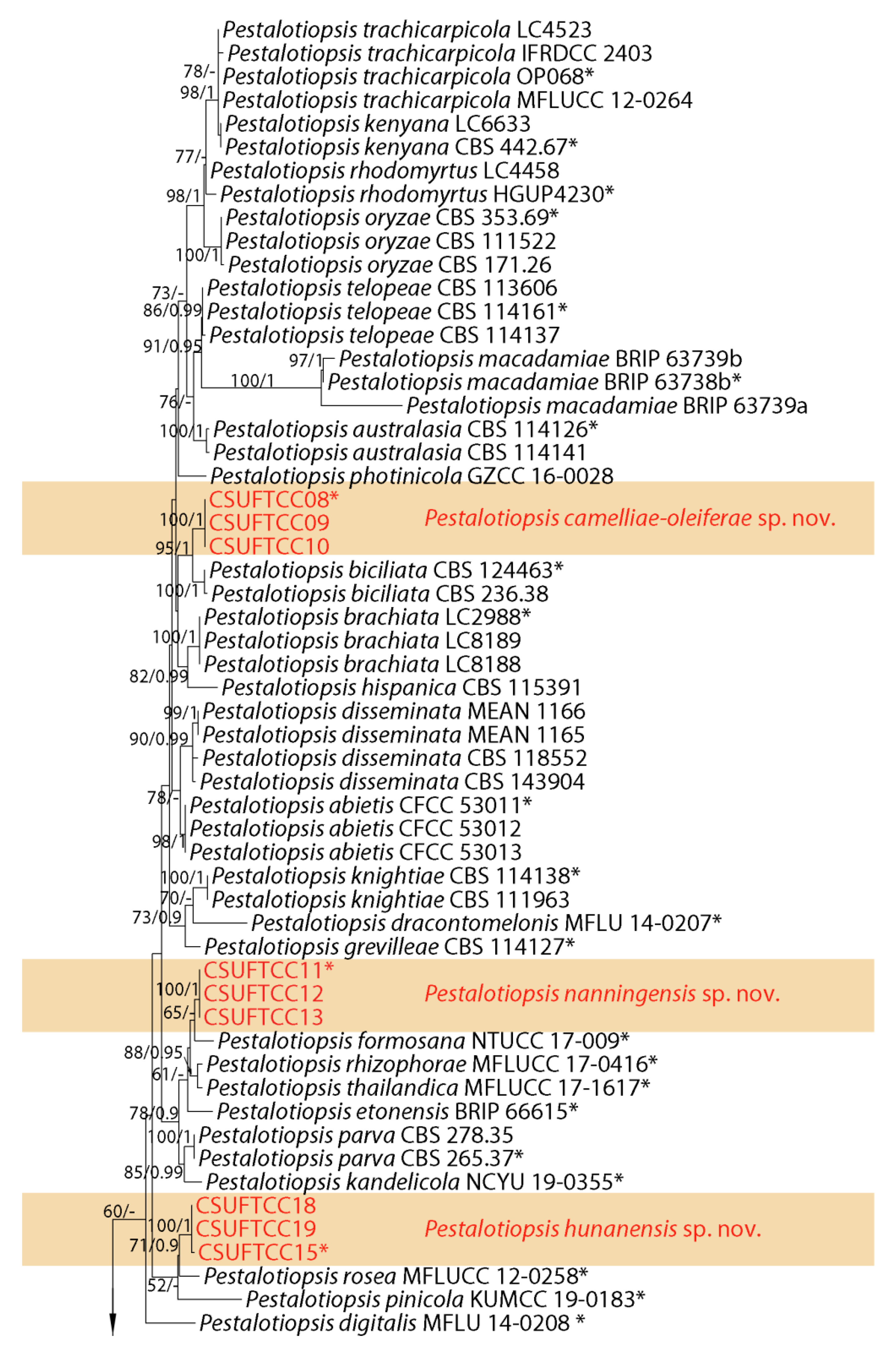
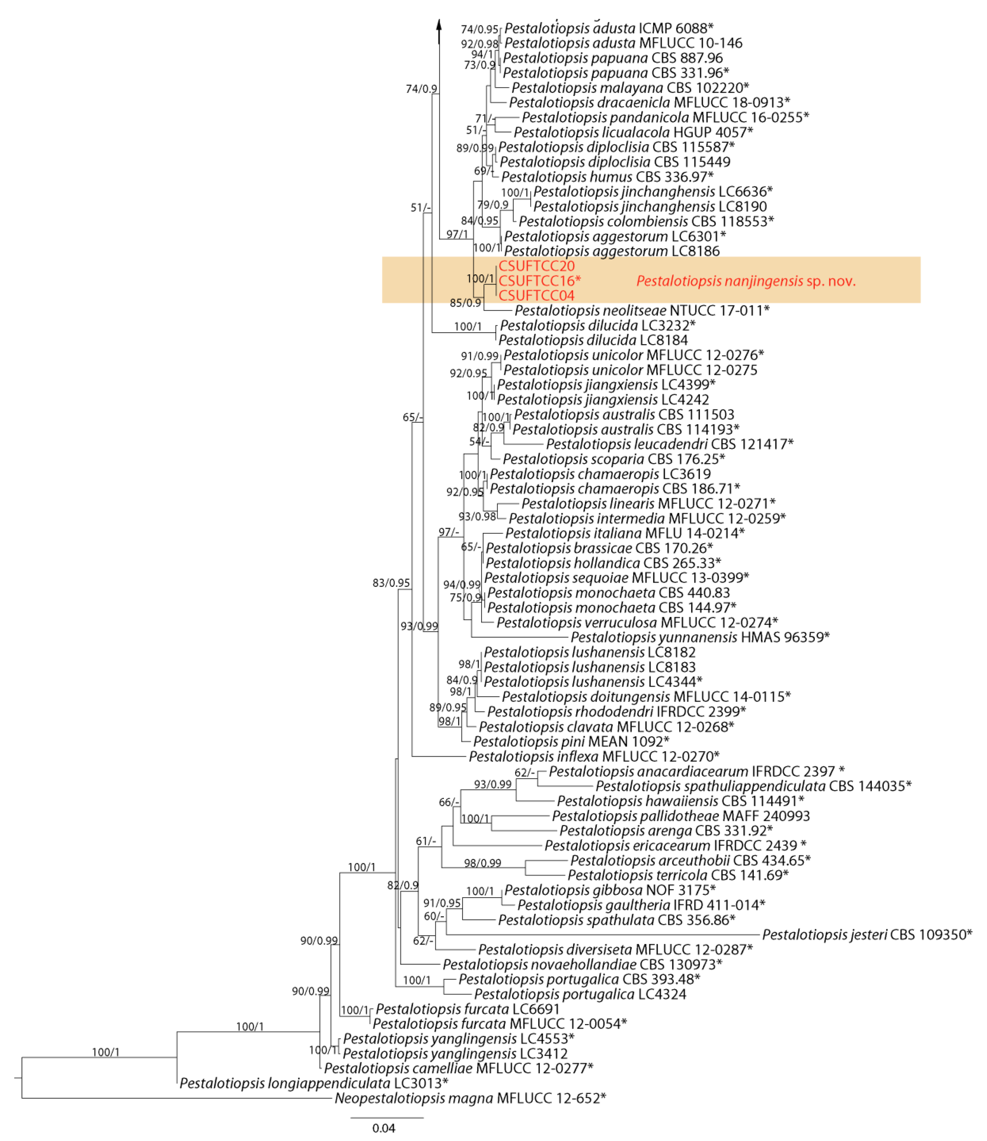
The second sequence datasets for the ITS, tef-1α and tub2 were analyzed in combination to infer the interspecific relationships within Pestalotiopsis. The combined species phylogeny of the Pestalotiopsis isolates consisted of 129 sequences, including the outgroup Neopestalotiopsis magna (culture MFLUCC 12-652). A total of 1557 characters including gaps (515 for ITS, 537 for tef-1α, and 505 for tub2) were included in the phylogenetic analysis. Similar tree topologies were obtained by ML and BI methods, and the best scoring ML tree is shown in Figure 2. ML bootstrap values and BI posterior probabilities (MLBS/BIPP) are given at nodes of the phylogram (Figure 2). The phylogenetic tree inferred from the concatenated alignment resolved the 12 Pestalotiopsis isolates from symptomatic leaves of Camellia oleifera into four well-supported monophyletic clades that represent four novel species of Pestalotiopsis (Figure 2).
3.2. Taxonomy
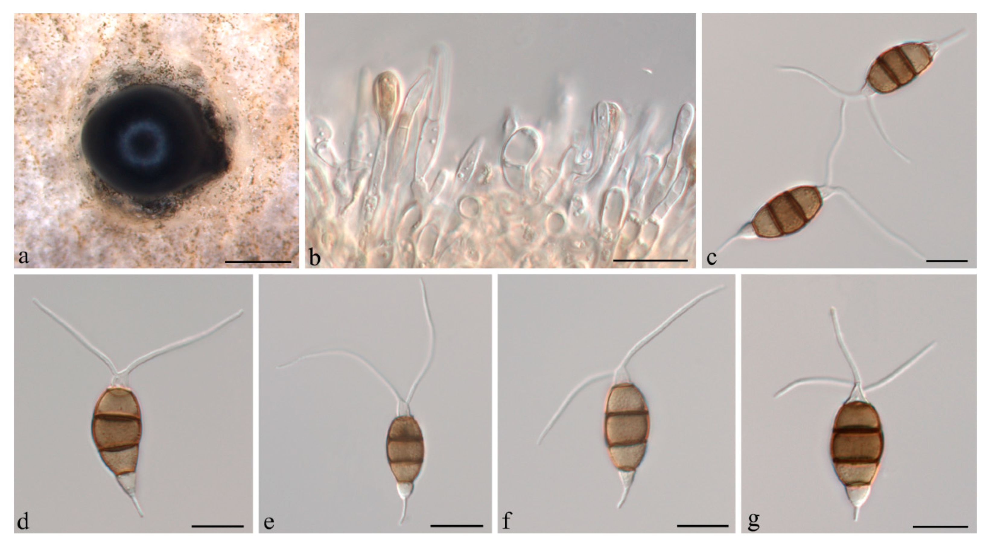
Neopestalotiopsis camelliae-oleiferae Q. Yang & H. Li, sp. nov. (Figure 3).
MycoBank: MB841476.
Etymology: Named after the host species, Camellia oleifera.
Holotype: CSUFT081.
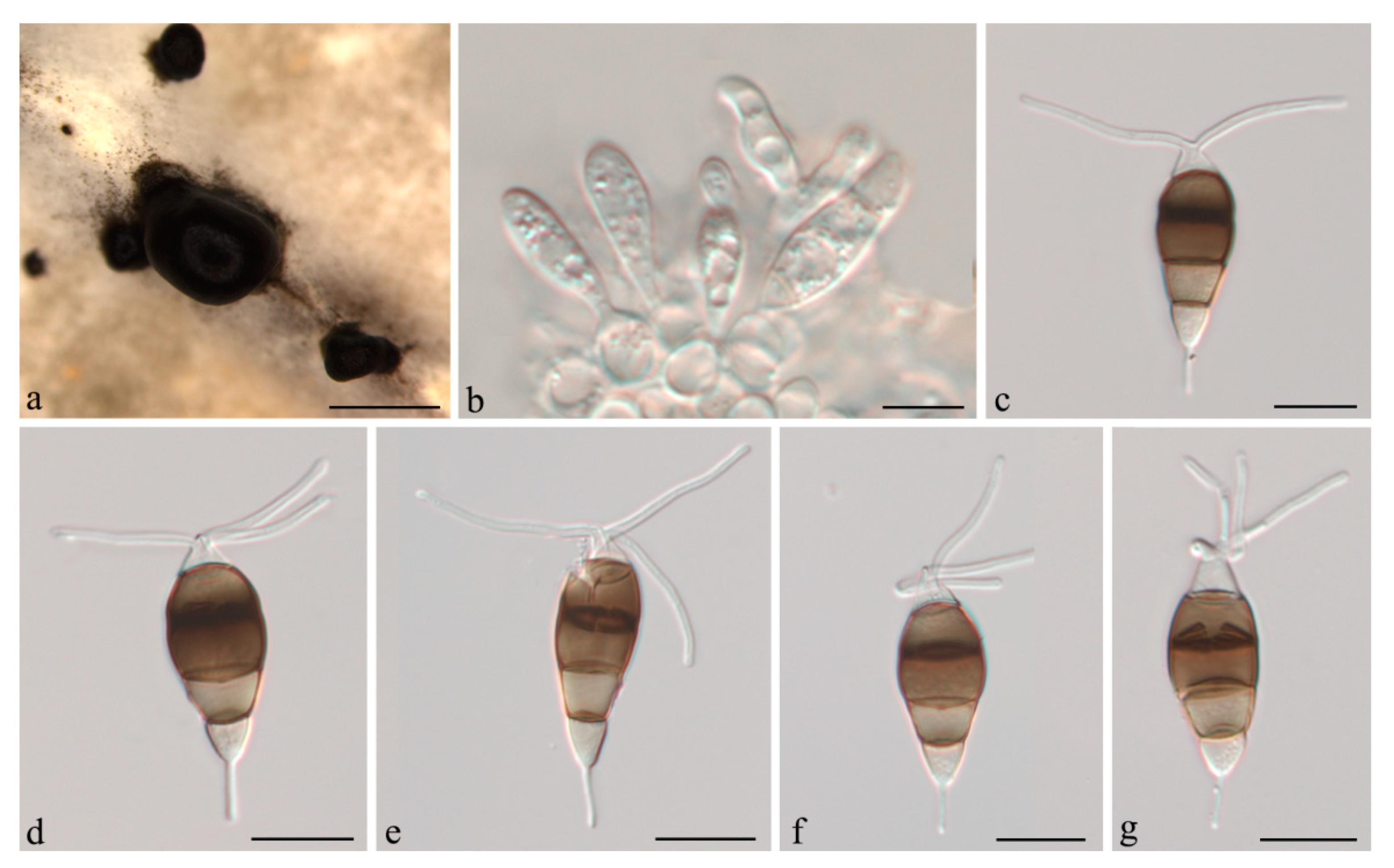
Description:Conidiomata acervular in culture on PDA, globose, 300–800 μm diam., solitary or aggregated in clusters, exuding black conidial masses. Conidiophores reduced to conidiogenous cells. Conidiogenous cells ampulliform, hyaline, smooth, annelidic. Conidia fusiform to clavate, straight or slightly curved, 22.5–24(−26.5) × (7–)8.5–10 μm, 4-septate; basal cell conical, 3.5–4.5 μm, hyaline or sometimes pale brown, smooth, thin-walled; with a single appendage filiform, unbranched, centric, (4.5–)6–8(−9) μm long; three median cells doliiform, 14–16(−18) μm long, smooth, versicoloured, septa darker than the rest of the cell (second cell from base pale brown, 4.5–5.5 μm long; third cell medium to dark brown, 5–5.5(−6.5) μm long; fourth cell medium to dark brown, 4.5–6 μm long); apical cell conical, 2.5–4.5 μm long, hyaline, smooth, thin-walled; with 2–3 apical tubular appendages unbranched, filiform, (13.5–)15.5–18.5(−20.5) μm long. Sexual morph not observed.
Culture characteristics: Colonies on PDA reaching 55 mm diameter after seven days at 25 °C. Colonies filamentous to circular, with dense aerial mycelium on surface, fruiting bodies black.
Material examined: CHINA, Jiangsu Province, Nanjing City, from leaf spots of Camellia oleifera, 25 Oct. 2020, H. Li (CSUFT081, holotype); ex-type living culture CSUFTCC81, living culture CSUFTCC82.
Notes:Neopestalotiopsis camelliae-oleiferae was collected from symptomatic leaves of C. oleifera in Jiangsu Province, China. Two isolates (CSUFTCC81 and CSUFTCC82) representing N. camelliae-oleiferae clustered in a well-support clade (ML/BI = 100/1). Neopestalotiopsis camelliae-oleiferae was sister to a clade containing N. longiappendiculata and N. vacciniicola. N. camelliae-oleiferae can be distinguished from N. longiappendiculata based on ITS, tef-1α and tub2 loci (3/449 in ITS, 3/450 in tef-1α , and 6/404 in tub2, no gaps). Morphologically, N. camelliae-oleiferae differs from N. longiappendiculata by wider conidia (8.5–10 vs. 7–7.8 μm); from N. vacciniicola by shorter apical tubular appendages (15.5–18.5 vs. 25.7–30.2 μm) [20]. Therefore, the collection in the present study is designated as a new species.
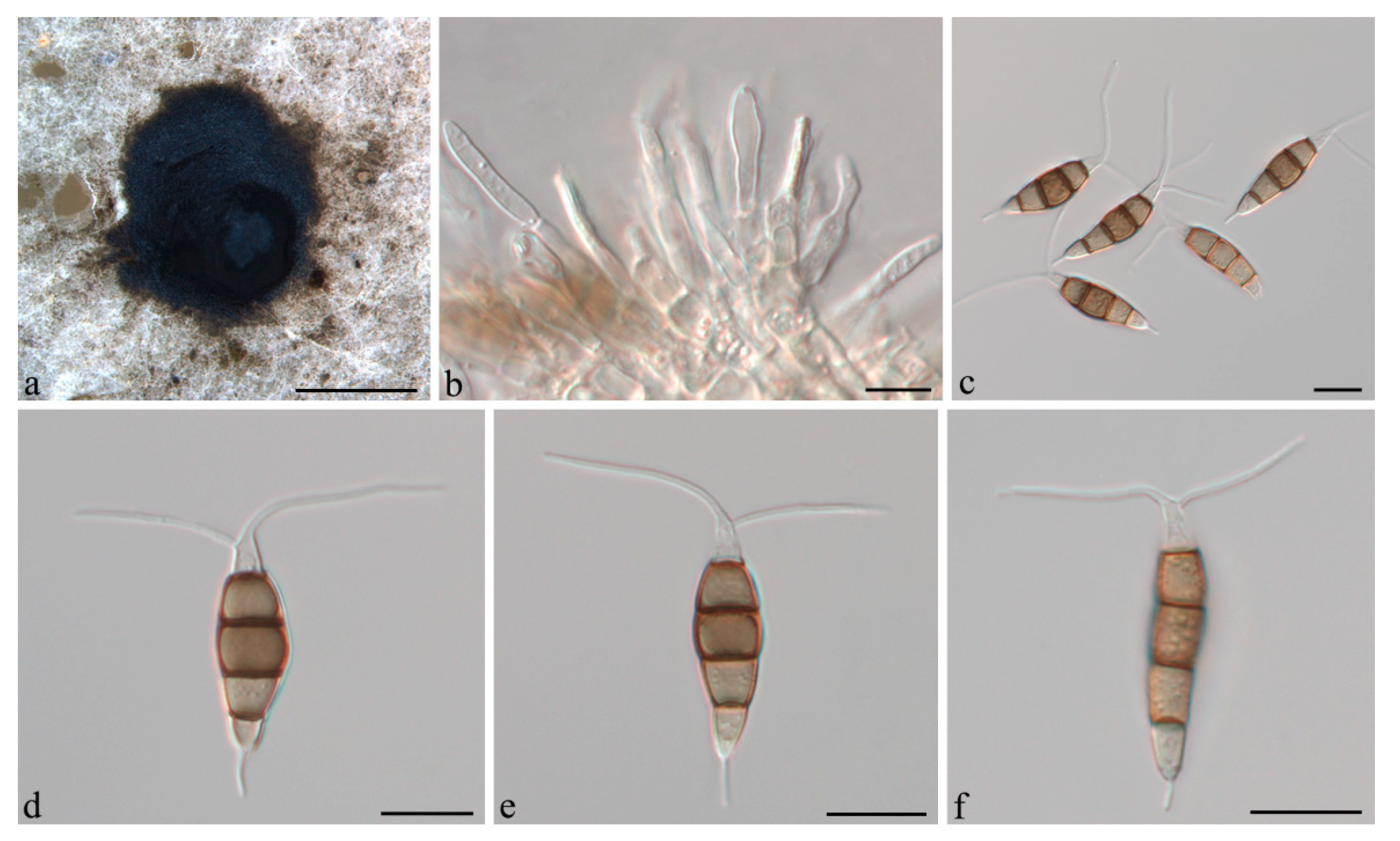
Neopestalotiopsis cubana Maharachch, K.D. Hyde & Crous, in Maharachchikumbura, Hyde, Groenewald, Xu & Crous, Stud. Mycol. 79: 138 (2014) (Figure 4).
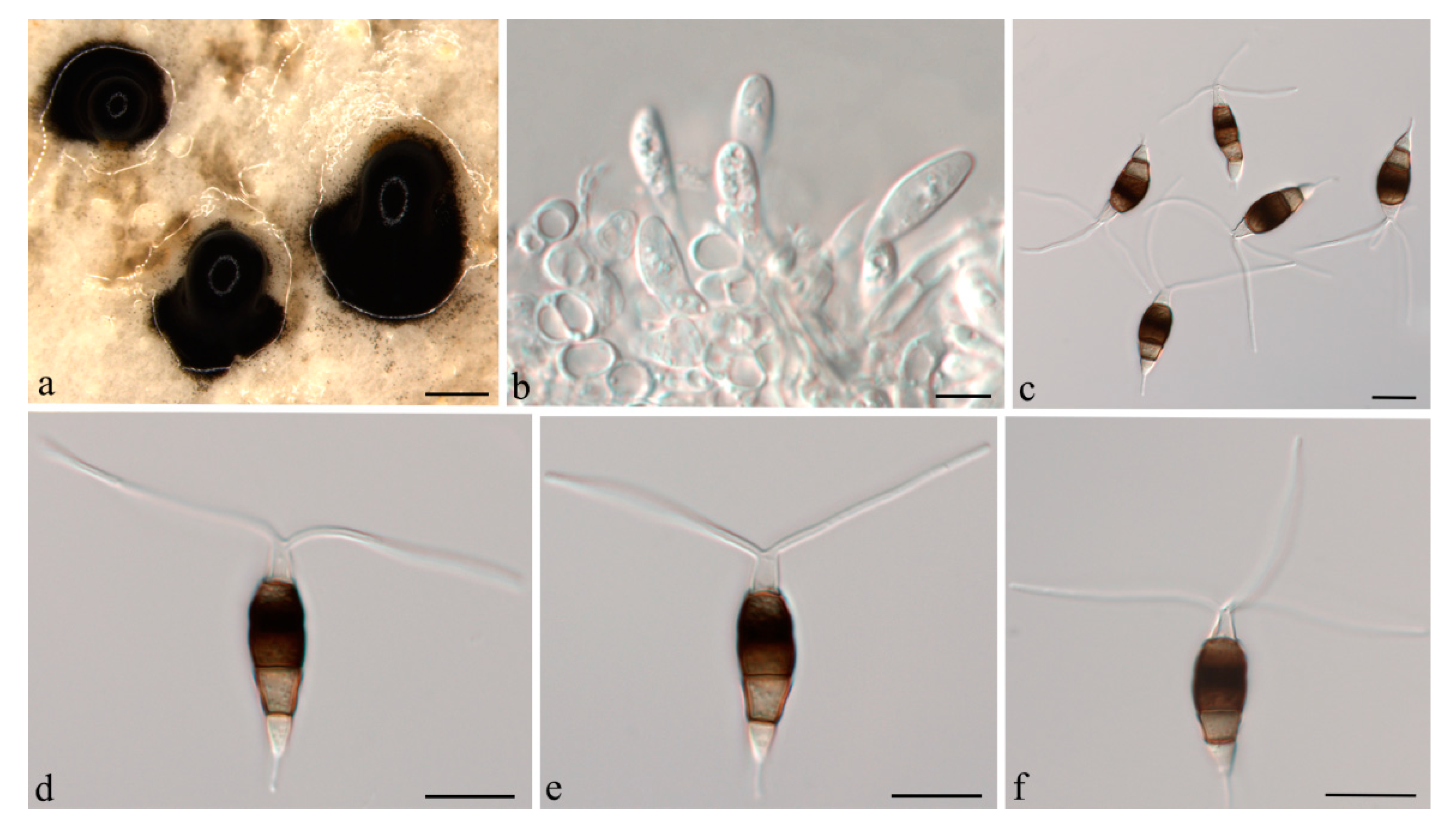
Description:Conidiomata acervular in culture on PDA, globose, 800–1350 μm diam., solitary or aggregated in clusters, exuding black conidial masses. Conidiophores reduced to conidiogenous cells. Conidiogenous cells ampulliform to cylindrical, hyaline, smooth, annelidic. Conidia fusoid to ellipsoidal, straight or slightly curved, (19.5–)21–25(−26.5) × (5.5–)6.5–8 μm, 4-septate; basal cell conical, 3.5–4.5 μm, hyaline or sometimes pale brown, smooth, thin-walled; with a single appendage filiform, unbranched, centric, 3–5.5 μm long; three median cells doliiform, 13.5–15(−16) μm long, smooth, versicoloured, septa darker than the rest of the cell (second cell from base pale brown, 3.5–5.5 μm long; third cell medium to dark brown, 4–5 μm long; fourth cell medium to dark brown, 3.5–4.5 μm long); apical cell conical, 3.5–4.5 μm long, hyaline, smooth, thin-walled; with 2–3 apical tubular appendages, unbranched, filiform, (21–)24–29(−31) μm long. Sexual morph not observed.
Culture characteristics: Colonies on PDA reaching 70 mm diameter after seven days at 25 °C. Colonies filamentous to circular, medium dense, aerial mycelium on surface flat or raised, pycnidia abundant, fruiting bodies black.
Material examined: CHINA, Hainan Province, Chengmai County, from leaf spots of Camellia oleifera, 9 Nov. 2020, H. Li (CSUFT042); living cultures CSUFTCC37 and CSUFTCC42.
Notes:Neopestalotiopsis cubana was originally described from leaf litter in Cuba [8]. In the present study, two isolates from leaves of symptomatic C. oleifera were congruent with N. cubana based on morphology and DNA sequences data (Figure 1). We therefore describe N. cubana as a known species for this clade.
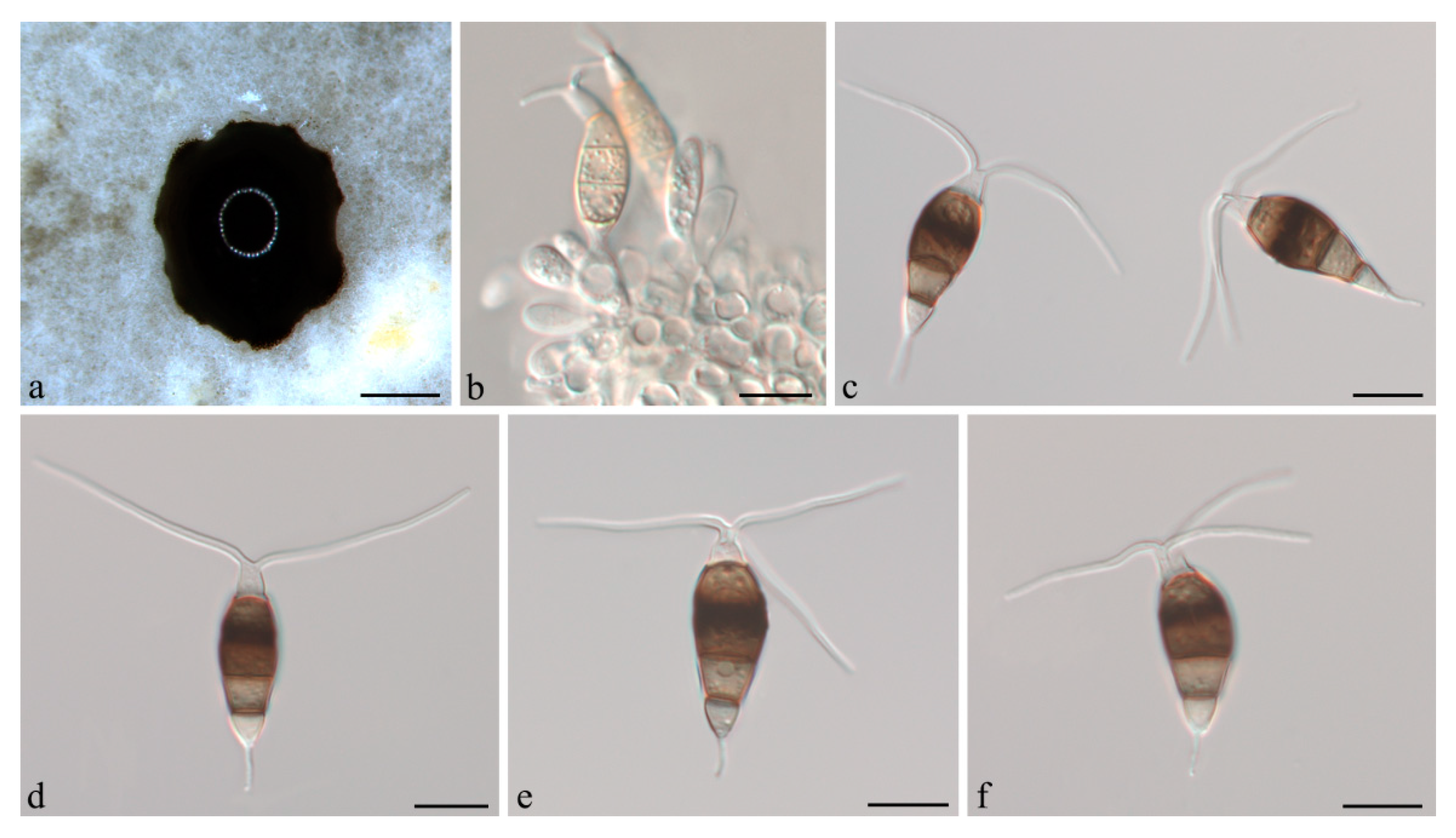
Neopestalotiopsis iberica E. Diogo, M.H. Bragança & A.J.L. Phillips, in Diogo, Gonçalves, Silva, Valente, Bragança & Phillips, Mycol. Progr. 20(11): 1449 (2021) (Figure 5).
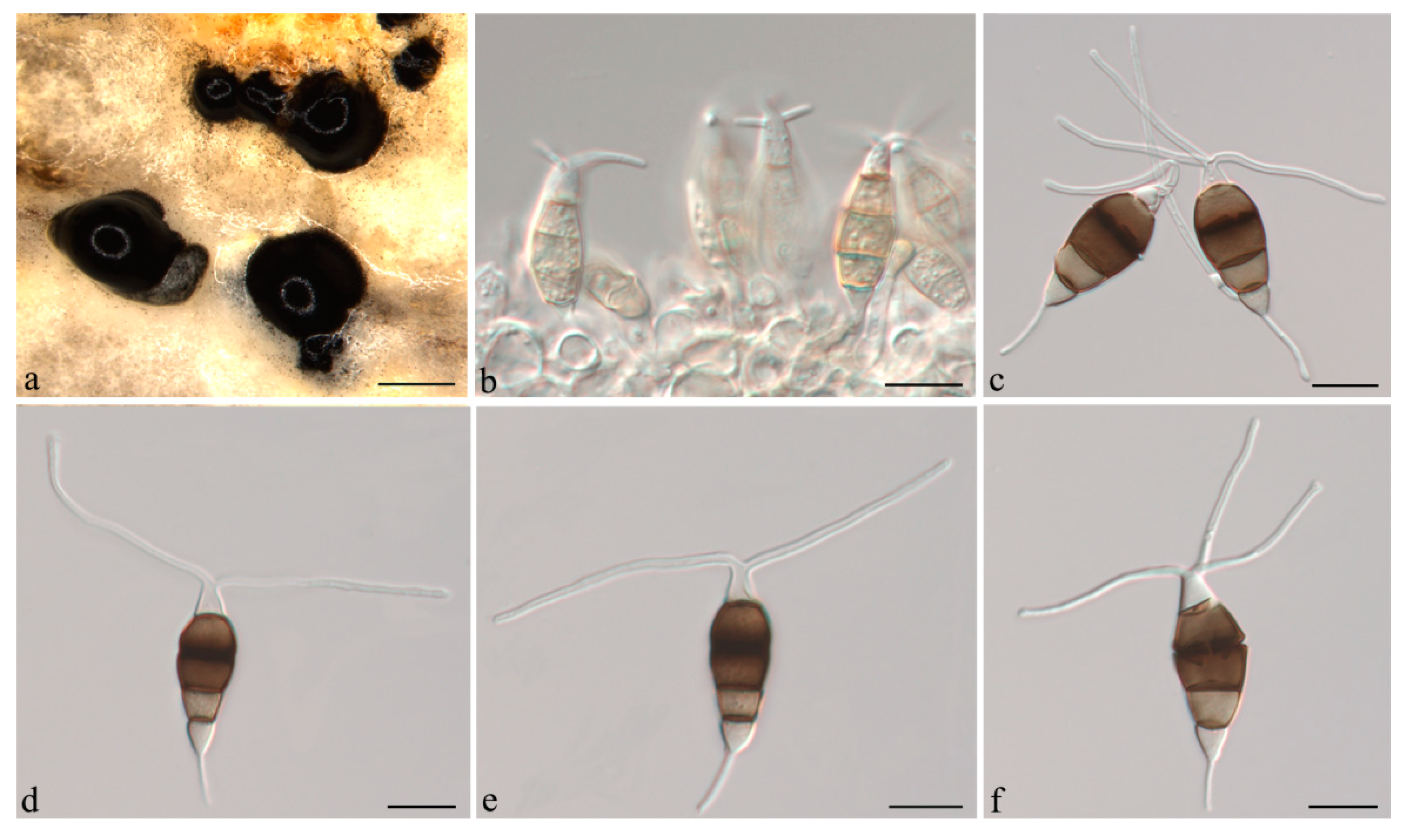
Description:Conidiomata acervular in culture on PDA, globose, 600–1500 μm diameter, solitary or aggregated in clusters, exuding black conidial masses. Conidiophores reduced to conidiogenous cells. Conidiogenous cells ampulliform, hyaline, smooth, annelidic. Conidia fusiform to ellipsoidal, straight or slightly curved, (21.5–)22.5–24(−26.5) × 7–9(−10.5) μm, 4-septate; basal cell conical, 3.5–4.5 μm, hyaline or sometimes pale brown, smooth, thin-walled; with a single appendage filiform, unbranched, centric, 2.5–4 μm long; three median cells doliiform, 12.5–14.5(−15.5) μm long, smooth, versicoloured, septa darker than the rest of the cell (second cell from base pale brown, 4.5–5 μm long; third cell medium to dark brown, 4.5–5.5(−6) μm long; fourth cell medium to dark brown, 4.5–5.5 μm long); apical cell conical, 2.5–4 μm long, hyaline, smooth, thin-walled; with 2–3 apical tubular appendages, unbranched, filiform, 24–26(−29.5) μm long. Sexual morph not observed.
Culture characteristics: Colonies on PDA reaching 70 mm diameter after seven days at 25 °C. Colonies filamentous to circular, medium dense, aerial mycelium on surface flat or raised, with filiform margin, fluffy, fruiting bodies black.
Material examined: CHINA, Jiangsu Province, Nanjing City, from leaf spots of Camellia oleifera, 25 Oct. 2020, H. Li (CSUFT091); living cultures LHNJ91, LHNJ92, and LHNJ93.
Notes:Neopestalotiopsis iberica was originally described from leaves and stems of Eucalyptus globulus in Portugal [30]. In the present study, three isolates from leaves of symptomatic C. oleifera were congruent with N. iberica based on morphology and DNA sequences data (Figure 1). We therefore describe N. iberica as a known species for this clade.
Pestalotiopsis camelliae-oleiferae Q. Yang & H. Li, sp. nov. (Figure 6).
MycoBank: MB841478.
Etymology: Named after the host species, Camellia oleifera.
Holotype: CSUFT008.
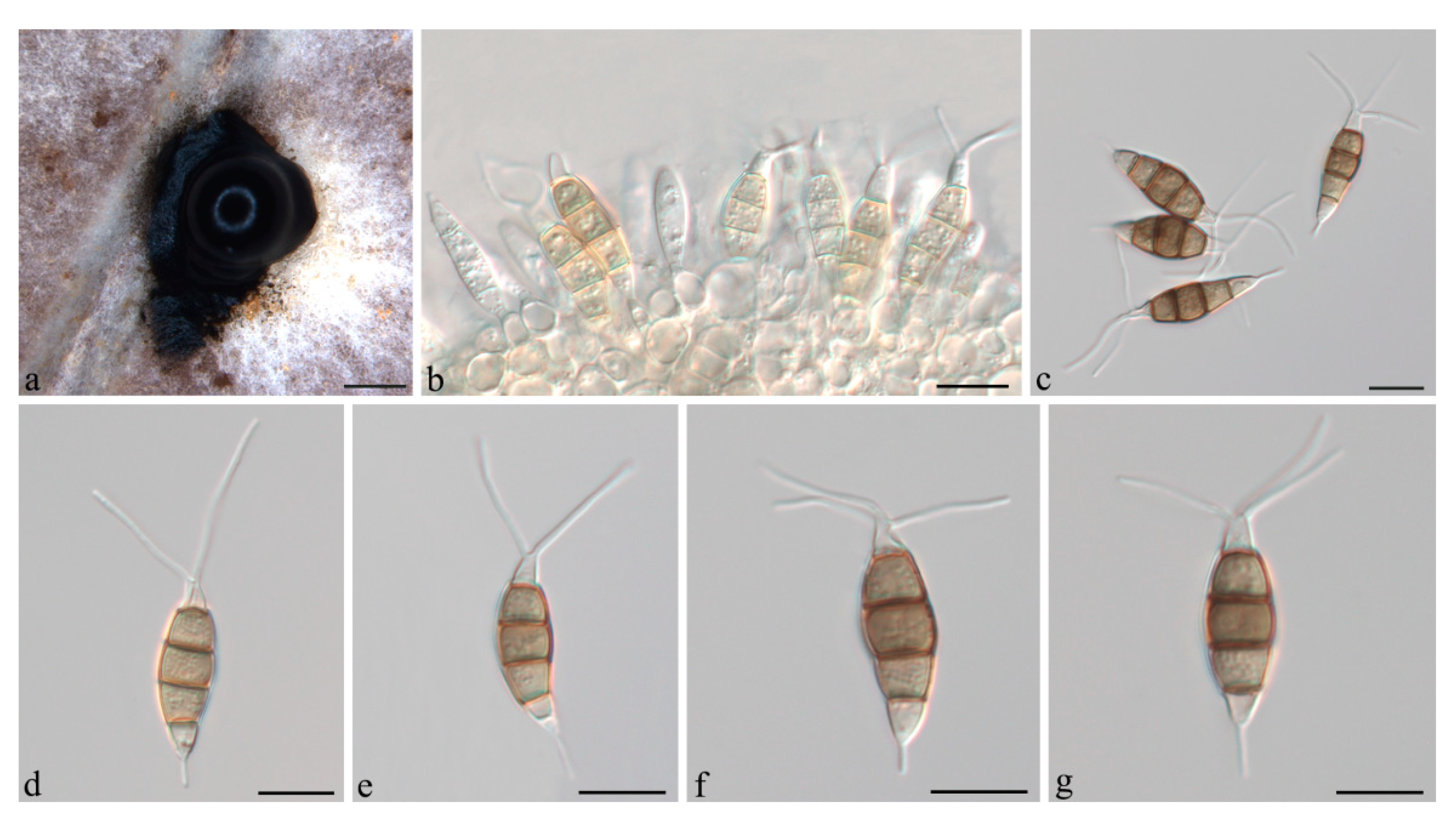
Description:Conidiomata acervular in culture on PDA, globose, 1.0–2.6 mm diameter, solitary or aggregated in clusters, exuding black conidial masses. Conidiophores reduced to conidiogenous cells. Conidiogenous cells discrete or integrated, cylindrical to subcylindrical, hyaline, smooth. Conidia fusoid, ellipsoid, straight or slightly curved, (19.5–)21.5–23(−25) × (5–)6–7 μm, 4-septate; basal cell conic to obconic with a truncate base, 3.5–5.5 μm, hyaline, smooth, thin-walled; with a single appendage filiform, unbranched, centric, 2.5–4.5 μm long; three median cells doliiform, 12.5–14 μm long, smooth, concolorous, brown, septa darker than the rest of the cell (second cell from base 4–4.5 μm long; third cell 4.5–5 μm long; fourth cell 3.5–4.5 μm long); apical cell conical, 2.5–4(−4.5) μm long, hyaline, smooth, thin-walled; with 2–3 apical tubular appendages, unbranched, filiform, (11–)12.5–14.5(−16) μm long. Sexual morph not observed.
Culture characteristics: Colonies on PDA reaching 70 mm diameter after seven days at 25 °C. Colonies filamentous to circular, medium dense, with white sparse mycelium, fruiting bodies black.
Material examined: CHINA, Hunan Province, Changsha City, from leaf spots of Camellia oleifera, 30 Aug. 2020, H. Li (CSUFT008, holotype); ex-type living culture CSUFTCC08, living cultures CSUFTCC09 and CSUFTCC10.
Notes:Pestalotiopsis camelliae-oleiferae was sister to P. biciliata in a well-supported clade (ML/BI = 100/1) (Figure 2). Pestalotiopsis camelliae-oleiferae can be distinguished from P. biciliata based on ITS, tef-1α and tub2 loci (4/500 in ITS, 1/473 intef-1α , and 6/443 in tub2, no gaps). Morphologically, P. camelliae-oleiferae differs from P. biciliata by shorter conidia (21.5–23 vs. 22–28 μm) [8]. Therefore, the collection in the present study is designated as a new species.
Pestalotiopsis hunanensis Q. Yang & H. Li, sp. nov. (Figure 7).
MycoBank: MB841480.
Etymology: In reference to the Hunan Province, from where the fungus was first collected.
Holotype: CSUFT015.
Description:Conidiomata acervular in culture on PDA, globose, 500–1000 μm diameter, solitary or aggregated in clusters, exuding black conidial masses. Conidiophores reduced to conidiogenous cells. Conidiogenous cells discrete or integrated, cylindrical to subcylindrical, hyaline, smooth, annelidic. Conidia fusoid, ellipsoid, straight or slightly curved, (20.5–)23–25(−26.5) × (7–)9–10.5 μm, 4-septate; basal cell conic to obconic with a truncate base, 4–5.5 μm, hyaline, smooth, thin-walled; with a single appendage filiform, unbranched, centric, 3–3.5 μm long; three median cells doliiform, (14–)15–18 μm long, smooth, concolorous, brown, septa darker than the rest of the cell (second cell from base 4–5 μm long; third cell 5–6.5 μm long; fourth cell 4.5–5.5 μm long); apical cell conical, 2.5–3 μm long, hyaline, smooth, thin-walled; with 2–3 apical tubular appendages, unbranched, filiform, (13.5–)15–22(−26.5) μm long. Sexual morph not observed.
Culture characteristics: Colonies on PDA reaching 50 mm diameter after seven days at 25 °C. Colonies filamentous to circular, with sparse aerial mycelium, fruiting bodies black.
Material examined: CHINA, Hunan Province, Xiangtan City, from leaf spots of Camellia oleifera, 7 Nov. 2020, H. Li (CSUFT015, holotype); ex-type living culture CSUFTCC15, living cultures CSUFTCC18 and CSUFTCC19.
Notes:Pestalotiopsis hunanensis was sister to P. rosae in a well-supported clade (ML/BI = 100/1) (Figure 2). Pestalotiopsis hunanensis can be distinguished from P. rosea based on ITS, tef-1α and tub2 loci (6/501 in ITS, 13/475 in tef-1α, and 7/446 in tub2, 12 gaps). Morphologically, P. hunanensis differs from P. rosae by lager conidia (23–25 × 9–10.5 vs. 17.5–21.8 × 5.7–7 μm) [6]. Therefore, the collection in the present study is designated as a new species.
Pestalotiopsis nanjingensis Q. Yang & H. Li, sp. nov. (Figure 8).
MycoBank: MB841481.
Etymology: In reference to the Nanjing City, from where the fungus was first collected.
Holotype: CSUFT016.
Description:Conidiomata acervular in culture on PDA, globose, 1000–1600 μm diameter, solitary or aggregated in clusters, exuding black conidial masses. Conidiophores reduced to conidiogenous cells. Conidiogenous cells discrete or integrated, cylindrical to subcylindrical, hyaline, smooth, annelidic. Conidia fusoid, ellipsoid, straight or slightly curved, (19.5–)22–25 × (4.5–)5–6.5 μm, 4-septate; basal cell conic to obconic with a truncate base, 4.5–5 μm, hyaline, smooth, thin-walled; with a single appendage filiform, unbranched, centric, 2.5–3.5 μm long; three median cells doliiform, 13–14.5(−16) μm long, smooth, concolorous, brown, septa darker than the rest of the cell (second cell from base 4.5–5.5 μm long; third cell 4.5–5.5 μm long; fourth cell 3.5–4.5 μm long); apical cell conical, 3.5–4 μm long, hyaline, smooth, thin-walled; with two apical tubular appendages, unbranched, filiform, (11–)13.5–18(−20) μm long. Sexual morph not observed.
Culture characteristics: Colonies on PDA reaching 60 mm diameter after seven days at 25 °C. Colonies filamentous to circular, medium dense, aerial mycelium on surface flat, fruiting bodies black.
Material examined: CHINA, Jiangsu Province, Nanjing city, from leaf spots of Camellia oleifera, 25 Oct. 2020, H. Li (CSUFT016, holotype); ex-type living culture CSUFTCC 16, living cultures CSUFTCC04 and CSUFTCC20.
Notes:Pestalotiopsis nanjingensis was sister to P. neolitseae in a well-supported clade (ML/BI = 100/1) (Figure 2). Pestalotiopsis nanjingensis can be distinguished from P. neolitseae based on ITS, tef-1α and tub2 loci (2/500 in ITS, 26/472 in tef-1α, and 2/442 in tub2, 5 gaps). Morphologically, P. nanjingensis differs from P. neolitseae by longer conidia (22–25 vs. 18–21 μm) and apical appendages (13.5–18 vs. 10–15 μm) [15]. Therefore, the collection in the present study is designated as a new species.
Pestalotiopsis nanningensis Q. Yang & H. Li, sp. nov. (Figure 9).
MycoBank: MB841479.
Etymology: In reference to the Nanning City, from where the fungus was first collected.
Holotype: CSUFT011.
Description:Conidiomata acervular in culture on PDA, globose, 750–1200 μm diameter, solitary or aggregated in clusters, exuding black conidial masses. Conidiophores reduced to conidiogenous cells. Conidiogenous cells discrete or integrated, cylindrical to subcylindrical, hyaline, smooth, annelidic. Conidia fusoid, ellipsoid, straight or slightly curved, (22–)24–26.5 × (6–)7–8(−9) μm, 4-septate; basal cell conical, 4.5–6 μm, hyaline, smooth, thin-walled; with a single appendage filiform, unbranched, centric, 4.5–6.5 μm long; three median cells doliiform, 13.5–15(−17) μm long, smooth, concolorous, brown, septa darker than the rest of the cell (second cell from base 4.5–5.5 μm long; third cell 5–6 μm long; fourth cell 4–5 μm long); apical cell conical, 3.5–4.5 μm long, hyaline, smooth, thin-walled; with 2–3 apical tubular appendages, unbranched, filiform, (13.5–)18–22.5(−26.5) μm long. Sexual morph not observed.
Culture characteristics: Colonies on PDA reaching 80 mm diameter after seven days at 25 °C. Colonies filamentous to circular, medium dense, white aerial mycelium on surface flat or raised.
Material examined: CHINA, Guangxi Province, Nanning City, from leaf spots of Camellia oleifera, 20 Oct. 2020, H. Li (CSUFT011, holotype); ex-type living culture CSUFTCC11, living cultures CSUFTCC12 and CSUFTCC13.
Notes: Pestalotiopsis nanningensis was sister to P. formosana in a well-supported clade (ML/BI = 100/1) (Figure 2). Pestalotiopsis nanningensis can be distinguished from P. formosana based on ITS and tef-1α loci (4/500 in ITS, 2/472 in tef-1α , and 1/442 in tub2, no gaps). Morphologically, P. nanningensis differs from P. formosana by lager conidia (24–26.5 × 7–8 vs. 18–22 × 6–7 μm) and longer apical appendages (18–22.5 vs. 11–16 μm) [15]. Therefore, the collection in the present study is designated as a new species.
3.3. Pathogenicity Assay
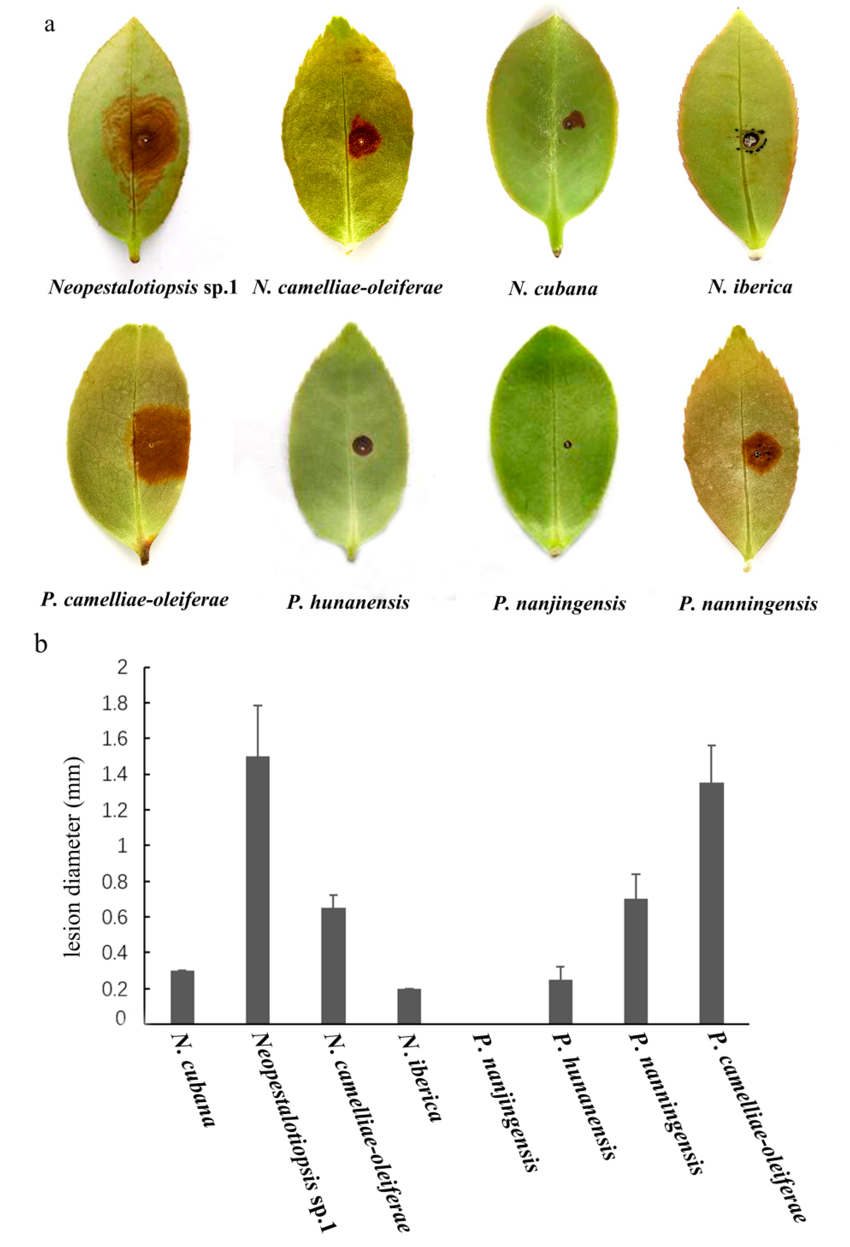
After five days, for the pathogenicity tests, N. camelliae-oleiferae, N. cubana, N. iberica Neopestalotiopsis sp.1, P. camelliae-oleiferae, P. hunanensis , and P. nanningensis developed brown lesions on wounded leaves (right), whereas the controls showed no symptoms (left). Neopestalotiopsis sp.1 had the highest virulence, while P. nanjingensis did not cause obvious symptoms (Figure 10). Koch’s postulates were fulfilled by reisolating the same fungi and verifying its colony and morphological characters.
4. Discussion
In this study, an investigation of C. oleifera diseases in China was carried out and Camellia leaf disease caused by pestalotioid fungi was observed as a common disease. Identification of our collections was conducted, based on isolates from symptomatic leaves of C. oleifera using three combined loci (ITS, tef-1α and tub2), as well as morphological characteristics. It includes N. cubana, N. iberica, as well as five new species named N. camelliae-oleiferae, P. camelliae-oleiferae, P. hunanensis, P. nanjingensis, and P. nanningensis.
The expanding cultivation of C. oleifera over the last several decades has attracted increasing attention from plant pathologists to infectious diseases on this crop. Therein, pestalotioid species are more frequently regarded as endophytes or latent pathogens causing diseases only on specific situations [4,6,12,63,64]. Understanding the diversity of pestalotioid species and the genetic variation within pathogen populations could help in developing sustainable disease management strategies.
Pestalotioid fungi (Pestalotiopsidaceae, Sordariomycetes) are species-rich asexual taxa, which are common pathogens that cause a variety of diseases, including leaf spots, shoot dieback, fruit rots and various post-harvest diseases [6,8,15,19,20,46,65]. As many peatalotioid species have overlapping morphological traits, sequence data is essential to resolve these three genera and introduce new species [8]. Combined gene sequence of ITS, tef-1α, and tub2 can provide a better resolution for Pestalotiopsis and Pseudopestalotiopsis. However, more genes are needed to provide better resolution and support in Neopestalotiopsis. Furthermore, this is the first systematic report of Neopestalotiopsis and Pestalotiopsis fungi associated with Camellia oleifera in China, which indicates that there may be a high undescribed diversity of fungi in this host.
Pathogenicity tests of eight pestalotioid species from Camellia oleifera showed that all species except for P. nanjingensis were capable of infecting wounded leaves. Neopestalotiopsis sp.1 and P. camelliae-oleiferae showed stronger virulence, with lesion diameters ranged from 14.7 to 17.8 mm on leaves of the Neopestalotiopsis sp.1 isolate (CSUFTCC61) and 13.5 to 15.5 mm on leaves of the P. camelliae-oleiferae isolate (CSUFTCC08). All pathogenicity tests were performed with a single C. camellia cultivar. Since different C. oleifera cultivars may have different resistance to pestalotioid species, more cultivars of C. oleifera should be studied for the variation of their resistance to pestalotioid pathogens. During the tests, the symptoms vary considerably with factors, such as relative humidity, temperature, and the inoculum concentration. In the future, field conditions with natural inoculum should be conducted rather than just in vitro artificial inoculation.
5. Conclusions
Seven peatalotioid species (two known species and five new species) were described and illustrated. This is the first systematic report of Neopestalotiopsis and Pestalotiopsis fungi associated with Camellia oleifera in China. The pathogenicity of these species on leaves were examined and showed that there were significant differences in the pathogenicity.
Author Contributions
Experiments, L.L.; Writing—original draft preparation, Q.Y.; Writing—review and editing, Q.Y. and H.L. All authors have read and agreed to the published version of the manuscript.
Funding
This research was funded by the introduction of talent research start-up fund project of CSUFT, grant number 2019YJ025 and the Research Foundation of Education Bureau of Hunan Province, grant number 19B608.
Institutional Review Board Statement
Not applicable.
Informed Consent Statement
Not applicable.
Data Availability Statement
All sequence data are available in NCBI GenBank following the accession numbers in the manuscript.
Acknowledgments
We are grateful for the assistance of Yuanhao He and Linxue Cao.
Conflicts of Interest
The authors declare no conflict of interest.
References
- Zhuang, R.L. Camellia oleifera, 2nd ed.; China Forestry Press: Beijing, China, 2008. [Google Scholar]
- Wang, W.J.; Chen, C.G.; Cheng, J. The medicinal active role of tea oil in health care. Food Nutr. China 2007, 9, 48–51. [Google Scholar]
- Wang, Y.; Chen, J.Y.; Xu, X.W.; Cheng, J.Y.; Zheng, L.; Huang, J.B.; Li, D.W. Identification and characterization of Colletotrichum species associated with anthracnose disease of Camellia oleifera in China. Plant Dis. 2020, 104, 474–482. [Google Scholar] [CrossRef] [PubMed]
- Yu, J.; Wu, Y.; He, Z.; Li, M.; Zhu, K.; Gao, B. Diversity and antifungal activity of endophytic fungi associated with Camellia oleifera. Mycobiology 2018, 46, 85–91. [Google Scholar] [CrossRef] [Green Version]
- Zhou, H.; Hou, C.L. Three new species of Diaporthe from China based on morphological characters and DNA sequence data analyses. Phytotaxa 2019, 422, 157–174. [Google Scholar] [CrossRef]
- Maharachchikumbura, S.S.N.; Guo, L.D.; Cai, L.; Chukeatirote, E.; Wu, W.P.; Sun, X.; Hyde, K.D. A multi-locus backbone tree for Pestalotiopsis, with a polyphasic characterization of 14 new species. Fungal Divers. 2012, 56, 95–129. [Google Scholar] [CrossRef] [Green Version]
- Maharachchikumbura, S.S.N.; Guo, L.D.; Chukeatirote, E.; Ekachai, C.; Bahkali, A.H.; Hyde, K.D. Pestalotiopsis—morphology, phylogeny, biochemistry and diversity. Fungal Divers. 2011, 50, 167–187. [Google Scholar] [CrossRef]
- Maharachchikumbura, S.S.N.; Hyde, K.D.; Groenewald, J.Z.; Xu, J.; Crous, P.W. Pestalotiopsis revisited. Stud. Mycol. 2014, 79, 121–186. [Google Scholar] [CrossRef] [PubMed] [Green Version]
- Jeewon, R.; Liew, E.C.Y.; Hyde, K.D. Phylogenetic relationships of Pestalotiopsis and allied genera inferred from ribosomal DNA sequences and morphological characters. Mol. Phylogenet. Evol. 2002, 25, 378–392. [Google Scholar] [CrossRef]
- Jeewon, R.; Liew, E.C.Y.; Simpson, J.A.; Hodgkiss, I.J.; Hyde, K.D. Phylogenetic significance of morphological characters in the taxonomy of Pestalotiopsis species. Mol. Phylogenet. Evol. 2003, 27, 372–383. [Google Scholar] [CrossRef]
- Maharachchikumbura, S.S.N.; Laringnonl, P.; Hyde, K.D.; Al-Sady, A.; Liu, Z. Characterization of Neopestalotiopsis, Pestalotiopsis and Truncatella species associated with grapevine trunk diseases in France. Phytopathol. Mediterr. 2016, 55, 380–390. [Google Scholar]
- Liu, F.; Hou, L.W.; Raza, M.; Cai, L. Pestalotiopsis and allied genera from Camellia, with description of 11 new species from China. Sci. Rep. 2017, 7, 1–19. [Google Scholar] [CrossRef] [PubMed] [Green Version]
- Nozawa, S.; Yamaguchi, K.; Van Hop, D.; Phay, N.; Ando, K.; Watanabe, K. Identification of two new species and asexual morph from the genus Pseudopestalotiopsis. Mycoscience 2017, 58, 328–337. [Google Scholar] [CrossRef]
- Akinsanmi, O.A.; Nisa, S.; Jeff-Ego, O.S.; Shivas, R.G.; Drenth, A. Dry flower disease of macadamia in Australia caused by Neopestalotiopsis macadamiae sp. nov. and Pestalotiopsis macadamiae sp. nov. Plant Dis. 2017, 101, 45–53. [Google Scholar] [CrossRef] [Green Version]
- Ariyawansa, H.A.; Hyde, K.D. Hyde. Additions to Pestalotiopsis in Taiwan. Mycosphere 2018, 9, 999–1013. [Google Scholar] [CrossRef]
- Jiang, N.; Bonthond, G.; Fan, X.L.; Tian, C.M. Neopestalotiopsis rosicola sp. nov. causing stem canker of Rosa chinensis in China. Mycotaxon 2018, 133, 271–283. [Google Scholar] [CrossRef]
- Tsai, I.; Maharachchikumbura, S.S.N.; Hyde, K.D.; Ariyawansa, H.A. Molecular phylogeny, morphology and pathogenicity of Pseudopestalotiopsis species of Ixora in Taiwan. Mycol. Prog. 2018, 17, 941–952. [Google Scholar] [CrossRef]
- Tibpromma, S.; Hyde, K.D.; McKenzie, E.H.C.; Bhat, D.J.; Phillips, A.J.L.; Wanasinghe, D.N.; Samarakoon, M.C.; Jayawardena, R.; Dissanayake, A.J.; Tennakoon, D.S.; et al. Fungal diversity notes 840–928: Microfungi associated with Pandanaceae. Fungal Divers. 2018, 93, 1–160. [Google Scholar] [CrossRef]
- Norphanphoun, C.; Jayawardena, R.S.; Chen, Y.; Wen, T.C.; Meepol, W.; Hyde, K.D. Morphological and phylogenetic characterization of novel pestalotioid species associated with mangroves in Thailand. Mycosphere 2019, 10, 531–578. [Google Scholar] [CrossRef]
- Diogo, E.; Gonçalves, C.I.; Silva, A.C.; Valente, C.; Bragança, H.; Phillips, A.J. Five new species of Neopestalotiopsis associated with diseased Eucalyptus spp. in Portugal. Mycol. Prog. 2021, 20, 1441–1456. [Google Scholar] [CrossRef]
- Prasannath, K.; Shivas, R.G.; Galea, V.J.; Akinsanmi, O.A. Neopestalotiopsis species associated with flower diseases of Macadamia integrifolia in Australia. J. Fungi 2021, 7, 771. [Google Scholar] [CrossRef]
- Crous, P.W.; Gams, W.; Stalpers, J.A.; Robert, V.; Stegehuis, G. MycoBank: An online initiative to launch mycology into the 21st century. Stud. Mycol. 2004, 50, 19–22. [Google Scholar]
- Doyle, J.J.; Doyle, J.L. Isolation of plant DNA from fresh tissue. Focus 1990, 12, 13–15. [Google Scholar]
- Katoh, K.; Toh, H. Parallelization of the MAFFT multiple sequence alignment program. Bioinformatics 2010, 26, 1899–1900. [Google Scholar] [CrossRef]
- Hall, T. BioEdit: A user-friendly biological sequence alignment editor and analysis program for Windows 95/98/NT. Nucleic Acids Symp. Series 1999, 41, 95–98. [Google Scholar]
- Miller, M.A.; Pfeiffer, W.; Schwartz, T. Creating the CIPRES Science Gateway for Inference of Large Phylogenetic Trees; Institute of Electrical and Electronics Engineers: New Orleans, LA, USA, 2010. [Google Scholar]
- Ronquist, F.; Huelsenbeck, J.P. MrBayes 3: Bayesian phylogenetic inference under mixed models. Bioinformatics 2003, 19, 1572–1574. [Google Scholar] [CrossRef] [Green Version]
- Jiang, N.; Voglmayr, H.; Bian, D.R.; Piao, C.G.; Wang, S.K.; Li, Y. Morphology and Phylogeny of Gnomoniopsis (Gnomoniaceae, Diaporthales) from Fagaceae Leaves in China. J. Fungi 2021, 7, 792. [Google Scholar] [CrossRef] [PubMed]
- Darriba, D.; Taboada, G.L.; Doallo, R.; Posada, D. jModelTest 2: More models, new heuristics and parallel computing. Nat. Methods 2012, 9, 772. [Google Scholar] [CrossRef] [PubMed] [Green Version]
- Kumar, V.; Cheewangkoon, R.; Gentekaki, E.; Maharachchikumbura, S.S.; Brahmanage, R.S.; Hyde, K.D. Neopestalotiopsis alpapicalis sp. nov. a new endophyte from tropical mangrove trees in Krabi Province (Thailand). Phytotaxa 2019, 393, 251–262. [Google Scholar] [CrossRef]
- Bezerra, J.D.P.; Machado, A.R.; Firmino, A.L.; Rosado, A.W.C.; Souza, C.A.F.D.; Souza-Motta, C.M.D.; Freire, K.T.L.D.S.; Paiva, L.M.; Magalhães, O.M.C.; Pereira, O.L.; et al. Mycological diversity description I. Acta Bot. Bras. 2018, 32, 656–666. [Google Scholar] [CrossRef] [Green Version]
- Liu, X.; Tibpromma, S.; Zhang, F.; Xu, J.; Chethana, K.W.T.; Karunarathna, S.C.; Mortimer, P.E. Neopestalotiopsis cavernicola sp. nov. from Gem Cave in Yunnan Province, China. Phytotaxa 2021, 512, 1–27. [Google Scholar] [CrossRef]
- Song, Y.; Geng, K.; Hyde, K.D.; Zhao, W.; Wei, J.G.; Kang, J.C.; Wang, Y. Two new species of Pestalotiopsis from Southern China. Phytotaxa 2013, 126, 22–30. [Google Scholar] [CrossRef]
- Ma, X.Y.; Maharachchikumbura, S.S.; Chen, B.W.; Hyde, K.D.; Mckenzie, E.H.; Chomnunti, P.; Kang, J.C. Endophytic pestalotiod taxa in Dendrobium orchids. Phytotaxa 2019, 419, 268–286. [Google Scholar] [CrossRef]
- Crous, P.W.; Wingfield, M.J.; Le Roux, J.J.; Richardson, D.M.; Strasberg, D.; Shivas, R.G.; Alvarado, P.; Edwards, J.; Moreno, G.; Sharma, R.; et al. Fungal Planet description sheets: 371–399. Persoonia 2015, 35, 264. [Google Scholar] [CrossRef]
- Ul Haq, I.; Ijaz, S.; Khan, N.A. Genealogical concordance of phylogenetic species recognition-based delimitation of Neopestalotiopsis species associated with leaf spots and fruit canker disease affected guava plants. Pak. J. Agric. Sci. 2021, 58, 1301–1313. [Google Scholar]
- Freitas, E.F.S.; Da Silva, M.; Barros, M.V.P.; Kasuya, M.C.M. Neopestalotiopsis hadrolaeliae sp. nov., a new endophytic species from the roots of the endangered orchid Hadrolaelia jongheana in Brazil. Phytotaxa 2019, 416, 211–220. [Google Scholar] [CrossRef]
- Huanluek, N.; Jjayawardena, R.S.; Maharachchikumbura, S.S.N.; Harishchandra, D.L. Additions to pestalotioid fungi in Thailand: Neopestalotiopsis hydeana sp. nov. and Pestalotiopsis hydei sp. nov. Phytotaxa 2021, 479, 23–43. [Google Scholar] [CrossRef]
- Ayoubi, N.; Soleimani, M.J. Strawberry fruit rot caused by Neopestalotiopsis iranensis sp. nov., and N. mesopotamica. Curr. Microbiol. 2016, 72, 329–336. [Google Scholar] [CrossRef] [PubMed]
- Song, Y.; Maharachchikumbura, S.S.; Jiang, Y.L.; Hyde, K.D.; Wang, Y. Pestalotiopsis keteleeria sp. nov., isolated from Keteleeria pubescens in China. Chiang Mai J. Sci. 2014, 41, 885–893. [Google Scholar]
- Maharachchikumbura, S.S.; Guo, L.D.; Chukeatirote, E.; Hyde, K.D. Improving the backbone tree for the genus Pestalotiopsis; addition of P. steyaertii and P. magna sp. nov. Mycol. Prog. 2014, 13, 617–624. [Google Scholar] [CrossRef]
- Crous, P.W.; Wingfield, M.J.; Chooi, Y.H.; Gilchrist, C.L.M.; Lacey, E.; Pitt, J.I.; Roets, F.; Swart, W.J.; Cano-Lira, J.F.; Valenzuela-Lopez, N.; et al. Fungal Planet description sheets: 1042–1111. Persoonia 2020, 44, 301–459. [Google Scholar] [CrossRef]
- Silvério, M.L.; Cavalcanti, M.A.Q.; Silva, G.A.; Oliveira, R.J.V.; Bezerra, J.L. A new epifoliar species of Neopestalotiopsis from Brazil. Agrotropica 2016, 28, 151–158. [Google Scholar] [CrossRef]
- Crous, P.W.; Summerell, B.A.; Swart, L.; Denman, S.; Taylor, J.E.; Bezuidenhout, C.M.; Palm, M.E.; Marincowitz, S.; Groenewald, J.Z. Fungal pathogens of Proteaceae. Persoonia 2011, 27, 20–45. [Google Scholar] [CrossRef] [Green Version]
- Yang, Q.; Zeng, X.Y.; Yuan, J.; Zhang, Q.; He, Y.K.; Wang, Y. Two new species of Neopestalotiopsis from southern China. Biodivers. Data J. 2021, 9, e70446. [Google Scholar] [CrossRef]
- Santos, J.; Hilário, S.; Pinto, G.; Alves, A. Diversity and pathogenicity of pestalotioid fungi associated with blueberry plants in Portugal, with description of three novel species of Neopestalotiopsis. Eur. J. Plant Pathol. 2021, 161, 1–17. [Google Scholar] [CrossRef]
- Jayawardena, R.S.; Liu, M.; Maharachchikumbura, S.S.N.; Zang, W.; Xing, Q.K.; Hyde, K.D.; Nilthong, S.; Li, X.; Yan, J. Neopestalotiopsis vitis sp. nov. causing grapevine leaf spot in China. Phytotaxa 2016, 258, 63–74. [Google Scholar] [CrossRef]
- Gu, M.; Hu, D.; Han, B.; Jiang, N.; Tian, C.M. Pestalotiopsis abietis sp. nov. from Abies fargesii in China. Phytotaxa 2021, 509, 93–105. [Google Scholar] [CrossRef]
- Liu, J.K.; Hyde, K.D.; Jones, E.G.; Ariyawansa, H.A.; Bhat, D.J.; Boonmee, S.; Maharachchikumbura, S.; McKenzie, E.H.C.; Phookamsak, R.; Phukhamsakda, C.; et al. Fungal diversity notes 1–110: Taxonomic and phylogenetic contributions to fungal species. Fungal Divers. 2015, 72, 1–197. [Google Scholar] [CrossRef]
- Silva, A.C.; Diogo, E.; Henriques, J.; Ramos, A.P.; Sandoval-Denis, M.; Crous, P.W.; Bragança, H. Pestalotiopsis pini sp. nov., an Emerging Pathogen on Stone Pine (Pinus pinea L.). Forests 2020, 11, 805. [Google Scholar] [CrossRef]
- Chaiwan, N.; Wanasinghe, D.N.; Mapook, A.; Jayawardena, R.S.; Norphanphoun, C.; Hyde, K.D. Novel species of Pestalotiopsis fungi on Dracaena from Thailand. Mycology 2020, 11, 306–315. [Google Scholar] [CrossRef]
- Zhang, Y.; Maharachchikumbura, S.S.; Tian, Q.; Hyde, K.D. Pestalotiopsis species on ornamental plants in Yunnan Province, China. Sydowia 2013, 65, 113–128. [Google Scholar]
- Maharachchikumbura, S.S.N.; Chukeatirote, E.; Guo, L.-D.; Crous, P.W.; McKenzie, E.H.C.; Hyde, K.D. Pestalotiopsis species associated with Camellia sinensis (tea). Mycotaxon 2013, 123, 47–61. [Google Scholar] [CrossRef]
- Watanabe, K.; Nozawa, S.; Hsiang, T.; Callan, B. The cup fungus Pestalopezia brunneopruinosa is Pestalotiopsis gibbosa and belongs to Sordariomycetes. PLoS ONE 2018, 13, e0197025. [Google Scholar] [CrossRef] [PubMed] [Green Version]
- Hyde, K.D.; Jeewon, R.; Chen, Y.J.; Bhunjun, C.S.; Calabon, M.S.; Jiang, H.B.; Lin, C.G.; Norphanphoun, C.; Sysouphanthong, P.; Pem, D.; et al. The numbers of fungi: Is the descriptive curve flattening? Fungal Divers. 2020, 103, 219–271. [Google Scholar] [CrossRef]
- Liu, F.; Bonthond, G.; Groenewald, J.Z.; Cai, L.; Crous, P.W. Sporocadaeceae, a family of coelomycetous fungi with appendage-bearing conidia. Stud. Mycol. 2019, 92, 287–415. [Google Scholar] [CrossRef]
- Geng, K.; Zhang, B.; Hyde, K.D.; Kang, J.C.; Wang, Y. A new species of Pestalotiopsis from leaf spots of Licuala grandis from Hainan, China. Phytotaxa 2013, 88, 49–54. [Google Scholar] [CrossRef] [Green Version]
- Watanabe, K.; Motohashi, K.; Ono, Y. Description of Pestalotiopsis pallidotheae: A new species from Japan. Mycoscience 2010, 51, 182–188. [Google Scholar] [CrossRef]
- Chen, Y.Y.; Maharachchikumbura, S.S.N.; Liu, J.K.; Hyde, K.D.; Nanayakkara, R.R.; Zhu, G.S.; Liu, Z.Y. Fungi from Asian Karst formations I. Pestalotiopsis photinicola sp. nov., causing leaf spots of Photinia serrulata. Mycosphere 2017, 8, 103–110. [Google Scholar]
- Tibpromma, S.; Mortimer, P.E.; Karunarathna, S.C.; Zhan, F.; Xu, J.; Promputtha, I.; Yan, K. Morphology and multi-gene phylogeny reveal Pestalotiopsis pinicola sp. nov. and a new host record of Cladosporium anthropophilum from edible pine (Pinus armandii) seeds in Yunnan province, China. Pathogens 2019, 8, 285. [Google Scholar] [CrossRef] [Green Version]
- Li, G.J.; Hyde, K.D.; Zhao, R.L.; Hongsanan, S.; Abdel-Aziz, F.A.; Abdel-Wahab, M.A.; Alvarado, P.; Alves-Silva, G.; Ammirati, J.F.; Ariyawansa, H.A.; et al. Fungal diversity notes 253–366: Taxonomic and phylogenetic contributions to fungal taxa. Fungal Divers. 2016, 78, 1–237. [Google Scholar] [CrossRef]
- Zhang, Y.; Maharachchikumbura, S.S.; Mckenzie, E.H.; Hyde, K.D. A novel species of Pestalotiopsis causing leaf spots of Trachycarpus fortunei. Cryptogamie Mycol. 2012, 33, 311–318. [Google Scholar] [CrossRef]
- Wei, J.G.; Phan, C.K.; Wang, L.; Xu, T.; Luo, J.T.; Sun, X.; Guo, L.D. Pestalotiopsis yunnanensis sp. nov., an endophyte from Podocarpus macrophyllus (Podocarpaceae) based on morphology and ITS sequence data. Mycol. Prog. 2013, 12, 563–568. [Google Scholar] [CrossRef]
- Wei, J.G.; Xu, T.; Guo, L.D.; Liu, A.R.; Pan, X.H. Endophytic Pestalotiopsis species associated with plants of Podocarpaceae, Theaceae and Taxaceae in southern China. Fungal Divers. 2007, 24, 55–74. [Google Scholar]
- Sessa, L.; Abreo, E.; Lupo, S. Diversity of fungal latent pathogens and true endophytes associated with fruit trees in Uruguay. J. Phytopathol. 2018, 166, 633–647. [Google Scholar] [CrossRef]
Figure 1.
Phylogram generated from RAxML analysis based on combined ITS, tef-1α and tub2 sequence data of Neopestalotiopsis isolates. The tree was rooted to Pestalotiopsis trachicarpicola (OP068). The scale bar indicates 0.04 nucleotide changes per site. Isolates from this study are marked in red and the identified species is marked in yellow. Ex-type strains are labeled with *.


Figure 2.
Phylogram generated from RAxML analysis based on combined ITS, tef-1α and tub2 sequence data of Pestalotiopsis isolates. The tree was rooted to Neopestalotiopsis magna (MFLUCC 12-652). The scale bar indicates 0.04 nucleotide changes per site. Isolates from this study are marked in red and the identified species is marked in yellow. Ex-type strains are labeled with *.


Figure 3.
Neopestalotiopsis camelliae-oleiferae (CSUFTCC81). (a) Conidioma formed on PDA, (b) conidiogenous cells, and (c–g) conidia. Scale bars: (a) = 1 mm, (b–g) = 10 μm.
Figure 3.
Neopestalotiopsis camelliae-oleiferae (CSUFTCC81). (a) Conidioma formed on PDA, (b) conidiogenous cells, and (c–g) conidia. Scale bars: (a) = 1 mm, (b–g) = 10 μm.

Figure 4.
Neopestalotiopsis cubana (CSUFTCC37). (a) Conidiomata formed on PDA, (b) conidiogenous cells, and (c–f) conidia. Scale bars: (a) = 500 μm, (b–f) = 10 μm.
Figure 4.
Neopestalotiopsis cubana (CSUFTCC37). (a) Conidiomata formed on PDA, (b) conidiogenous cells, and (c–f) conidia. Scale bars: (a) = 500 μm, (b–f) = 10 μm.

Figure 5.
Neopestalotiopsis iberica (CSUFTCC91). (a) Conidiomata formed on PDA, (b) conidiogenous cells, and (c–f) conidia. Scale bars: (a) = 1 mm, (b–f) = 10 μm.
Figure 5.
Neopestalotiopsis iberica (CSUFTCC91). (a) Conidiomata formed on PDA, (b) conidiogenous cells, and (c–f) conidia. Scale bars: (a) = 1 mm, (b–f) = 10 μm.

Figure 6.
Pestalotiopsis camelliae-oleiferae (CSUFTCC08). (a) Conidioma formed on PDA, (b) conidiogenous cells, and (c–g) conidia. Scale bars: (a) = 1 mm, (b–g) = 10 μm.
Figure 6.
Pestalotiopsis camelliae-oleiferae (CSUFTCC08). (a) Conidioma formed on PDA, (b) conidiogenous cells, and (c–g) conidia. Scale bars: (a) = 1 mm, (b–g) = 10 μm.

Figure 7.
Pestalotiopsis hunanensis (CSUFTCC15). (a) Conidioma formed on PDA, (b) conidiogenous cells, and (c–g) conidia. Scale bars: (a) = 1 mm, (b–g) = 10 μm.
Figure 7.
Pestalotiopsis hunanensis (CSUFTCC15). (a) Conidioma formed on PDA, (b) conidiogenous cells, and (c–g) conidia. Scale bars: (a) = 1 mm, (b–g) = 10 μm.

Figure 8.
Pestalotiopsis nanjingensis (CSUFTCC16). (a) Conidioma formed on PDA, (b) conidiogenous cells, and (c–f) conidia. Scale bars: (a) = 1 mm, (b–f) = 10 μm.
Figure 8.
Pestalotiopsis nanjingensis (CSUFTCC16). (a) Conidioma formed on PDA, (b) conidiogenous cells, and (c–f) conidia. Scale bars: (a) = 1 mm, (b–f) = 10 μm.

Figure 9.
Pestalotiopsis nanningensis (CSUFTCC10). (a) Conidioma formed on PDA, (b) conidiogenous cells, and (c–f) conidia. Scale bars: (a) = 500 μm, (b–f) = 10 μm.
Figure 9.
Pestalotiopsis nanningensis (CSUFTCC10). (a) Conidioma formed on PDA, (b) conidiogenous cells, and (c–f) conidia. Scale bars: (a) = 500 μm, (b–f) = 10 μm.

Figure 10.
Pathogenicity of eight pestalotioid species from tea-oil leaves. (a) Induced symptoms on tea-oil leaves after 5 days. (b). The virulence of the isolates was evaluated by measuring the diameters of the necrotic lesions on infected tea-oil leaves 5 days after wounding.
Figure 10.
Pathogenicity of eight pestalotioid species from tea-oil leaves. (a) Induced symptoms on tea-oil leaves after 5 days. (b). The virulence of the isolates was evaluated by measuring the diameters of the necrotic lesions on infected tea-oil leaves 5 days after wounding.


Table 1.
Isolates and GenBank accession numbers of sequences used in this study.
| Species | Isolate | Host/Substrate | Location | GenBank Accessions Numbers | |||
|---|---|---|---|---|---|---|---|
| ITS | tub2 | tef-1α | References | ||||
| Neopestalotiopsis acrostichi | MFLUCC 17-1754 * | Acrostichum aureum | Thailand | MK764272 | MK764338 | MK764316 | [19] |
| MFLUCC 17-1755 | Acrostichum aureum | Thailand | MK764273 | MK764339 | MK764317 | [19] | |
| N. alpapicalis | MFLUCC 17-2544 * | Rhizophora mucronata | Thailand | MK357772 | MK463545 | MK463547 | [30] |
| MFLUCC 17-2545 | Symptomatic Rhizophora | Thailand | MK357773 | MK463546 | MK463548 | [30] | |
| N. aotearoa | CBS 367.54 * | Canvas | New Zealand | KM199369 | KM199454 | KM199526 | [6] |
| N. asiatica | MFLUCC 12-0286 * | Prunus dulcis | China | JX398983 | JX399018 | JX399049 | [8] |
| N. australis | CBS 114159 * | Telopea sp. | Australia | KM199348 | KM199432 | KM199537 | [8] |
| N. brachiata | MFLUCC 17-1555 * | Rhizophora apiculata | Thailand | MK764274 | MK764340 | MK764318 | [19] |
| N. brasiliensis | COAD 2166 * | Psidium guajava | Brazil | MG686469 | MG692400 | MG692402 | [31] |
| N. camelliae-oleiferae | CSUFTCC81 * | Camellia oleifera | China | OK493585 | OK562360 | OK507955 | This study |
| CSUFTCC82 | Camellia oleifera | China | OK493586 | OK562361 | OK507956 | This study | |
| N. cavernicola | KUMCC 20-0269 * | Cave | China | MW545802 | MW557596 | MW550735 | [32] |
| N. chiangmaiensis | MFLUCC 18-0113 * | Pandanus sp. | Thailand | NA | MH412725 | MH388404 | [18] |
| N. chrysea | MFLUCC 12-0261 * | Dead leaves | China | JX398985 | JX399020 | JX399051 | [6] |
| MFLUCC 12-0262 | Dead leaves | China | JX398986 | JX399021 | JX399052 | [6] | |
| N. clavispora | MFLUCC 12-0281 * | Magnolia sp. | China | JX398979 | JX399014 | JX399045 | [6] |
| MFLUCC 12-0280 | Magnolia sp. | China | JX398978 | JX399013 | JX399044 | [6] | |
| N. cocoës | MFLUCC 15-0152 * | Cocos nucifera | Thailand | NR 156312 | NA | KX789689 | [19] |
| N. coffeae-arabicae | HGUP4015 | Coffea arabica | China | KF412647 | KF412641 | KF412644 | [33] |
| HGUP4019 * | Coffea arabica | China | KF412649 | KF412643 | KF412646 | [33] | |
| N. cubana | CBS 600.96 * | Leaf litter | Cuba | KM199347 | KM199438 | KM199521 | [8] |
| CSUFTCC37 | Camellia oleifera | China | OK493583 | OK562358 | OK507953 | This study | |
| CSUFTCC42 | Camellia oleifera | China | OK493584 | OK562359 | OK507954 | This study | |
| N. dendrobii | MFLUCC 14-0106 * | Dendrobium cariniferum | Thailand | MK993571 | MK975835 | MK975829 | [34] |
| MFLUCC 14-0099 | Dendrobium cariniferum | Thailand | MK993570 | MK975834 | MK975828 | [34] | |
| N. drenthii | BRIP 72263a | Macadamia integrifolia | Australia | MZ303786 | MZ312679 | MZ344171 | [21] |
| BRIP 72264a * | Macadamia integrifolia | Australia | MZ303787 | MZ312680 | MZ344172 | [21] | |
| N. egyptiaca | CBS 1401628 | Mangifera indica | Egypt | KP943747 | KP943746 | KP943748 | [35] |
| N. ellipsospora | MFLUCC 12-02838 | Dead plant material | China | JX398980 | JX399016 | JX399047 | [6] |
| N. eucalyptorum | CBS 147684 * | Eucalyptus globulus | Portugal | MW794108 | MW802841 | MW805397 | [20] |
| N. eucalypticola | CBS 264.37 * | Eucalyptus globulus | NA | KM199376 | KM199431 | KM199551 | [8] |
| N. foedans | CGMCC 3.9123 * | Mangrove plant | China | JX398987 | JX399022 | JX399053 | [6] |
| CGMCC 3.9178 | Neodypsis decaryi | China | JX398989 | JX399024 | JX399055 | [6] | |
| N. formicarum | CBS 362.72 * | Dead ant | Cuba | KM199358 | KM199455 | KM199517 | [8] |
| CBS 115.83 | Plant debris | Cuba | KM199344 | KM199444 | KM199519 | [8] | |
| N. guajavae | FMBCC 11.1 * | Guava | Pakistan | MF783085 | MH460871 | MH460868 | [36] |
| N. guajavicola | FMBCC 11.4 * | Guava | Pakistan | MH209245 | MH460873 | MH460870 | [36] |
| N. hadrolaeliae | EHJ6a | Cattleya jongheana | Brazil | MK454709 | MK465120 | MK465122 | [37] |
| N. hispanica | CBS 147686 * | Eucalyptus globulus | Portugal | MW794107 | MW802840 | MW805399 | [20] |
| N. honoluluana | CBS 114495 * | Telopea sp. | USA | KM199364 | KM199457 | KM199548 | [8] |
| CBS 111535 | Telopea sp. | USA | KM199363 | KM199461 | KM199546 | [8] | |
| N. hydeana | MFLUCC 20-0132 * | Artocarpus heterophyllus | Thailand | MW266069 | MW251119 | MW251129 | [38] |
| N. iberica | CSUFTCC91 | Camellia oleifera | China | OK493587 | OK562362 | OK507957 | This study |
| CSUFTCC92 | Camellia oleifera | China | OK493588 | OK562363 | OK507958 | This study | |
| CSUFTCC93 | Camellia oleifera | China | OK493589 | OK562364 | OK507959 | This study | |
| CBS 147688 * | Eucalyptus globulus | Portugal | MW794111 | MW802844 | MW805402 | [20] | |
| N. iraniensis | CBS 137768 * | Fragaria ananassa | Iran | KM074048 | KM074057 | KM074051 | [39] |
| CBS 137767 | Fragaria ananassa | Iran | KM074045 | KM074056 | KM074053 | [39] | |
| N. javaensis | CBS 257.31 * | Cocos nucifera | Indonesia | KM199357 | KM199457 | KM199548 | [8] |
| N. keteleerie | MFLUCC 13-0915 * | Keteleeria pubescens | China | KJ503820 | KJ503821 | KJ503822 | [40] |
| N. longiappendiculata | CBS 147690 * | Eucalyptus globulus | Portugal | MW794110 | MW802845 | MW805404 | [20] |
| N. lusitanica | CBS 147692 * | Eucalyptus globulus | Portugal | MW794112 | MW802843 | MW805406 | [20] |
| N. macadamiae | BRIP 63737c * | Macadamia integrifolia | Australia | KX186604 | KX186654 | KX186629 | [14] |
| BRIP 63742a | Macadamia integrifolia | Australia | KX186599 | KX186657 | KX186627 | [14] | |
| N. maddoxii | BRIP 72266a * | Macadamia integrifolia | Australia | MZ303782 | MZ312675 | MZ344167 | [14] |
| N. magna | MFLUCC 12-0652 * | Pteridium sp. | France | KF582795 | KF582793 | KF582791 | [41] |
| N. mesopotamica | CBS 336.86 * | Pinus brutia | Iraq | KM199362 | KM199441 | KM199555 | [8] |
| CBS 299.74 | Eucalyptus sp. | Turkey | KM199361 | KM199435 | KM199541 | [8] | |
| N. musae | MFLUCC 15-0776 * | Musa sp. | Thailand | KX789683 | KX789686 | KX789685 | [19] |
| N. natalensis | CBS 138.41 * | Acacia mollissima | South Africa | KM199377 | KM199466 | KM199552 | [8] |
| N. nebuloides | BRIP 66617 * | Sporobolus elongatus | Australia | MK966338 | MK977632 | MK977633 | [42] |
| N. olumideae | BRIP 72273a * | Macadamia integrifolia | Australia | MZ303790 | MZ312683 | MZ344175 | [21] |
| N. pandanicola | KUMCC 17-0175 | Pandanus sp. | China | NA | MH412720 | MH388389 | [18] |
| N. pernambucana | URM7148-01 * | Vismia guianensis | Brazil | KJ792466 | NA | KU306739 | [43] |
| URM7148-02 | Vismia guianensis | Brazil | KJ792467 | NA | KU306740 | [43] | |
| N. perukae | FMBCC 11.3 * | Guava | Pakistan | MH209077 | MH460876 | MH523647 | [36] |
| N. petila | MFLUCC 17-1738 | Rhizophora mucronata | Thailand | MK764275 | MK764341 | MK764319 | [19] |
| MFLUCC 17-1737 * | Rhizophora mucronata | Thailand | MK764276 | MK764342 | MK764320 | [19] | |
| N. phangngaensis | MFLUCC 18-0119 * | Pandanus sp. | Thailand | MH388354 | MH412721 | MH388390 | [18] |
| N. piceana | CBS 254.32 | Cocos nucifera | Indonesia | KM199372 | KM199452 | KM199529 | [8] |
| CBS 394.48 * | Picea sp. | UK | KM199368 | KM199453 | KM199527 | [8] | |
| N. protearum | CBS 114178 * | Leucospermum cuneiforme cv. “Sunbird” | Zimbabwe | JN712498 | KM199463 | LT853201 | [44] |
| N. psidii | FMBCC 11.2 * | Guava | Pakistan | MF783082 | MH477870 | MH460874 | [36] |
| N. rhapidis | GUCC 21501 * | Rhododendron simsii | China | MW931620 | MW980441 | MW980442 | [45] |
| N. rhizophorae | MFLUCC 17-1550 * | Rhizophora mucronata | Thailand | MK764277 | MK764343 | MK764321 | [19] |
| MFLUCC 17-1551 | Rhizophora mucronata | Thailand | MK764278 | MK764344 | MK764322 | [19] | |
| N. rhododendri | GUCC 21504 * | Rhododendron simsii | China | MW979577 | MW980443 | MW980444 | [45] |
| GUCC 21505 | Rhododendron simsii | China | MW979576 | MW980445 | MW980446 | [45] | |
| N. rosae | CBS 101057 * | Rosa sp. | New Zealand | KM199359 | KM199429 | KM199523 | [8] |
| CBS 124745 | Paeonia suffruticosa | USA | KM199360 | KM199430 | KM199524 | [8] | |
| N. rosicola | CFCC 51992 * | Rosa chinensis | China | KY885239 | KY885245 | KY885243 | [15] |
| CFCC 51993 | Rosa chinensis | China | KY885240 | KY885246 | KY885244 | [15] | |
| N. samarangensis | CBS 115451 | Unidentified tree | China | KM199365 | KM199447 | KM199556 | [8] |
| N. saprophytica | MFLUCC 12-0282 * | Magnolia sp. | China | JX398982 | JX399017 | JX399048 | [8] |
| N. scalabiensis | MUM 21.34 * | Vaccinium corymbosum | Portugal | MW969748 | MW934611 | MW959100 | [46] |
| N. sichuanensis | CFCC 54338 * | Castanea mollissima | China | MW166231 | MW218524 | MW199750 | [16] |
| SM15-1C | Castanea mollissima | China | MW166232 | MW218525 | MW199751 | [16] | |
| N. sonneratae | MFLUCC 17-1745 * | Sonneronata alba | Thailand | MK764279 | MK764345 | MK764323 | [19] |
| MFLUCC 17-1744 | Sonneronata alba | Thailand | MK764280 | MK764346 | MK764324 | [19] | |
| Neopestalotiopsis sp.1 | CSUFTCC61 | Camellia oleifera | China | OK493590 | OK562365 | OK507960 | This study |
| CSUFTCC62 | Camellia oleifera | China | OK493591 | OK562366 | OK507961 | This study | |
| CSUFTCC63 | Camellia oleifera | China | OK493592 | OK562367 | OK507962 | This study | |
| N. steyaertii | IMI 192475 * | Eucalyptus viminalis | Australia | KF582796 | KF582794 | KF582792 | [8] |
| N. surinamensis | CBS 450.74 * | Soil under Elaeis guineensis | Suriname | KM199351 | KM199465 | KM199518 | [8] |
| N. thailandica | MFLUCC 17-1730 * | Rhizophora mucronata | Thailand | MK764281 | MK764347 | MK764325 | [19] |
| MFLUCC 17-1731 | Rhizophora mucronata | Thailand | MK764282 | MK764348 | MK764326 | [19] | |
| N. umbrinospora | MFLUCC 12-0285 * | Unidentified plant | China | JX398984 | JX399019 | JX399050 | [6] |
| N. vaccinii | MUM 21.36 * | Vaccinium corymbosum | Portugal | MW969747 | MW934610 | MW959099 | [46] |
| N. vacciniicola | MUM 21.35 * | Vaccinium corymbosum | Portugal | MW969751 | MW934614 | MW959103 | [46] |
| N. vheenae | BRIP 72293a * | Macadamia integrifolia | Australia | MZ303792 | MZ312685 | MZ344177 | [21] |
| N. vitis | MFLUCC 15-1265 * | Vitis vinifera cv. “Summer black” | China | KU140694 | KU140685 | KU140676 | [47] |
| MFLUCC 15-1270 | Vitis vinifera cv. “Kyoho” | China | KU140699 | KU140690 | KU140681 | [47] | |
| N. zakeelii | BRIP 72282a * | Macadamia integrifolia | Australia | MZ303789 | MZ312682 | MZ344174 | [21] |
| N. zimbabwana | CBS 111495 * | Leucospermum cunciforme | Zimbabwe | JX556231 | KM199456 | KM199545 | [8] |
| Pestalotiopsis abietis | CFCC 53011 * | Abies fargesii | China | MK397013 | MK622280 | MK622277 | [48] |
| CFCC 53012 | Abies fargesii | China | MK397014 | MK622281 | MK622278 | [48] | |
| CFCC 53013 | Abies fargesii | China | MK397015 | MK622282 | MK622279 | [48] | |
| P. adusta | ICMP 6088 * | Refrigerator door | Fiji | JX399006 | JX399037 | JX399070 | [6] |
| MFLUCC 10-146 | Syzygium sp. | Thailand | JX399007 | JX399038 | JX399071 | [6] | |
| P. aggestorum | LC6301 * | Camellia sinensis | China | KX895015 | KX895348 | KX895234 | [12] |
| LC8186 | Camellia sinensis | China | KY464140 | KY464160 | KY464150 | [12] | |
| P. anacardiacearum | IFRDCC 2397 * | Mangifera indica | China | KC247154 | KC247155 | KC247156 | [8] |
| P. arceuthobii | CBS 434.65 * | Arceuthobium campylopodum | USA | KM199341 | KM199427 | KM199516 | [8] |
| P. arenga | CBS 331.92 * | Arenga undulatifolia | Singapore | KM199340 | KM199426 | KM199515 | [8] |
| P. australasia | CBS 114126 * | Knightia sp. | New Zealand | KM199297 | KM199409 | KM199499 | [8] |
| CBS 114141 | Protea sp. | New South Wales | KM199298 | KM199410 | KM199501 | [8] | |
| P. australis | CBS 111503 | Protea neriifolia × susannae cv. “Pink Ice” | South Africa | KM199331 | KM199382 | KM199557 | [8] |
| CBS 114193 * | Grevillea sp. | New South Wales | KM199332 | KM199383 | KM199475 | [8] | |
| P. biciliata | CBS 124463 * | Platanus × hispanica | Slovakia | KM199308 | KM199399 | KM199505 | [8] |
| CBS 236.38 | Paeonia sp. | Italy | KM199309 | KM199401 | KM199506 | [8] | |
| P. brachiata | LC2998 * | Camellia sp. | China | KX894933 | KX895265 | KX895150 | [12] |
| LC8188 | Camellia sp. | China | KY464142 | KY464162 | KY464152 | [12] | |
| LC8189 | Camellia sp. | China | KY464143 | KY464163 | KY464153 | [12] | |
| P. brassicae | CBS 170.26 * | Brassica napus | New Zealand | KM199379 | NA | KM199558 | [8] |
| P. camelliae | MFLUCC 12-0277 * | Camellia japonica | China | JX399010 | JX399041 | JX399074 | [6] |
| P. camelliae-oleiferae | CSUFTCC08 * | Camellia oleifera | China | OK493593 | OK562368 | OK507963 | In this study |
| CSUFTCC09 | Camellia oleifera | China | OK493594 | OK562369 | OK507964 | In this study | |
| CSUFTCC10 | Camellia oleifera | China | OK493595 | OK562370 | OK507965 | In this study | |
| P. chamaeropis | CBS 186.71 * | Chamaerops humilis | Italy | KM199326 | KM199391 | KM199473 | [6] |
| LC3619 | Camellia sp. | China | KX894991 | KX895322 | KX895208 | [12] | |
| P. clavata | MFLUCC 12-0268 * | Buxus sp. | China | JX398990 | JX399025 | JX399056 | [6] |
| P. colombiensis | CBS 118553 * | Eucalyptus eurograndis | Colombia | KM199307 | KM199421 | KM199488 | [8] |
| P. digitalis | MFLU 14-0208 * | Digitalis purpurea | New Zealand | KP781879 | KP781883 | NA | [49] |
| P. dilucida | LC3232 * | Camellia sinensis | China | KX894961 | KX895293 | KX895178 | [12] |
| LC8184 | Camellia sinensis | China | KY464138 | KY464158 | KY464148 | [12] | |
| P. diploclisiae | CBS 115449 | Psychotria tutcheri | China | KM199314 | KM199416 | KM199485 | [8] |
| CBS 115587 * | Diploclisia glaucescens | China | KM199320 | KM199419 | KM199486 | [8] | |
| P. disseminata | CBS 118552 | Eucalyptus botryoides | New Zealand | MH553986 | MH554652 | MH554410 | [12] |
| CBS 143904 | Persea americana | New Zealand | MH554152 | MH554825 | MH554587 | [12] | |
| MEAN 1165 | Pinus pinea | Portugal | MT374687 | MT374712 | MT374699 | [50] | |
| MEAN 1166 | Pinus pinea | Portugal | MT374688 | MT374713 | MT374700 | [50] | |
| P. diversiseta | MFLUCC 12-0287 * | Rhododendron sp. | China | JX399009 | JX399040 | JX399073 | [6] |
| P. doitungensis | MFLUCC 14-0115 * | Dendrobium sp. | Thailand | MK993574 | MK975837 | MK975832 | [34] |
| P. dracaenicla | MFLUCC 18-0913 * | Dracaena sp. | Thailand | MN962731 | MN962733 | MN962732 | [51] |
| P. dracontomelonis | MFLU 14-0207 * | Dracontomelon dao | Thailand | NA | NA | KP781880 | [49] |
| P. ericacearum | IFRDCC 2439 * | Rhododendron delavayi | China | KC537807 | KC537821 | KC537814 | [52] |
| P. etonensis | BRIP 66615 * | Sporobolus jacquemontii | Australia | MK966339 | MK977634 | MK977635 | [42] |
| P. formosana | NTUCC 17-009 * | On dead grass | China | MH809381 | MH809385 | MH809389 | [15] |
| P. furcata | MFLUCC 12-0054 * | Camellia sinensis | Thailand | JQ683724 | JQ683708 | JQ683740 | [53] |
| LC6691 | Camellia sinensis | China | KX895030 | KX895363 | KX895248 | [12] | |
| P. gaultheria | IFRD 411-014 * | Gaultheria forrestii | China | KC537805 | KC537819 | KC537812 | [8] |
| P. gibbosa | NOF 3175 * | Gaultheria shallon | Canada | LC311589 | LC311590 | LC311591 | [54] |
| P. grevilleae | CBS 114127 * | Grevillea sp. | Australia | KM199300 | KM199407 | KM199504 | [8] |
| P. hawaiiensis | CBS 114491 * | Leucospermum sp. | Hawaii | KM199339 | KM199428 | KM199514 | [8] |
| P. hollandica | CBS 265.33 * | Sciadopitys verticillata | Netherlands | KM199328 | KM199388 | KM199481 | [8] |
| P. hispanica | CBS 115391 * | Protea cv. ‘Susara’ | Spain | MH553981 | MH554640 | MH554399 | [8] |
| P. humus | CBS 336.97 * | Soil | Papua New Guinea | KM199317 | KM199420 | KM199484 | [8] |
| P. hunanensis | CSUFTCC15 * | Camellia oleifera | China | OK493599 | OK562374 | OK507969 | In this study |
| CSUFTCC18 | Camellia oleifera | China | OK493600 | OK562375 | OK507970 | In this study | |
| CSUFTCC19 | Camellia oleifera | China | OK493601 | OK562376 | OK507971 | In this study | |
| P. inflexa | MFLUCC 12-0270 * | Unidentified tree | China | JX399008 | JX399039 | JX399072 | [6] |
| P. intermedia | MFLUCC 12-0259 * | Unidentified tree | China | JX398993 | JX399028 | JX399059 | [6] |
| P. italiana | MFLU 14-0214 * | Cupressus glabra | Italy | KP781878 | KP781882 | KP781881 | [49] |
| P. jesteri | CBS 109350 * | Fragraea bodenii | Papua New Guinea | KM199380 | KM199468 | KM199554 | [8] |
| P. jiangxiensis | LC4242 | Eurya sp. | China | KX895035 | KX895327 | KX895213 | [12] |
| LC4399 * | Camellia sp. | China | KX895009 | KX895341 | KX895227 | [12] | |
| P. jinchanghensis | LC6636 * | Camellia sinensis | China | KX895028 | KX895361 | KX895247 | [12] |
| LC8190 | Camellia sinensis | China | KY464144 | KY464164 | KY464154 | [12] | |
| P. kandelicola | NCYU 19-0355 * | Kandelia candel | China | MT560723 | MT563100 | MT563102 | [55] |
| P. kenyana | CBS 442.67 * | Coffea sp. | Kenya | KM199302 | KM199395 | KM199502 | [8] |
| LC6633 | Camellia sinensis | China | KX895027 | KX895360 | KX895246 | [8] | |
| P. knightiae | CBS 111963 | Knightia sp. | New Zealand | KM199311 | KM199406 | KM199495 | [8] |
| CBS 114138 * | Knightia sp. | New Zealand | KM199310 | KM199408 | KM199497 | [8] | |
| P. leucadendri | CBS 121417 * | Leucadendron sp. | South Africa | MH553987 | MH554654 | MH554412 | [56] |
| P. licualacola | HGUP 4057 * | Licuala grandis | China | KC492509 | KC481683 | KC481684 | [57] |
| P. linearis | MFLUCC 12-0271 * | Trachelospermum sp. | China | JX398992 | JX399027 | JX399058 | [6] |
| P. longiappendiculata | LC3013 * | Camellia sinensis | China | KX894939 | KX895271 | KX895156 | [12] |
| P. lushanensis | LC4344 * | Camellia sp. | China | KX895005 | KX895337 | KX895223 | [12] |
| LC8182 | Camellia sp. | China | KY464136 | KY464156 | KY464146 | [12] | |
| LC8183 | Camellia sp. | China | KY464137 | KY464157 | KY464147 | [12] | |
| P. macadamiae | BRIP 63738b * | Macadamia integrifolia | Australia | KX186588 | KX186680 | KX186621 | [14] |
| BRIP 63739a | Macadamia integrifolia | Australia | KX186589 | KX186681 | KX186622 | [14] | |
| BRIP 63739b | Macadamia integrifolia | Australia | KX186587 | KX186679 | KX186620 | [14] | |
| P. malayana | CBS 102220 * | Macaranga triloba | Malaysia | KM199306 | KM199411 | KM199482 | [8] |
| P. monochaeta | CBS 144.97 * | Quercus robur | Netherlands | KM199327 | KM199386 | KM199479 | [8] |
| CBS 440.83 | Taxus baccata | Netherlands | KM199329 | KM199387 | KM199480 | [8] | |
| P. nanjingensis | CSUFTCC16 * | Camellia oleifera | China | OK493602 | OK562377 | OK507972 | This study |
| CSUFTCC20 | Camellia oleifera | China | OK493603 | OK562378 | OK507973 | This study | |
| CSUFTCC04 | Camellia oleifera | China | OK493604 | OK562379 | OK507974 | This study | |
| P. nanningensis | CSUFTCC10 * | Camellia oleifera | China | OK493596 | OK562371 | OK507966 | This study |
| CSUFTCC11 | Camellia oleifera | China | OK493597 | OK562372 | OK507967 | This study | |
| CSUFTCC12 | Camellia oleifera | China | OK493598 | OK562373 | OK507968 | This study | |
| P. neolitseae | NTUCC 17-011 * | On leaf of Neolitsea villosa | Taiwan | MH809383 | MH809387 | MH809391 | [15] |
| P. novaehollandiae | CBS 130973 * | Banksia grandis | Australia | KM199337 | KM199425 | KM199511 | [8] |
| P. oryzae | CBS 111522 | Telopea sp. | USA | KM199294 | KM199394 | KM199493 | [8] |
| CBS 171.26 | NA | Italy | KM199304 | KM199397 | KM199494 | [8] | |
| CBS 353.69 * | Oryza sativa | Denmark | KM199299 | KM199398 | KM199496 | [8] | |
| P. pandanicola | MFLUCC 16-0255 * | Pandanus sp. | Thailand | MH388361 | MH412723 | MH388396 | [18] |
| P. papuana | CBS 331.96 * | Coastal soil | Papua New Guinea | KM199321 | KM199413 | KM199491 | [8] |
| CBS 887.96 | Cocos nucifera | Papua New Guinea | KM199318 | KM199415 | KM199492 | [8] | |
| P. pallidotheae | MAFF 240993 * | Pieris japonica | Japan | NR111022 | LC311584 | LC311585 | [58] |
| P. parva | CBS 265.37 * | Delonix regia | NA | KM199312 | KM199404 | KM199508 | [8] |
| CBS 278.35 | Leucothoe fontanesiana | NA | KM199313 | KM199405 | KM199509 | [8] | |
| P. photinicola | GZCC 16-0028 * | Photinia serrulata | China | KY092404 | KY047663 | KY047662 | [59] |
| P. portugalica | CBS 393.48 * | NA | Portugal | KM199335 | KM199422 | KM199510 | [8] |
| LC4324 | Camellia chekiangoleosa | China | KX895001 | KX895333 | KX895219 | [12] | |
| P. pini | MEAN 1092 * | Pinus pinea | Portugal | MT374680 | MT374705 | MT374693 | [50] |
| P. pinicola | KUMCC 19-0183 * | Pinus armandii | China | MN412636 | MN417507 | MN417509 | [60] |
| P. rhododendri | IFRDCC 2399 * | Rhododendron sinogrande | China | KC537804 | KC537818 | KC537811 | [52] |
| P. rhodomyrtus | HGUP4230 * | Rhodomyrtus tomentosa | China | KF412648 | KF412642 | KF412645 | [33] |
| LC4458 | Camellia sinensis | China | KX895010 | KX895342 | KX895228 | [12] | |
| P. rhizophorae | MFLUCC 17-0416 * | Rhizophora apiculata | Thailand | MK764283 | MK764349 | MK764327 | [19] |
| P. rosea | MFLUCC 12-0258 * | Pinus sp. | China | JX399005 | JX399036 | JX399069 | [6] |
| P. scoparia | CBS 176.25 * | Chamaecyparis sp. | NA | KM199330 | KM199393 | KM199478 | [8] |
| P. sequoiae | MFLUCC 13-0399 * | Sequoia sempervirens | Italy | KX572339 | NA | NA | [61] |
| P. spathulata | CBS 356.86 * | Gevuina avellana | Chile | KM199338 | KM199423 | KM199513 | [8] |
| P. spathuliappendiculata | CBS 144035 * | Phoenix canariensis | Australia | MH554172 | MH554845 | MH554607 | [56] |
| P. telopeae | CBS 114137 | Protea sp. | Australia | KM199301 | KM199469 | KM199559 | [8] |
| CBS 114161 * | Telopea sp. | Australia | KM199296 | KM199403 | KM199500 | [8] | |
| CBS 113606 | Telopea sp. | Australia | KM199295 | KM199402 | KM199498 | [8] | |
| P. terricola | CBS 141.69 * | Soil | Pacific Islands | MH554004 | MH554680 | MH554438 | [56] |
| P. thailandica | MFLUCC 17-1616 * | Rhizophora apiculata | Thailand | MK764285 | MK764351 | MK764329 | [19] |
| P. trachicarpicola | IFRDCC 2403 | Podocarpus macrophyllus | China | KC537809 | KC537823 | KC537816 | [52] |
| LC4523 | Camellia sinensis | China | KX895011 | KX895344 | KX895230 | [12] | |
| MFLUCC 12-0264 | Chrysophyllum sp. | China | JX399004 | JX399035 | JX399068 | [6] | |
| OP068 * | Trachycarpus fortunei | China | JQ845947 | JQ845945 | JQ845946 | [62] | |
| P. unicolor | MFLUCC 12-0276 * | Rhododendron sp. | China | JX398999 | JX399030 | NA | [6] |
| MFLUCC 12-0275 | unidentified tree | China | JX398998 | JX399029 | JX399063 | [6] | |
| P. verruculosa | MFLUCC 12-0274 * | Rhododendron sp. | China | JX398996 | NA | JX399061 | [6] |
| P. yanglingensis | LC4553 * | Camellia sinensis | China | KX895012 | KX895345 | KX895231 | [12] |
| LC3412 | Camellia sinensis | China | KX894980 | KX895312 | KX895197 | [12] | |
| P. yunnanensis | HMAS 96359 * | Podocarpus macrophyllus | China | AY373375 | NA | NA | [63] |
BRIP: Queensland Plant Pathology Herbarium, Brisbane, Australia; CBS: Culture Collection of the Westerdijk Fungal Biodiversity Institute, Utrecht, The Netherlands; CFCC: China Forestry Culture Collection Center, Beijing, China; CGMCC: China General Microbiological Culture Collection Center, Institute of Microbiology, Chinese Academy of Sciences, Beijing, China; COAD: Coleção Octávio Almeida Drummond, Universidade Federal de Viçosa, Brazil; CSUFTCC: Central South University of Forestry and Technology Culture Collection, Hunan, China; FMB: Fungal Molecular Biology Laboratory, Department of Plant Pathology, University of Agriculture Faisalabad, Pakistan; GZCC: Guizhou Academy of Agricultural Sciences Culture Collection, Guizhou, China; HGUP: Plant Pathology Herbarium of Guizhou University, Guizhou, China; HMAS: Mycological Herbarium, Institute of Microbiology, Chinese Academy of Sciences, Beijing, China; ICMP: International Collection of Micro-organisms from Plants, Landcare Research, Private Bag 92170, Auckland, New Zealand; IFRDCC: International Fungal Research and Development Culture Collection; IMI: Culture Collection of CABI Europe UK Centre, Egham, UK; KNU: Kyungpook National University, Daegu, Korea; KUMCC: Kunming Institute of Botany Culture Collection, Yunnan, China; LC: working collection of Lei Cai, housed at the Institute of Microbiology, Chinese Academy of Sciences, Beijing, China; MAFF: Ministry of Agriculture, Forestry and Fisheries, Tsukuba, Ibaraki, Japan; MEAN: Instituto Nacional de Investigação Agrária e Veterinária I. P.; MFLUCC: Mae Fah Luang University Culture Collection, Chiang Rai, Thailand; MUM: Micoteca of Universidade do Minho, Portugal; NCYU: National Chiayi University, Chiayi, Taiwan; NOF: The Fungus Culture Collection of the Northern Forestry Centre, Alberta, Canada; NTUCC: the Department of Plant Pathology and Microbiology, National Taiwan University Culture Collection; URM: Culture Collection of the Universidade Federal de Pernambuco, Brazil. Ex-type strains are labeled with *. NA: Not available.
Publisher’s Note: MDPI stays neutral with regard to jurisdictional claims in published maps and institutional affiliations. |
© 2021 by the authors. Licensee MDPI, Basel, Switzerland. This article is an open access article distributed under the terms and conditions of the Creative Commons Attribution (CC BY) license (https://creativecommons.org/licenses/by/4.0/).
Share and Cite
MDPI and ACS Style
Li, L.; Yang, Q.; Li, H. Morphology, Phylogeny, and Pathogenicity of Pestalotioid Species on Camellia oleifera in China. J. Fungi 2021, 7, 1080. https://doi.org/10.3390/jof7121080
AMA Style
Li L, Yang Q, Li H. Morphology, Phylogeny, and Pathogenicity of Pestalotioid Species on Camellia oleifera in China. Journal of Fungi. 2021; 7(12):1080. https://doi.org/10.3390/jof7121080
Chicago/Turabian StyleLi, Lingling, Qin Yang, and He Li. 2021. "Morphology, Phylogeny, and Pathogenicity of Pestalotioid Species on Camellia oleifera in China" Journal of Fungi 7, no. 12: 1080. https://doi.org/10.3390/jof7121080
Note that from the first issue of 2016, this journal uses article numbers instead of page numbers. See further details here.