Article

Molecular and Morphological Assessment of Septoria Species Associated with Ornamental Plants in Yunnan Province, China

1
Department of Plant Pathology, College of Agriculture, Guizhou University, Guiyang 550025, China
2
Center of Excellence in Fungal Research, Mae Fah Luang University, Chiang Rai 57100, Thailand
3
Faculdade de Ciências, Biosystems and Integrative Sciences Institute (BioISI), Universidade de Lisboa, Campo Grande, 1749-016 Lisbon, Portugal
4
Institute of Plant Health, Zhongkai, University of Agriculture and Engineering, Haizhu District, Guangzhou 510225, China
*
Author to whom correspondence should be addressed.
J. Fungi 2021, 7(6), 483; https://doi.org/10.3390/jof7060483
Submission received: 5 April 2021 / Revised: 2 June 2021 / Accepted: 5 June 2021 / Published: 16 June 2021
(This article belongs to the Section Fungal Evolution, Biodiversity and Systematics)

Abstract

:
The Karst landform is the main geographic characteristic in South China. Such areas are rich in vegetation and especially suitable for growth of shrubs and herbaceous plants. In this study, 11 Septoria strains were obtained from different plants’ leaves collected in the Kunming Botanical Garden, Yunnan Province, China. Based on single-gene and multi-gene analyses of five gene loci (tef1, rpb2, tub2, ITS, and LSU) and four gene regions (without LSU), these strains were found to belong to three independent phylogenetic lineages representing five species, including four novel taxa, and one new record for China. Five single gene trees were also provided to evaluate the effectiveness of each gene for discriminating the species, as a result of which tub2 was found to have the most suitable DNA barcode for rapid identification. Morphological descriptions, illustrations, and comparisons are provided for a more comprehensive assessment. Genealogical Concordance Phylogenetic Species Recognition (GCPSR) with a pairwise homoplasy index (PHI) test was used to evaluate the conclusions of the phylogenetic analyses.

1. Introduction

Septoria Sacc., established by Saccardo in 1884, belongs to the Mycosphaerellaceae family of fungi and accommodates around 1000 species [1,2], although only 200 species have been confirmed by molecular data [2]. Many of these species cause leaf spot diseases of numerous cultivated and wild plants [3]. According to its morphology at the primary generic level, Septoria includes coelomycetous asexual morphs, which produce pycnidial conidiomata having holoblastic, hyaline, smooth, filiform-to-cylindrical multi-septate conidia [4,5,6,7,8,9]. On the basis of a polyphasic approach to taxon delimitation, Verkley et al. [3] pointed out that septoria-like fungi preserved in CBS were in fact distributed over three main clades and introduced a novel genus: Caryophylloseptoria Verkley, Quaedvlieg and Crous. Quaedvlieg et al. [10] re-defined Septoria as having pycnidial to acervular conidiomata and hyaline conidiophores that give rise to conidiogenous cells that proliferate both sympodially and percurrently to form hyaline, filiform conidia with transverse eusepta. Crous et al. [11] introduced Acervuloseptoria on account of its black, erumpent conidiomata, and the old name Septoria capensis G. Winter was transferred to this genus [12]. More DNA sequence data are necessary to support the morphological characters in this species identification [10].
In this study, 11 Septoria strains were obtained from different ornamental plants in a South China Karst region. Morphological comparisons, phylogenetic analyses based on five gene loci, DNA base-pair differences, and GCPSR evaluation confirmed that they formed three phylogenetic lineages representing five Septoria species comprising four novel species and one new Chinese record.

2. Materials and Methods

2.1. Fungus Collection and Isolation

The isolates included in this study were collected from the Kunming Botanical Garden, Yunnan Province, China, in 2018. Pure cultures were obtained by single-spore isolations following the methods of surface sterilization and incubation of specimens [13]. After 24 h of incubation, germinated conidia were transferred to the new potato-dextrose agar (PDA) medium and incubated at 25 °C. The holotype specimens were deposited in the Herbarium of the Department of Plant Pathology, Agricultural College, Guizhou University (HGUP). The type cultures were deposited in the Culture Collection at the Department of Plant Pathology, Agriculture College, Guizhou University, China (GUCC), and the Mae Fah Luang University Culture Collection (MFLUCC) in Thailand (Table 1).

2.2. Morphological Studies

Morphological characters were recorded from cultures that had been incubated for 2 to 3 weeks. For light microscopy, the relevant structures were mounted in Shear’s liquid, distilled water or lactic acid and examined with an Olympus BX53 microscope. Measurements of 30 conidia and other structures were made at a magnification of 1000× [14]. Taxonomic information of the new taxa was submitted to the MycoBank database (www.mycobank.org, accessed on 24 March 2021).

2.3. DNA Extraction, Amplification (PCR), and Sequencing

Methods outlined in [15] were followed for DNA extraction, amplification (PCR), sequencing, and phylogenetic analysis. Fresh fungal mycelia of strains were harvested using a sterile scalpel, and the genomic DNA was isolated using A BIOMIGA Fungus Genomic DNA Extraction Kit (GD2416) according to the manufacturer’s protocol. The DNA was amplified in a 25 μL reaction volume containing 2.5 μL 10× PCR buffer, 1 μL of each primer (10 μM), 1 μL template DNA, 0.25 μL Taq DNA polymerase (Promega, Madison, WI, USA), and 18.5 μL ddH2O. Five gene regions—loci β-tubulin (tub2), internal transcribed spacer (ITS), Translation elongation factor 1-alpha (tef1), 28S nrDNA (LSU), and RNA polymerase II second largest subunit (rpb2)—were targeted for Polymerase Chain Reaction (PCR) amplification and subsequent sequencing. The primers used and amplification conditions of the genes are listed in Table 2. The DNA sequences were submitted to GenBank and their accession numbers are provided in Table 1. The generated sequences for each locus and the reference sequences of ex-type or representative sequences of Septoria species downloaded from GenBank (Table 1) were aligned with the online version of MAFFT v. 7.307 [16,17].

2.4. Phylogenetic Analyses

The alignments were checked and manually improved where necessary using MEGA v. 5 [27]. Phylogenetic analyses were performed by maximum parsimony (MP), maximum likelihood (ML), and Bayesian methods for individual and combined locus datasets. Ambiguous regions were excluded from the analyses and gaps were treated as missing data. Maximum parsimony analysis was performed in PAUP v. 4.0b10 [28] using the heuristic search option with 100 random taxon additions and tree bisection and re-connection (TBR) as the branch-swapping algorithm with Maxtrees = 5000. Branches of zero length were collapsed and all multiple, and equally most parsimonious trees were saved. The robustness of the trees obtained was evaluated by 1000 bootstrap replications [29]. Other measures calculated included tree length (TL), consistency index (CI), retention index (RI), and rescaled consistency index (RC).
The resulting PHYLIP file was used to generate the ML tree on the CIPRES Science Gateway [30] using RAxML-HPC2 black box with 1000 bootstrap replicates and GTRGAMMA as the nucleotide substitution model. Bayesian analyses were launched with random starting trees for 10,000,000 generations. The heat parameter was set at 0.15 and trees were saved every 1000 generations until the average standard deviation of split frequencies reached 0.01 (stop value). Burn-in was set to 25% after which the likelihood values were considered to be stationary. All resulting trees were visualized with FigTree v. 1.4.3 (Institute of Evolutionary Biology, University of Edinburgh, UK) [31].

2.5. Genealogical Concordance Phylogenetic Species Recognition Analysis

The Genealogical Concordance Phylogenetic Species Recognition (GCPSR) concept with a pairwise homoplasy index (PHI) test was used to analyze the new species, their species boundaries, and their most closely related taxa as described by Quaedvlieg et al. [32]. The recombination level within phylogenetically closely related species was determined with the PHI test performed using SplitsTree4 [33,34]. The concatenated datasets (tef1, rpb2, tub2, ITS, and LSU) were used. The relationships between different taxa were visualized in splits graphs with both the Log-Det transformation and splits decomposition options. A pairwise homoplasy index below a 0.05 threshold (Fw < 0.05) indicated the presence of significant recombination in the dataset.

3. Results

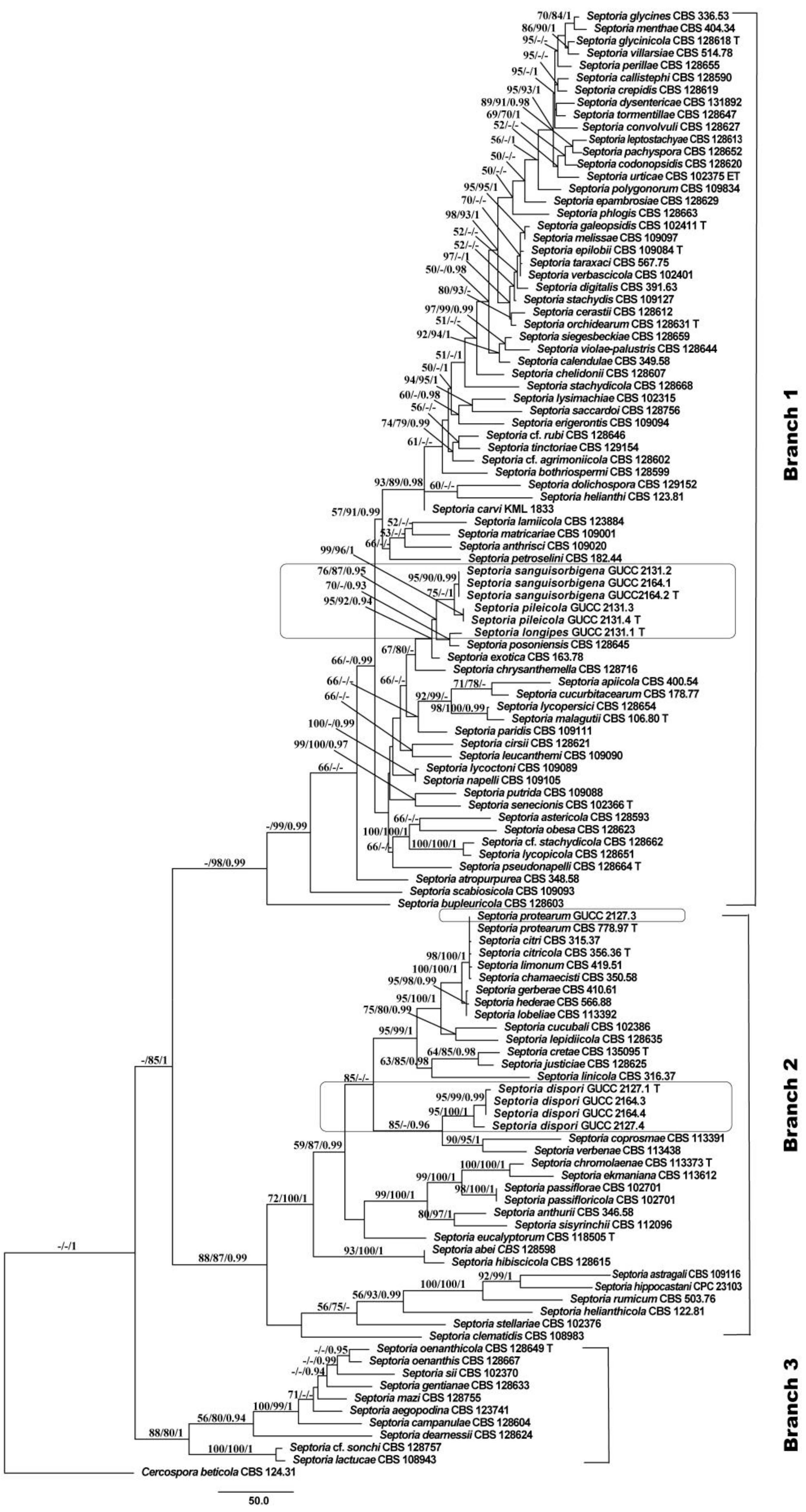
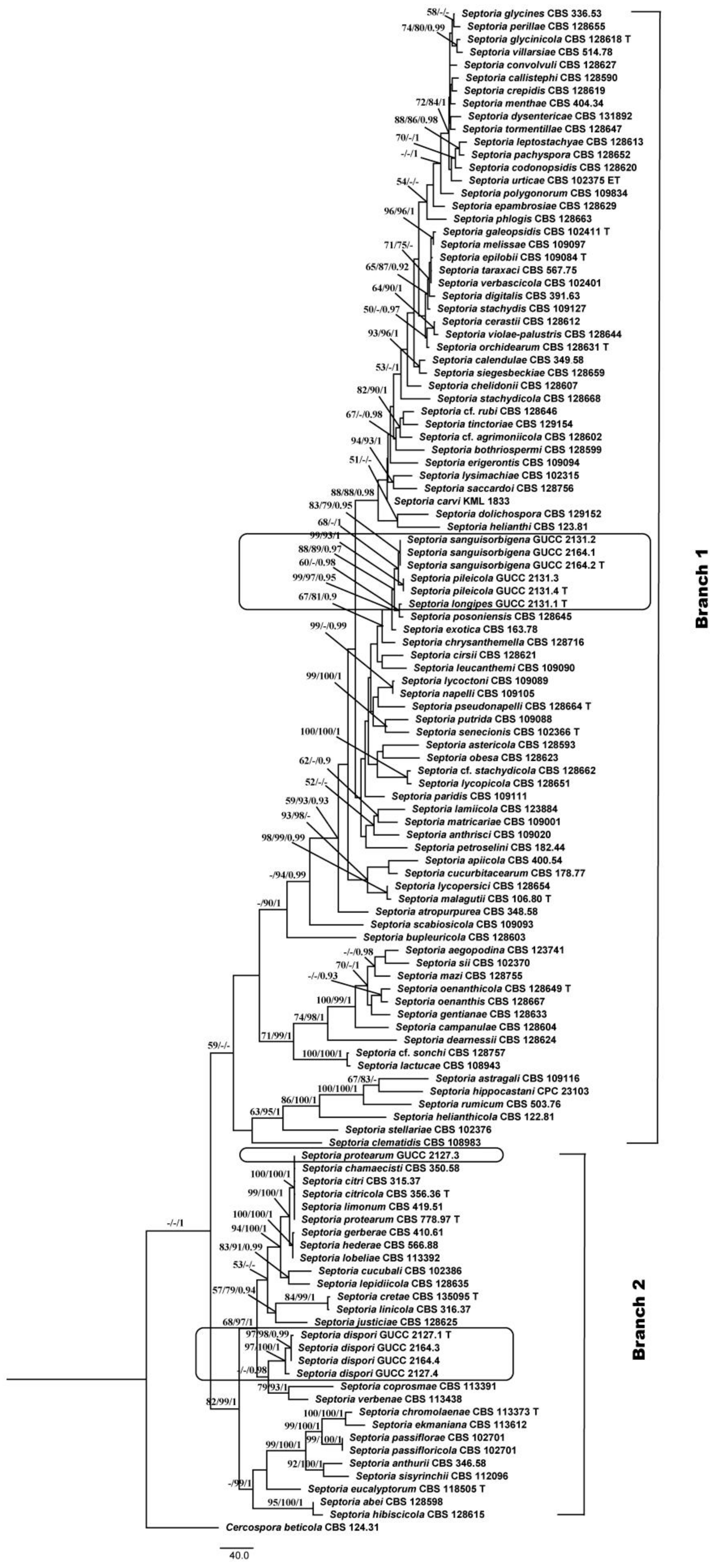
3.1. Phylogenetic Analyses

Eleven Septoria strains isolated from different plant hosts were sequenced. PCR products of 450–536 bp (tef1), 440–453 bp (tub2), 458–524 bp (ITS), 799–863 bp (LSU), and 718–1083 bp (rpb2) were obtained. By alignment with the single-gene region and then in combination in the order of tef1, rpb2, tub2, ITS, and LSU with Cercospora beticola (CBS 124.31), 2434 characters were obtained: tef1, 1–479; rpb2, 480–824; tub2, 825–1149; ITS, 1159–163; and LSU, 1636–2434. Among these characters, 1672 were constant, while 195 variable characters were parsimony-uninformative and 567 were parsimony informative. The parameters of the MP phylogenetic trees are shown in Table 3, and the procedure yielded a single most parsimonious tree (Figure 1). Similar topologies were obtained by MP, ML, and Bayesian methods. In the Septoria phylogenetic tree (Figure 1), all Septoria isolates were grouped together, but only the BI support was high (BPP = 1), while the three major clades received greater statistical support (Branch 1: ML/BI = 98%/0.99; Branches 2: MP/ML/BI = 88%/87%/0.99; Branch 3: MP/ML/BI = 88%/80%/1.00). Six strains (GUCC 2131.1, GUCC 2131.2, GUCC 2131.3, GUCC 2131.4, GUCC 2164.1, and GUCC 2164.2) were grouped in the clade that included S. posoniensis and S. exotica (MP: 95%, ML: 92% and BPP: 0.94) in Branch 1. In this group, five strains (GUCC 2131.2, GUCC 2131.3, GUCC 2131.4, GUCC 2164.1 and GUCC 2164.2) formed an independent branch adjacent to GUCC 2131.1 and S. posoniensis (MP: 76%, ML: 86%, and BPP: 0.95), but these five strains were split into two sub-branches: one containing GUCC 2131.2, GUCC2164.1, and GUCC2164.2, and the other containing GUCC 2131.3 and GUCC2131.4, with good support (MP: 75%; BPP: 1.00). Strain GUCC 2127.3 was aligned to the branch that included S. chamaecisti, S. citri, S. citricola, S. protearum, and S. limonum with high statistical support (MP: 98%, ML: 100% and BPP: 1) but small phylogenetic distances. Strains GUCC 2164.3, GUCC 2164.4, GUCC 2127.1, and GUCC 2127.4 formed a strongly supported group (MP: 95%; ML: 100%; BPP: 1.00) closely related to S. coprosmae and S. verbenae with good support values (MP: 85%; BPP: 0.96). In Branch 2, four strains clustered in a clade in which GUCC 2127.1, GUCC2164.3 and GUCC2164.4 formed a sub-group, were very close to GUCC 2127.4, supported by high statistical values (MP: 95%, ML: 100%, and BPP: 1).
We also compared the DNA base-pair differences in five different loci between our strains and related species (Supplementary Table S1). This revealed that the LSU gene region was too conserved for species-level identification, and the ITS had little value, but tef1, tub2, and rpb2 provided more than 80% of the DNA base-pair differences (Supplementary Table S1). We also built a phylogenetic tree based on four loci, excluding the LSU region (Figure 2), using the parameters for MP analysis in Table 3. The topology showed highly similar placements of our strains in the Septoria in Figure 1; however, in Figure 2 only two branches were formed and all members of Branch 3 were integrated with Branch 1. To evaluate the distinctive effectiveness of different DNA markers, five single gene trees were constructed (Supplementary Figures S1–S5) and all MP parameters were as indicated in Table 3. Through comparison, we found that only tub2 and tef1 included more parsimonious characters (50.7% and 49.2%), and the sequence of tub2 was shorter than that of tef1. Moreover, the topology originating from the tub2 gene region was more similar to Figure 2.

3.2. Genealogical Concordance Phylogenetic Species Recognition

In order to determine evolutionary independence, the GCPSR concept was applied to the GUCC 2164.2, GUCC 2131.4, GUCC 2131.1, and related taxa S. chrysanthemella (CBS 128716), S. exotica (CBS 163.78), and S. posoniensis (CBS 128645). A pairwise homoplasy index (PHI or Fw) less than 0.05 provided evidence of the presence of significant recombination within a dataset. According to the GCPSR analysis, our dataset showed PHI of 0.116, indicating no significant genetic recombination among our strains and related taxa. Hence, it was concluded that these taxa were significantly different from each other.
For GUCC 2164.3 and GUCC 2127.4 and related species S. coprosmae (CBS 113391) and S. verbenae (CBS 113438), the pairwise homoplasy index (PHI or Fw) was 1.173 × 10⁻⁸, which provided evidence for the presence of significant recombination within a dataset. The four strains could belong to a single species.

3.3. Taxonomy

(1)
Septoria sanguisorbigena Y.Y. An & Yong Wang bis, sp. nov. (Figure 3)
MycoBank MB 839125
Etymology: The name refers to the plant host, from which the fungus was collected.
Description in vitro: Colonies: on PDA 15–25 mm diameter after 2 weeks with a undulating even margin, restricted, irregularly pustulate; the surface almost black with low and finely felted diffuse, grey-to-white aerial mycelium. Conidiomata: pycnidial, epiphyllous, immersed, subglobose to globose, black, 120–250 µm diameter; ostiolum central, circular, initially 25–35 µm wide, later becoming more irregular and up to 100 µm wide, conidiomatal wall 20–40 µm thick, composed of an outer layer of angular-to-irregular cells mostly 4.5–10 µm diameter with pale to orange-brown walls and an inner layer of isodiametric, hyaline cells 7–20 μm diamater. Conidiogenous cells: hyaline, discrete, holoblastic, sympodially or percurrently proliferating, ampulliform, 4.5–8 × 1.5–2.5 µm (avg. = 5.6 × 2 µm, n = 30). Conidia: hyaline, filiform, straight to somewhat flexuous, the upper cell tapered into obtuse apex, relatively wide truncated base, (1–)3–5(–7) septate, not or only indistinctly constricted at the septa, contents granular or with minute oil-droplets around the septa and at the ends, 12.5–30 × 0.6–2 µm (avg. = 20.5 × 1.3 µm, n = 30). Sexual morph unknown.
Type: CHINA, Yunnan Province, Kunming Botanical Garden, from leaves of Sanguisorba officinalis L., February 2018, Y.Y. An (HGUP 2164.2, holotype); ex-type culture GUCC 2164.2; isotype culture MFLUCC 20-0185.
Other material examined: CHINA, Yunnan Province, Kunming Botanical Garden, from leaves of Sanguisorba officinalis, February 2018, Y.Y. An (HGUP 2164.2); from leaves of Pilea cadierei Gagnep. & Guillaumin, February 2018, Y.Y. An (HGUP 2131.2).
Notes: Phylogenetic analyses confirmed that three strains (GUCC 2131.2, GUCC 2164.1, and GUCC 2164.2) had a close relationship with S. chrysanthemella, S. exotica, S. longipes, S. pileicola, and S. posoniensis and this was supported by credible statistic values of the MP and ML methods (Figure 1). However, the independent branch only included those strains with high support values (MP: 95%, ML: 90%, and BPP: 0.99) adjacent to S. pileicola with moderate MP bootstrap but 1.00 BPP support. The new species had narrower conidia (0.6–2 µm) with 3–5 septa than those of S. pileicola (1.5–3.5 µm) with only 1–2 septa. In addition, this new taxon had obviously smaller conidia (12.5–30 × 0.6–2 µm) than S. chrysanthemella (34–66 × 2.5–3 µm) and S. longipes (17–46.5 × 1.5–2.5 µm) [35]. Septoria posoniensis and S. exotica have longer conidia, which was different to the new species [33,34]. DNA base differences indicated these three strains had nearly the identical sequence data (only two different bases on ITS region), but on protein-coding genes possessed more differences to distinguish them from related species (Supplementary Table S1). GCPSR test also provided a powerful proof to clarify them as different species.
(2)
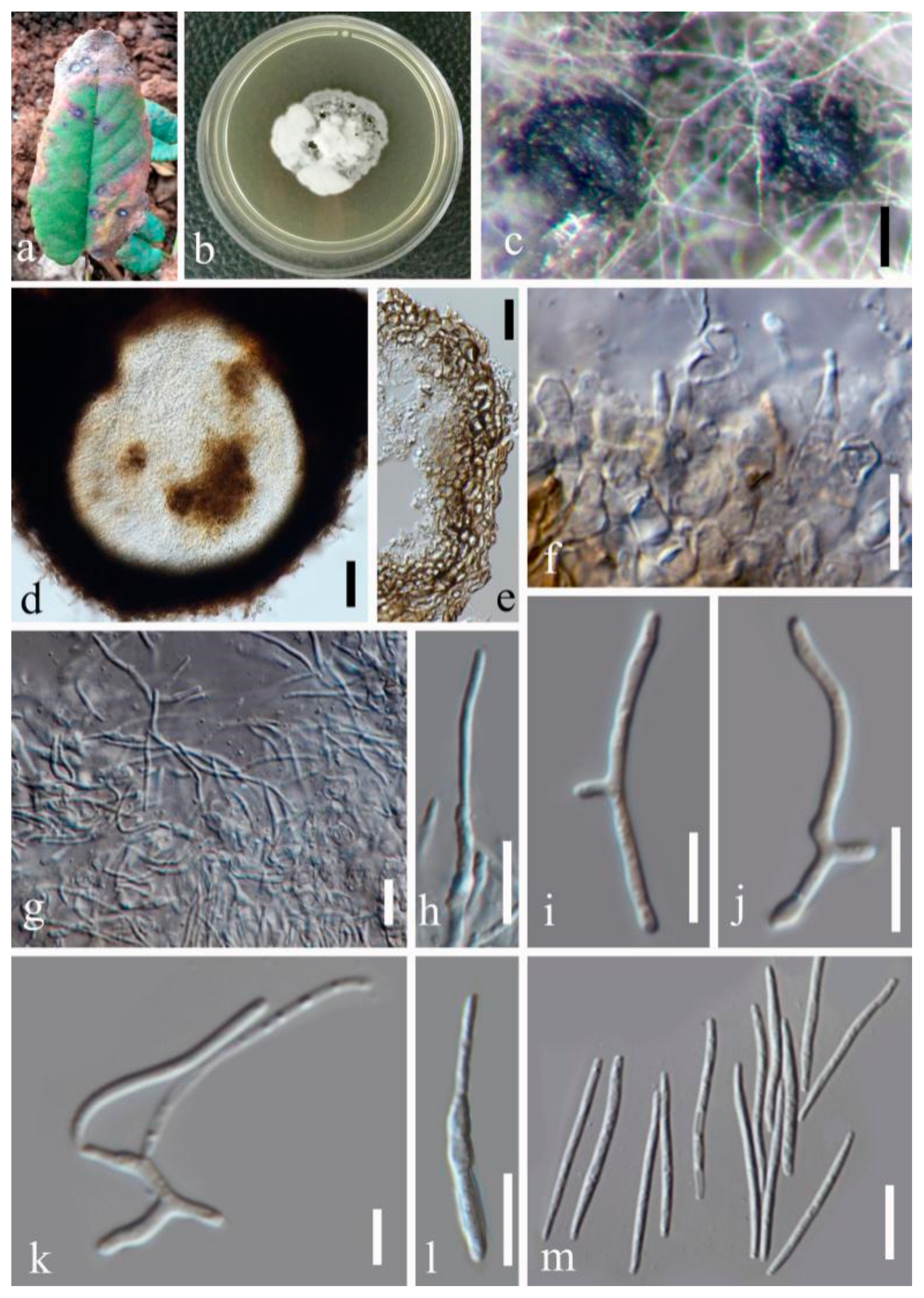
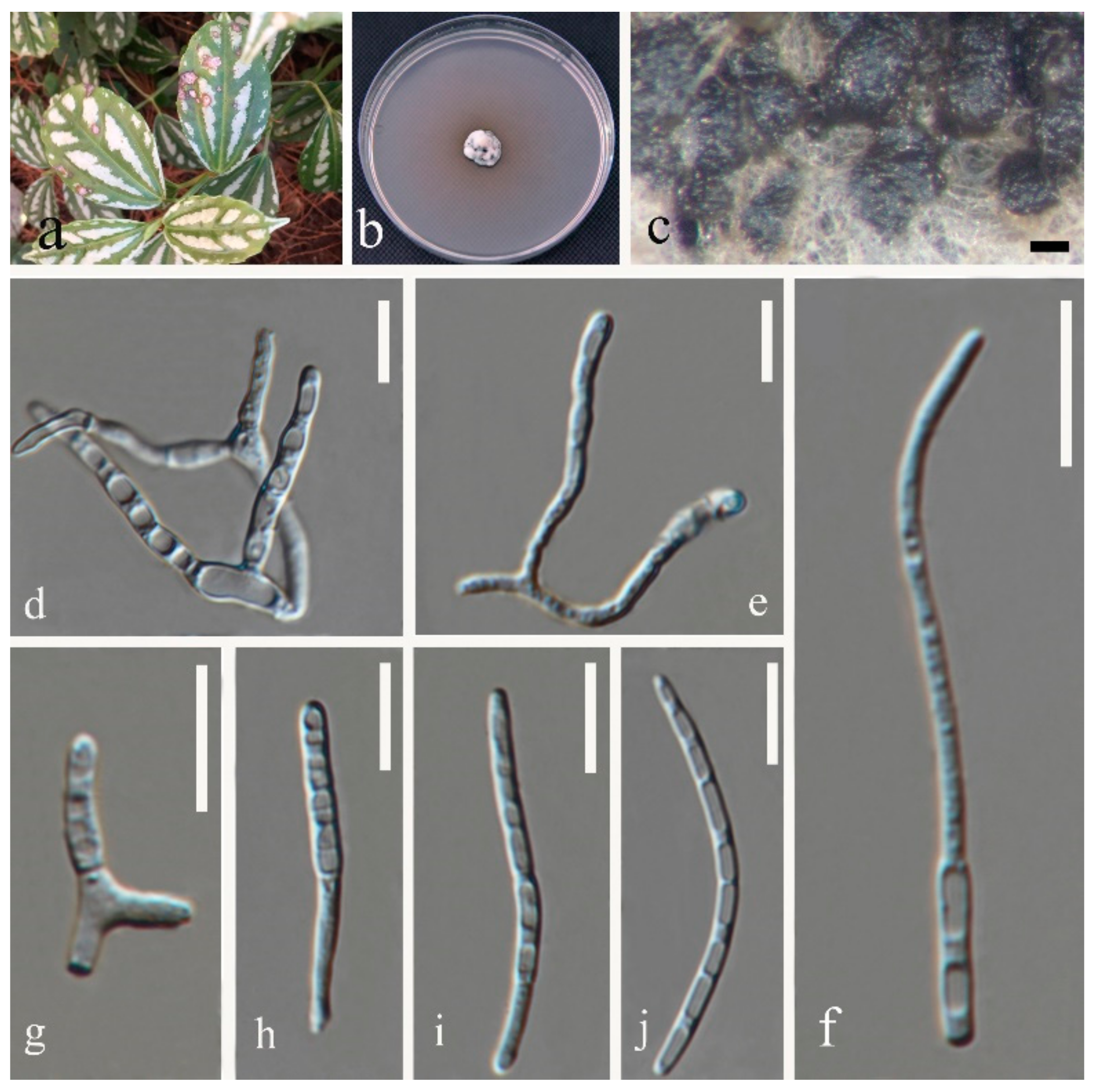
Septoria pileicola Y.Y. An & Yong Wang bis sp. nov. (Figure 4)
MycoBank MB 839126
Etymology: The name refers to the plant host from which the fungus was collected.
Description in vitro: Colonies: on PDA up to 10–15 mm diameter, with an even, glabrous, colourless margin in 2 weeks. Mycelium: greenish grey to dark slate-blue, immersed, throughout covered by well-developed, tufty whitish-grey aerial mycelium that later attains a reddish haze; reverse black, but margin paler; in the central part of the colony numerous pycnidia develop, releasing pale vinaceous to rosy-buff conidial ball. Conidiomata: pycnidial, epiphyllous but sometimes also visible from the underside of the lesion, one to a few in each leaf spot, subglobose to globose, brown to black, usually fully immersed, 80–120 µm diam. Ostiolum: central, initially circular and 15–30 µm wide, later becoming more irregular and up to 45 µm wide, surrounding cells concolorous to pale brown. Conidiogenous cells: hyaline, discrete, doliiform, or narrowly to broadly ampulliform, holoblastic, with a relatively narrow elongated neck, proliferating percurrently several times with distinct annellations, often also sympodially after a few percurrent proliferations, 5.5–12 × 2–3.5 µm. Conidia: cylindrical or filiform-cylindrical, straight to slightly curved, narrowly to broadly rounded at the apex, narrowing slightly or more distinctly to a truncate base, (0–)1–2-septate, not or slightly constricted around the septa, hyaline, contents with a few minute oil-droplets and granular material in each cell in the rehydrated state, 8.5–30 × 1.5–3.5 µm. Sexual morph unknown.
Type: CHINA, Yunnan Province, Kunming Botanical Garden, from leaves of Pilea cadierei Gagnep. & Guillaumin, February 2018, Y.Y. An (HGUP 2131.4, holotype); ex-type culture GUCC 2131.4; isotype culture MFLUCC 20-0184.
Other material examined: CHINA, Yunnan Province, Kunming Botanical Garden, from leaves of Pilea cadierei, February 2018, Y.Y. An (HGUP 2131.3).
Note: Phylogenetic analyses based on five gene regions showed that Septoria pileicola strains GUCC 2131.3 and GUCC 2131.4 are closely related to S. chrysanthemella, S. exotica, S. longipes, S. posoniensis, and S. sanguisorbigena (Figure 1), but formed a subclade with S. sanguisorbigena. After morphological comparisons, we found that Septoria pileicola can be distinguished from S. sanguisorbigena by its wider conidia, and from S. posoniensis and S. exotica by its shorter conidia with obviously fewer septa [36,37]. For S. chrysanthemella and S. longipes, the species had apparently shorter conidia [35]. The two strains of Septoria pileicola had nearly the identical sequences (only one different ITS base pair); however, the tub2 gene provided enough base distinction to separate it from related species (Supplementary Table S1) according to the guidelines of Jeewon and Hyde [38]. The PHI value was 0.116 (>0.05), indicating no significant genetic recombination among S. pileicola, S. sanguisorbigena, S. chrysanthemella, S. exotica, and S. posoniensis. Thus, they should belong to different species [39].
(3)
Septoria longipes Y.Y. An & Yong Wang bis sp. nov. (Figure 5)
MycoBank MB 839127
Etymology: The name refers to the long conidia of this species.
Description in vitro: Colonies: on PDA 11–15 mm diameter, with an even, light brown to dark-brown margin in 2 weeks; immersed mycelium grey to dark slate-blue in the center, black near the margin. Aerial mycelium: well-developed, white to snow white, covering the colony surface. Conidiomata: pycnidial, numerous, mostly epiphyllous, semi-immersed, black, mostly 80–200 µm diameter, with a central, first narrow, later wider opening, releasing pale white cirrhi of conidia. Conidiomatal wall: one or two layers of brown-walled, angular cells, lined by a layer of hyaline cells. Conidiogenous cells: hyaline, discrete, holoblastic, sympodially or percurrently proliferating, ampulliform, 8–16 × 1.5–5.5 µm. Conidia: filiform to filiform-cylindrical, straight, flexuous or curved, attenuated gradually to the narrowly rounded to pointed apex, attenuated gradually or more abruptly to the narrowly truncate base, (0–)3–5(–8)-septate, 17–46.5 × 1.5–2.5 µm. Sexual morph unknown.
Type: CHINA, Yunnan Province, Kunming Botanical Garden, from leaves of Pilea cadierei Gagnep. & Guillaumin, February 2018, Y.Y. An (HGUP 2131.1, holotype); ex-type culture GUCC 2131.1)
Notes: Only one strain (GUCC 2131.1) of this taxon was available. It clustered with S. posoniensis supported by MP (70%) and Bayesian (0.93) analyses and is closely related to S. chrysanthemella, S. exotica, S. pileicola, and S. sanguisorbigena. Morphological comparisons indicated that GUCC 2131.1 differed from S. posoniensis by conidia by more septa, and from S. chrysanthemella (4–10 × 5–6 µm) by larger conidiogenous cells (8–16 × 1.5–5.5 µm) [35,36]. This species produced longer conidia than S. pileicola and S. sanguisorbigena. It was confirmed that two protein-coding genes, except for tef1, provided enough base distinction with related species (Supplementary Table S1). GCPSR test also supported them as different species.
(4)
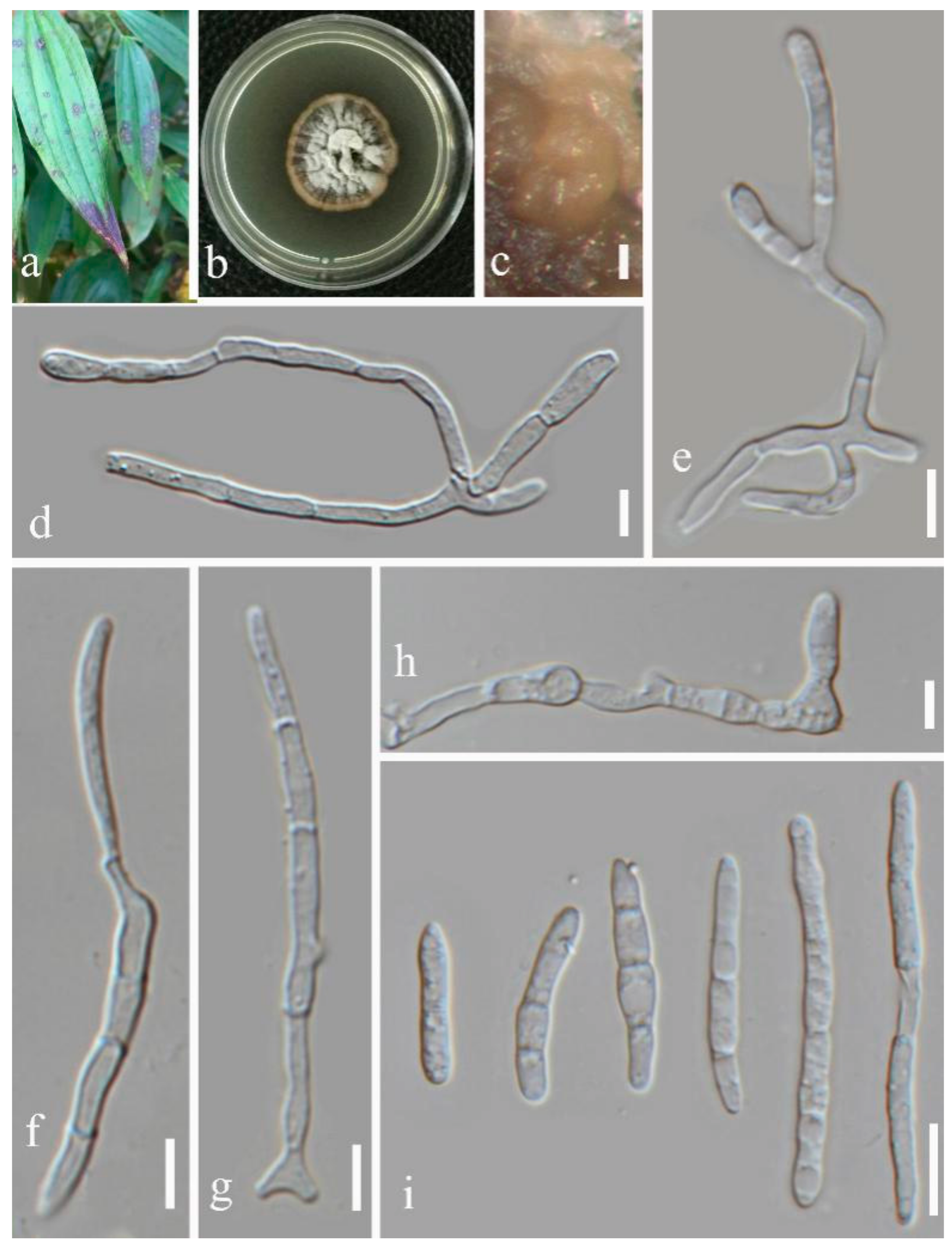
Septoria dispori Y.Y. An & Yong Wang bis sp. nov. (Figure 6)
MycoBank MB 839128
Etymology: The name refers to the plant host from which the fungus was collected.
Description in vitro: Colonies: on PDA 2.0–3.5 mm diameter, with an even to slightly ruffled, glabrous, dull yellow margin in 2 weeks, spreading, remaining almost plane, immersed mycelium yellowish brown to brown; aerial mycelium well-developed, goose feather flocculent on the surface of the colony; numerous conidiomatal initials developing at the surface, mature ones releasing cirrhi of conidia that first are milky white, later salmon, sometimes merging to form slimy masses covering areas of the colony surface. Conidiogenous cells: hyaline, broadly or elongated ampulliform, normally with a distinct neck, hyaline, holoblastic, proliferating percurrently, annellations indistinct, 10–15 ×1.5–2.5 µm. Conidia: cylindrical to filiform-cylindrical, slightly to strongly curved, rarely somewhat flexuous, narrowly rounded to pointed at the apex, attenuated gradually or more abruptly towards a narrowly truncate base, 3–5–8-septate, later with secondary septa dividing the cells, sometimes breaking up into smaller fragments in the cirrhus, not or slightly constricted around the septa, hyaline, 14–41.5 × 1.5–2.5 µm. Sexual morph unknown.
Type: CHINA, Yunnan Province, Botanical Garden of Kunming country, from leaves of Disporum bodinieri (Levl. et Vaniot.) Wang et Y. C. Tang, February 2018, Y.Y. An (HGUP 2127.1, holotype); ex-type culture GUCC 2127.1.
Other material examined: CHINA, Yunnan Province, Kunming Botanical Garden, from leaves of Disporum bodinieri, February 2018, Y.Y. An (HGUP 2127.4); from leaves of Sanguisorba officinalis L., February 2018, Y.Y. An (HGUP 2164.3 and HGUP 2164.4).
Note: Four strains (GUCC 2127.1, GUCC 2127.4, GUCC 2164.3, and GUCC 2164.4) of Septoria dispori clustered together with high statistical support (MP: 95%, ML: 100%, BPP: 1.00) adjacent to S. coprosmae and S. verbenae. Thus, we consider these four strains to be a single species. Septoria coprosmae produced spermatogonia of an Asteromella-state, but this species did not [40]. Conidia of S. verbenae possessed fewer septa than those of Septoria dispori [41]. GUCC 2127.4 showed some phylogenetic distance from the other three strains, however DNA base comparison (Supplementary Table S1) revealed only 11 bases that had tub2 differences. The PHT test confirmed significant recombination between strains GUCC 2164. 4 and GUCC 2164.3 and they were morphologically similar. Thus by combining the above evidence, we established the four strains as a new taxon.
(5)
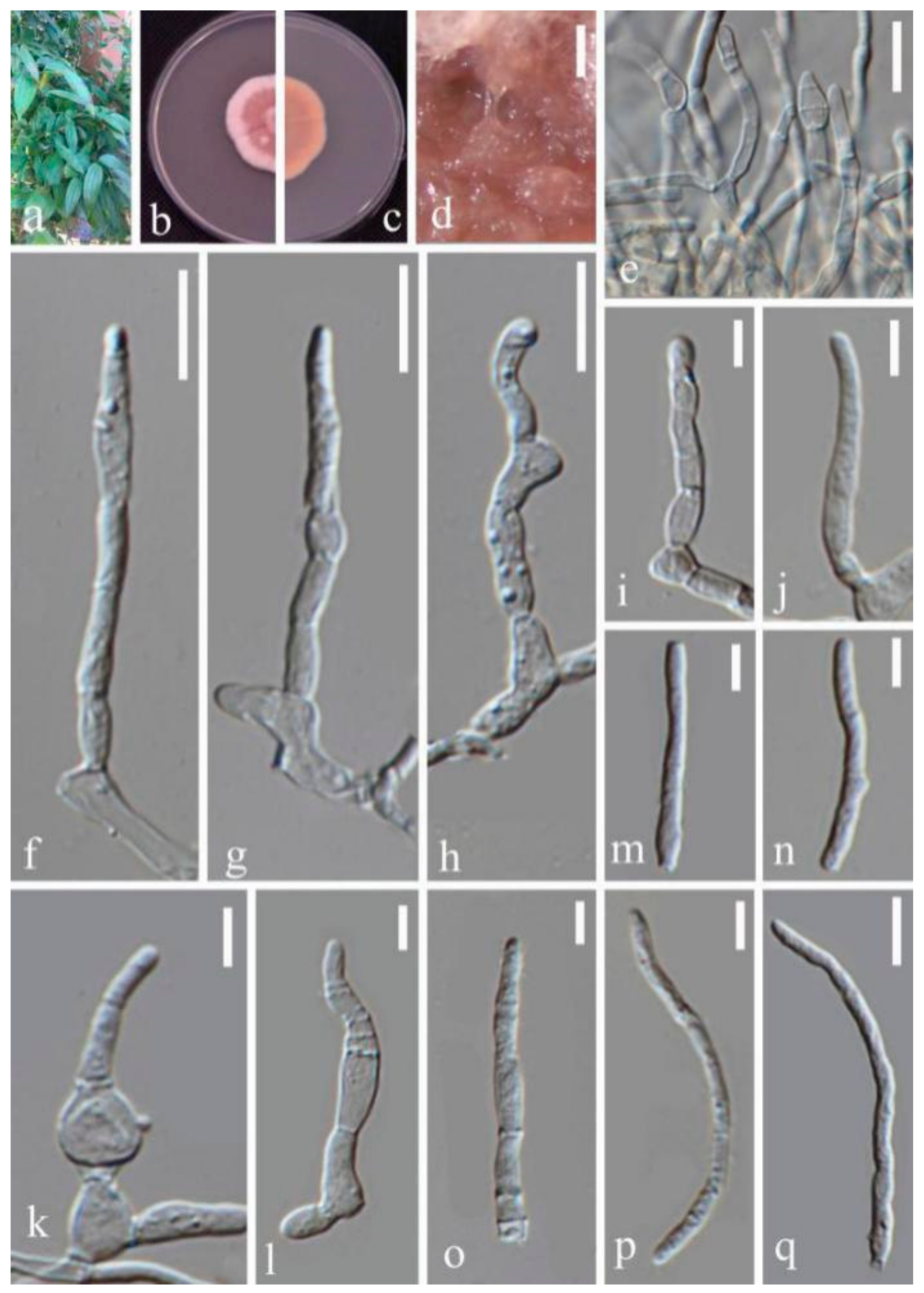
Septoria protearum Viljoen & Crous, in Swart, Crous, Denman & Palm, S. Afr. J. Bot. 64(2): 144 (1998) (Figure 7)
Description in vitro: Colonies: on PDA 15–25 mm with an even, glabrous white margin in 2 weeks, plane spreading, immersed. Mycelium: pink, lacking aerial hypha. Conidiomata developing after 1 week, mostly immersed and releasing whitish conidial slime. Conidiogenous cells: hyaline, cylindrical, broadly to narrowly ampulliform, with a distinct neck of variable length, holoblastic, with several distinct percurrent proliferations, more rarely also sympodial after a sequence of percurrent proliferations of the same cell, 5–10(–13.5) × 2.5–3(–3.5) µm. Conidia: filiform, straight, more often irregularly curved, 0–4 septate, not or only inconspicuously constricted around the septa, hyaline, 16–25 × 2.5–3.5 µm. Sexual morph unknown.
Material examined: CHINA, Yunnan Province, Kunming Botanical Garden, from leaves of Disporum bodinieri (Levl. et Vaniot.) Wang et Y. C. Tang, February 2018, Y.Y. An (HGUP 2127.3), living culture GUCC 2127.3.
Notes: DNA base comparison (Supplementary Table S1), revealed that sequences of strain GUCC 2127.3 were identical to the ex-type strain of S. protearum (CBS 778.97) in four gene regions. Conidial shape and size range of S. protearum (12–22 × 1.5–2 µm) were similar to the present strain [42]. The number of conidial septa of the two strains was also the same (0–4 septa). Thus, we conclude that GUCC 2127.3 is S. protearum.

4. Discussion

Verkley et al. [3] pointed out that for the identification of the Septoria species, morphological description must be integrated with sequences analyses. Quaedvlieg et al. [10] treated species in Septoria within a modern taxonomic framework and pointed out that Septoria spp. formed a well-defined phylogenetic clade. Regarding morphology, the species concept was to produce pycnidial, ostiolate conidiomata; conidiophores reduced to conidiogenous cells that proliferate sympodially; and hyaline, filiform conidia with transverse eusepta that fit the original concept of [4]. We followed this system and applied morphological and phylogenetic approaches to the present study. After comparing the topologies of five single-gene and two multi-gene trees (Figure 1 and Figure 2 and Supplementary Figures S1–S5), we showed that Septoria forms two branches (Branch 1 and Branch 2), mainly because only the phylogenetic trees based on the LSU region and five DNA fragments (including LSU) supported three branches, whereas the conserved LSU sequences included the least parsimonious characters (31/799) (Table 3). In morphology, all species in Branch 3 produced filiform or fusiform, sub-straight to slightly curved conidia mainly with 3 septa, which was not a unique characteristic. Thus, we proposed exclusion of the LSU region for multi-gene analyses of Septoria at the species level, but always as the primary DNA barcode with more parsimonious characters (43/486), the ITS fragment was conserved in the present phylogenetic analysis.
The S. protearum complex accommodated eight members: S. citri, S. citicola, S. chamaecisti, S. gerberae, S. hederae, S. lobelia, S. limonum, and S. protearum, according to Verkley et al. [3]. Apart from S. protearum, the other species were old names without ex-type cultures, and thus no sequences were available. The base comparison of DNA sequences originated from Verkley et al. [3], who indicated that among these eight species there were only approximately 10 base-pair differences on the rpb2 fragment (434 characters) of S. gerberae, S. hederae, and S. lobelia compared to the other five species, while for the other four gene regions, their sequences were nearly identical (≤1 base difference) (Supplementary Table S1). On the other hand, in the literature these seven species are depicted only by simple descriptions often without drawings or photographs, which does not strongly support them as different taxa. Comparing with the sequences from Verkley et al. [3] and in the absence of type materials, we were more willing to believe that they belonged to the same species, S. protearum.
Our 11 strains isolated from Disporum bodinieri, Pilea cadierei, Sanguisorba officinalis all from the Botanical Garden of Kunming county represented five Septoria species and included four novel species supported by morphology and phylogeny. Septoria sanguisorbigena was obtained from two plant hosts (Sanguisorba officinalis and P. cadierei), and S. dispori was also on two hosts (D. bodinieri and Sanguisorba officinalis). Septoria pileicola and S. longipes were only discovered on one host (P. cadierei). Our S. protearum strain was on D. bodinieri. Verkley et al. [3] recalled that trans-family host jumping must be a major force driving the evolution of Septoria. Our results support this hypothesis as we found the same species on different hosts. However, our findings revealed that the Septoria species did not show any host specialization, which differs from the view of Verkley et al. [3].

5. Conclusions

In this study, our 11 Septoria strains represented five species including four novel taxa, and one new record for China by morphological comparison and multi-gene analyses. The Septoria species are pathogens often causing leaf spot diseases of many plant hosts worldwide [10]. Based on previous studies, relatively sufficient reference sequences are available for rapid identification of Septoria pathogens. By comparing the parsimonious-informative characters of different DNA fragments (Supplementary Table S1), we showed that either tef1 or tub2 is suitable as a secondary DNA barcode, and that the latter was more discriminating than the former. Moreover, the DNA fragment of tub2 (≈300 bp) was shorter than that of tef1 (≈450 bp) with a high PCR amplification success rate. Consequently, a standardized approach including morphological characters and phylogenetic analysis is needed for the correct and precise identification of Septoria isolates.

Supplementary Materials

The following are available online at https://www.mdpi.com/article/10.3390/jof7060483/s1. Figure S1: The phylogenetic tree based on ITS region. Figure S2: The phylogenetic tree based on LSU region. Figure S3: The phylogenetic tree based on rpb2 region. Figure S4: The phylogenetic tree based on tef1 region. Figure S5: The phylogenetic tree based on tub2 region. Cercospora beticola (CBS 124.31) was used as outgroup taxon. MP and ML above 50% and BPP above 0.90 were placed close to topological nodes and separated by “/”, otherwise were labeled with “-”. Taxa from this study are highlighted in green. Table S1: DNA base difference between our Septoria strains and related species.

Author Contributions

Morphological and phylogenetic analyses, Y.-Y.A.; GCPSR analyses, M.C.D.; data curation, X.-Y.Z.; writing—original draft preparation, Y.W.; writing—review and editing, K.D.H. and A.J.L.P. All authors have read and agreed to the published version of the manuscript.

Funding

The study was funded by the National Natural Science Foundation of China (No. 31972222, 31560489), Program of Introducing Talents of Discipline to Universities of China (111 Program, D20023), Talent project of Guizhou Science and Technology Cooperation Platform ((2017)5788-5 and (2019)5641), Guizhou Science, Technology Department of International Cooperation Base project ((2018)5806), Guizhou Science and Technology Innovation Talent Team Project ((2020)5001), and UIDB/04046/2020 and UIDP/04046/2020 Centre grants from FCT, Portugal (to BioISI).

Institutional Review Board Statement

Not applicable.

Informed Consent Statement

Not applicable.

Data Availability Statement

The data presented in this study are available in Supplementary Figures S1–S5 and Table S1.

Acknowledgments

The authors would like to thank anonymous reviewers for helpful comments.

Conflicts of Interest

The authors declare no conflict of interest.

References

  1. Index Fungorum 2020. Available online: http://www.indexfungorum.org/names/Names.asp (accessed on 8 October 2020).
  2. Wijayawardene, N.N.; Hyde, K.D.; Al-Ani, L.K.T.; Tedersoo, L.; Haelewaters, D.; Rajeshkumar, K.C.; Zhao, R.L.; Aptroot, A.; Leontyev, D.V.; Saxena, R.K.; et al. Outline of Fungi and fungus-like taxa. Mycosphere 2020, 11, 1060–1456. [Google Scholar] [CrossRef]
  3. Verkley, G.J.M.; Quaedvlieg, W.; Shin, H.D.; Crous, P.W. A new approach to species delimitation in Septoria. Stud. Mycol. 2013, 75, 213–305. [Google Scholar] [CrossRef] [Green Version]
  4. Sutton, B.C. The Coelomycetes. Fungi Imperfecti with Pycnidia, Acervuli and Stromata; Commonwealth Mycological Institute: Kew, UK, 1980. [Google Scholar]
  5. Constantinescu, O. Taxonomic revision of Septoria-like fungi parasitic on Betulaceae. Trans. Br. Mycol. Soc. 1984, 83, 383–398. [Google Scholar] [CrossRef]
  6. Sutton, B.C.; Pascoe, I.G. Septoria species on Acacia. Trans. Br. Mycol. Soc. 1987, 89, 521–532. [Google Scholar] [CrossRef]
  7. Sutton, B.C.; Pascoe, I.G. Some Septoria species on native Australian plants. Stud. Mycol. 1989, 31, 177–186. [Google Scholar]
  8. Farr, D.F. Septoria species on Cornus. Mycologia 1991, 83, 611–623. [Google Scholar] [CrossRef]
  9. Farr, D.F. Species of Septoria on the Fabaceae, subfamily Faboidae, tribe Genistae. Sydowia 1992, 44, 13–31. [Google Scholar]
  10. Quaedvlieg, W.; Verkley, G.J.M.; Shin, H.D.; Barretto, R.W.; Alfenas, A.C.; Swart, W.J.; Groenewald, J.Z.; Crous, P.W. Sizing up Septoria. Stud. Mycol. 2013, 75, 307–390. [Google Scholar] [CrossRef] [Green Version]
  11. Crous, P.W.; Shivas, R.G.; Quaedvlieg, W.; van der Bank, M.; Zhang, Y.; Summerell, B.A.; Guarro, J.; Wingfield, M.J.; Wood, A.R.; Alfenas, A.C.; et al. Fungal Planet Description Sheets: 214–280. Persoonia 2014, 32, 184–306. [Google Scholar] [CrossRef]
  12. Crous, P.W.; Schumacher, R.K.; Wingfield, M.J.; Lombard, L.; Giraldo, A.; Christensen, M.; Gardiennet, A.; Nakashima, C.; Pereira, O.; Smith, A.J.; et al. Fungal Systematics and Evolution: FUSE 1. Sydowia 2015, 67, 81–118. [Google Scholar]
  13. Senanayake, I.C.; Rathnayaka, A.R.; Marasinghe, D.S.; Calabon, M.; Vedprakash, H.G. Morphological approaches in studying fungi: Collection, examination, isolation, sporulation and preservation. Mycosphere 2020, 11, 2678–2754. [Google Scholar] [CrossRef]
  14. Braun, U.; Nakashima, C.; Bakhshi, M.; Zare, R.; Shin, H.D.; Alves, R.F.; Sposito, M.B. Taxonomy and phylogeny of cercosporoid ascomycetes on Diospyros spp. with special emphasis on Pseudocercospora spp. Fungal Syst. Evol. 2020, 6, 95–127. [Google Scholar] [CrossRef]
  15. Dissanayake, A.J.; Bhunjun, C.S.; Maharachchikumbura, S.S.N.; Liu, J.K. Applied aspects of methods to infer phylogenetic relationships amongst fungi. Mycosphere 2020, 11, 2652–2676. [Google Scholar] [CrossRef]
  16. Kuraku, S.; Zmasek, C.M.; Nishimura, O.; Katoh, K. Leaves facilitates on-demand exploration of metazoan gene family trees on MAFFT sequence alignment server with enhanced interactivity. Nucleic Acids Res. 2013, 41, W22–W28. [Google Scholar] [CrossRef] [PubMed] [Green Version]
  17. Katoh, K.; Rozewicki, J.; Yamada, K.D. MAFFT online service: Multiple sequence alignment, interactive sequence choice and visualization. Brief. Bioinform. 2017, 20, 1160–1166. [Google Scholar] [CrossRef] [Green Version]
  18. Carbone, I.; Kohn, L.M. A method for designing primer sets for speciation studies in fifilamentous ascomycetes. Mycologia 1999, 91, 553–556. [Google Scholar] [CrossRef]
  19. O’Donnell, K.; Kistler, H.C.; Cigelnik, E.; Ploetz, R.C. Multiple evolutionary origins of the fungus causing Panama disease of banana: Concordant evidence from nuclear and mitochondrial gene genealogies. Proc. Natl. Acad. Sci. USA 1998, 95, 2044–2049. [Google Scholar] [CrossRef] [Green Version]
  20. O’Donnell, K.; Cigelnik, E. Two divergent intragenomic rDNA ITS2 types within a monophyletic lineage of the fungus Fusarium are nonorthologous. Mol. Phylogenetics Evol. 1997, 7, 103–116. [Google Scholar] [CrossRef] [PubMed]
  21. Stukenbrock, E.H.; Quaedvlieg, W.; Javan-Nikhah, M.; Zala, M.; Crous, P.W.; McDonald, B.A. Zymoseptoria ardabilia and Z. pseudotritici, two progenitor species of the Septoria tritici leaf blotch fungus Z. tritici (synonym: Mycosphaerella graminicola). Mycologia 2012, 104, 1397–1407. [Google Scholar] [CrossRef] [Green Version]
  22. Liu, Y.; Whelen, S.; Hall, B. Phylogenetic relationships among ascomycetes: Evidence from an RNA polymerse II subunit. Mol. Biol. Evol. 1999, 16, 1799–1808. [Google Scholar] [CrossRef]
  23. Quaedvlieg, W.; Kema, G.H.J.; Groenewald, J.Z.; Verkley, G.J.M.; Seifbarghi, S.; Razavi, M.; Gohari, A.M.; Mehrabi, R.; Crous, P.W. Zymoseptoria gen. nov.: A new genus to accommodate Septoria-like species occurring on graminicolous hosts. Persoonia 2011, 26, 57–69. [Google Scholar] [CrossRef] [PubMed] [Green Version]
  24. Crous, P.W.; Braun, U.; Wingfield, M.J.; Wood, A.R.; Shin, H.D.; Summerell, B.A.; Alfenas, A.C.; Cumagun, C.J.; Groenewald, J.Z. Phylogeny and taxonomy of obscure genera of microfungi. Persoonia 2009, 22, 139–161. [Google Scholar] [CrossRef] [Green Version]
  25. Vilgalys, R.; Hester, M. Rapid genetic identifification and mapping of enzymatically amplifified ribosomal DNA from several Cryptococcus species. J. Bacteriol. 1990, 172, 4238–4246. [Google Scholar] [CrossRef] [PubMed] [Green Version]
  26. White, T.J.; Bruns, T.; Lee, S.; Taylor, J.W. Amplifification and direct sequencing of fungal ribosomal RNA genes for phylogenetics. In PCR Protocols: A Guide to Methods and Applications; Innis, M.A., Gelfland, D.H., Sininsky, J.J., White, T.J., Eds.; Academic Press: San Diego, CA, USA, 1990; pp. 315–322. [Google Scholar]
  27. Tamura, K.; Peterson, D.; Peterson, N.; Stecher, G.; Nei, M.; Kumar, S. MEGA5: Molecular evolutionary genetics analysis using maximum likelihood, evolutionary distance, and maximum parsimony methods. Mol. Biol. Evol. 2011, 28, 2731–2739. [Google Scholar] [CrossRef] [PubMed] [Green Version]
  28. Swofford, D.L. PAUP—Phylogenetic Analysis Using Parsimony and Other Methods; Version 4; Sinauer Associates: Sunderland, MA, USA, 2003. [Google Scholar]
  29. Hillis, D.M.; Bull, J.J. An empirical test of bootstrapping as a method for assessing confidence in phylogenetic analysis. Syst. Biol. 1993, 42, 182–192. [Google Scholar] [CrossRef]
  30. CIPRES Science Gateway. Available online: https://www.phylo.org/portal2/login.action (accessed on 22 June 2020).
  31. FigTree. Available online: http://tree.bio.ed.ac.uk/software/figtree (accessed on 22 June 2020).
  32. Quaedvlieg, W.; Binder, M.; Groenewald, J.Z.; Summerell, B.A.; Carnegie, A.J.; Burgess, T.I.; Crous, P.W. Introducing the consolidated species concept to resolve species in the Teratosphaeriaceae. Persoonia 2014, 33, 1–40. [Google Scholar] [CrossRef] [PubMed] [Green Version]
  33. Huson, M.G. Physical properties of wool fibers in electrolyte solutions. Text. Res. J. 1998, 68, 595–605. [Google Scholar] [CrossRef]
  34. Huson, D.H.; Bryant, D. Application of phylogenetic networks in evolutionary studies. Mol. Biol. Evol. 2006, 23, 254–267. [Google Scholar] [CrossRef]
  35. Saccardo, P.A. Supplementum Universale, Pars. III. Sylloge Fungorum 1895, 11, 1–753. [Google Scholar]
  36. Bäumler, J.A. Mycologisches aus Pressburg. Hedwigia 1885, 24, 75. [Google Scholar]
  37. Spegazzini, C. Fungi argentini. Pugillus secundus (Continuacion). An. Soc. Cient. Argent. 1880, 10, 5–33. [Google Scholar]
  38. Jeewon, R.; Hyde, K.D. Establishing species boundaries and new taxa among fungi: Recommendations to resolve taxonomic ambiguities. Mycosphere 2016, 7, 1669–1677. [Google Scholar] [CrossRef]
  39. Chethana, K.W.T.; Zhou, Y.; Zhang, W.; Liu, M.; Xing, Q.K.; Hyde, K.D.; Yan, J.Y.; Li, X.H. Coniella vitis sp. nov. is the common pathogen of white rot in Chinese vineyards. Plant Dis. 2017, 101, 2123–2136. [Google Scholar] [CrossRef]
  40. Cooke, M.C. Some exotic fungi. Grevillea 1886, 14, 129–130. [Google Scholar]
  41. Saccardo, P.A. Sylloge Fungorum: Sylloge Sphaeropsidearum et Melanconiearum; Sumptibus auctoris: Patavii, Italy, 1884; Volume 3, pp. 1–860. [Google Scholar]
  42. Swart, L.; Crous, P.W.; Denman, S.; Palm, M.E. Fungi occurring on Proteaceae. I. S. Afr. J. Bot. 1998, 64, 137–146. [Google Scholar] [CrossRef] [Green Version]
Figure 1. Maximum Parsimony (MP) topology of Septoria generated from a combination of tef1, rpb2, tub2, ITS, and LSU sequences. Cercospora beticola (CBS 124.31) was used as outgroup taxon. MP and ML above 50% and BPP above 0.90 were placed close to topological nodes and separated by “/”, otherwise were labeled with “-”.
Jof 07 00483 g001
Figure 2. Maximum parsimony (MP) topology of Septoria generated from a combination of tef1, rpb2, tub2, and ITS sequences. Cercospora beticola (CBS 124.31) was used as an outgroup taxon. MP and ML above 50% and BPP above 0.90 were placed close to topological nodes and separated by “/”; otherwise, they were labeled with “-”.
Jof 07 00483 g002
Figure 3. Septoria sanguisorbigena (GUCC 2164.2) (a) Leaf spot symptoms on the host. (b) Colony on PDA culture. (c) Conidiomata formed on PDA culture. (d) Section through a conidioma. (e) Peridium. (fk) Conidiogenous cells, (l,m) Conidia. Scale bars: (c) = 25 µm, (d) = 50 µm, (e) = 20 µm, (fj) = 10 µm, (k) = 5 µm, (l,m) = 10 µm.
Jof 07 00483 g003
Figure 4. Septoria pileicola (GUCC 2131.4): (a) Leaf spot symptoms on the host. (b) Colonies on PDA culture. (c,d) Conidiomata on PDA culture. (e,f) Section though conidioma. (gk) Conidiogenous cells. (l,m) conidia. Scale bars: (c,d) = 125 µm, (eg) = 10 µm, (hm) = 10 µm.
Jof 07 00483 g004
Figure 5. Septoria longipes (GUCC 2131.1) (a) Leaf spot symptoms on the host. (b) Colony on PDA. (c) Conidiomata on PDA culture. (dg) Conidiophores, Conidiogenous cells and conidia. (hj) Conidia. Scale bars: (c) = 20 µm. (dj) = 10 µm.
Jof 07 00483 g005
Figure 6. Septoria dispori (GUCC 2127.1) (a) Leaf spot symptoms on the host. (b) Colony on PDA. (c) Conidiomata on PDA culture. (d,e) Conidiophores. (fh) Conidiogenous cells and conidia. (i) Conidia. Scale bars: (c) = 20 µm, (d) = 10 µm (e) = 5 µm, (fi) = 10 µm.
Jof 07 00483 g006
Figure 7. Septoria protearum (GUCC 2127.3) (a. Leaf spot symptoms on the host. (b,c) Colony on PDA. (b) From above; c. from below). (d) Mycelium. (e) Conidiophores. (fl) Conidiogenous cells and conidia. (mq) Conidia. Scale bars: (d) = 125 µm. (eh) = 10 µm. (i) = 5 µm. (jq) = 10 µm.
Jof 07 00483 g007
Table 1. Strains numbers and GenBank accession numbers for phylogenetic study.
SpeciesIsolate No.GenBank Accession No.
tef1tub2rpb2LSUITS
Cercospora beticolaCBS 124.31KF253246KF252780KF252304KF251802KF251298
Septoria aegopodinaCBS 123741KF253282KF252807KF251838KF251334
S. anthrisciCBS 109020KF253286KF252811KF252340KF251843KF251339
S. anthuriiCBS 346.58KF253288KF252813KF252342KF251845KF251341
S. apiicolaCBS 400.54KF253292KF252817KF252346KF251849KF251345
S. astericolaCBS 128593KF253294KF252819KF252348KF251851KF251347
S. astragaliCBS 109116KF253298KF252823KF252352KF251855KF251351
S. atropurpureaCBS 348.58KF253299KF252824KF252353KF251856KF251352
S. bothriospermiCBS 128599KF253301KF252826KF252355KF251858KF251354
S. bupleuricolaCBS 128603KF253303KF252828KF252357KF251860KF251356
S. calendulaeCBS 349.58KF253304KF252829KF252358KF251861KF251357
S. callistephiCBS 128590KF253305KF252830KF252359KF251862KF251358
S. campanulaeCBS 128604KF253308KF252833KF252362KF251865KF251361
S. carviKML 1833KX453687
S. cerastiiCBS 128612KF253311KF252836KF252365KF251868KF251364
S. cf. agrimoniicolaCBS 128602KF253284KF252809KF252338KF251841KF251337
S. cf. rubiCBS 128646KF253314KF252839KF252368KF251871KF251367
S. cf. sonchiCBS 128757KF253500KF253020KF252546KF252057KF251552
S. cf. stachydicolaCBS 128662KF253513KF253034KF252559KF252071KF251566
S. chamaecistiCBS 350.58KF253318KF252843KF252372KF251875KF251371
S. chelidoniiCBS 128607KF253319KF252844KF252373KF251876KF251372
S. chromolaenaeCBS 113373 TKF253321KF252846KF252375KF251878KF251374
S. chrysanthemellaCBS 128716KF253325KF252850KF252379KF251882KF251378
S. cirsiiCBS 128621KF253328KF252853KF252382KF251885KF251381
S. citriCBS 315.37KF253465KF252511KF252021KF251516
S. citricolaCBS 356.36 TKF253329KF252854KF252383KF251886KF251382
S. clematidisCBS 108983KF253330KF252855KF252384KF251887KF251383
S. codonopsidisCBS 128620KF253333KF252858KF252387KF251890KF251386
S. convolvuliCBS 128627KF253336KF252861KF252390KF251893KF251389
S. coprosmaeCBS 113391KF253255KF252787KF252313KF251812KF251308
S. crepidisCBS 128619KF253338KF252863KF252392KF251895KF251391
S. cretaeCBS 135095 TKF252720KF251736KF251233
S. cruciataeCBS 123747KF253340KF252865KF252394KF251897KF251393
S. cucubaliCBS 102386KF253344KF252869KF252398KF251901KF251397
S. cucurbitacearumCBS 178.77KF253346KF252400KF251903KF251399
S. dearnessiiCBS 128624KF253347KF252871KF252401KF251904KF251400
S. digitalisCBS 391.63KF253349KF252873KF252403KF251906KF251402
S. disporiGUCC 2127.1 TMT996515MT984348MT993632MT985366MT974584
S. disporiGUCC 2164.3MT996523MT984357MT993641MT985375MT974593
S. disporiGUCC 2164.4MT996524MT984358MT993642MT985376MT974594
S. disporiGUCC 2127.4MT996517MT984350MT993634MT985368MT974586
S. dolichosporaCBS 129152KF253350KF252874KF251907KF251403
S. dysentericaeCBS 131892KF253353KF252877KF252406KF251910KF251406
S. ekmanianaCBS 113612KF253355KF252879KF251912KF251408
S. epambrosiaeCBS 128629KF253356KF252880KF252407KF251913KF251409
S. epilobiiCBS 109084 TKF253358KF252882KF252409KF251915KF251411
S. erigerontisCBS 109094KF253360KF252884KF252411KF251917KF251413
S. eucalyptorumCBS 118505 TKF253365KF252889KF252415KF251921KF251417
S. exoticaCBS 163.78KF253366KF252890KF252416KF251922KF251418
S. galeopsidisCBS 102411 TKF253372KF252896KF252422KF251928KF251424
S. gentianaeCBS 128633KF253374KF252898KF252424KF251930KF251426
S. gerberaeCBS 410.61KF253468KF252988KF252514KF252024KF251519
S. glycinesCBS 336.53KF253377KF252901KF251933KF251429
S. glycinicolaCBS 128618 TKF253378KF252902KF252427KF251934KF251430
S. hederaeCBS 566.88KF253470KF252990KF252515KF252026KF251521
S. helianthiCBS 123.81KF253379KF252903KF252428KF251935KF251431
S. helianthicolaCBS 122.81KF253380KF252904KF252429KF251936KF251432
S. hibiscicolaCBS 128615KF253382KF252906KF252431KF251938KF251434
S. hippocastaniCPC 23103KF253510KF253031KF252556KF252068KF251563
S. justiciaeCBS 128625KF253385KF252909KF252434KF251941KF251437
S. lactucaeCBS 108943KF253387KF252911KF252436KF251943KF251439
S. lamiicolaCBS 123884KF253397KF252921KF252446KF251953KF251449
S. lepidiicolaCBS 128635KF253398KF252922KF252447KF251954KF251450
S. leptostachyaeCBS 128613KF253399KF252923KF252448KF251955KF251451
S. leucanthemiCBS 109090KF253403KF252927KF252452KF251959KF251455
S. limonumCBS 419.51KF253407KF252931KF252456KF251963KF251459
S. linicolaCBS 316.37KF253408KF252932KF252457KF251964KF251460
S. lobeliaeCBS 113392KF253460KF252981KF252507KF252016KF251511
S. longipesGUCC 2131.1 TMT984351MT993635MT985369MT974587
S. lycoctoniCBS 109089KF253409KF252933KF252458KF251965KF251461
S. lycopersiciCBS 128654KF253410KF252934KF252459KF251966KF251462
S. lycopicolaCBS 128651KF253412KF252936KF252461KF251968KF251464
S. lysimachiaeCBS 102315KF253413KF252937KF252462KF251969KF251465
S. malagutiiCBS 106.80 TKF253418KF252467KF251974KF251470
S. matricariaeCBS 109001KF253420KF252943KF252469KF251976KF251472
S. maziCBS 128755KF253422KF252945KF252471KF251978KF251474
S. melissaeCBS 109097KF253423KF252946KF252472KF251979KF251475
S. menthaeCBS 404.34KF253424KF252947KF251980KF251476
S. napelliCBS 109105KF253426KF252949KF252474KF251982KF251478
S. obesaCBS 128623KF253429KF252952KF252477KF251985KF251481
S. oenanthicolaCBS 128649 TKF253433KF252954KF252239KF251737KF251234
S. oenanthisCBS 128667KF253432KF252953-KF251989KF251485
S. orchidearumCBS 128631 TKF253434KF252955KF252482KF251990KF251486
S. pachysporaCBS 128652KF253437KF252958KF252485KF251993KF251488
S. paridisCBS 109111KF253438KF252959KF252486KF251994KF251489
S. passifloricolaCBS 102701KF253442KF252963KF252490KF251998KF251493
S. perillaeCBS 128655KF253444KF252965KF252491KF252000KF251495
S. petroseliniCBS 182.44KF253446KF252967KF252493KF252002KF251497
S. phlogisCBS 128663KF253448KF252969KF252495KF252004KF251499
S. pileicolaGUCC 2131.3MT996519MT984353MT993637MT985371MT974589
S. pileicolaGUCC 2131.4 TMT996520MT984354MT993638MT985372MT974590
S. polygonorumCBS 109834KF253453KF252974KF252500KF252009KF251504
S. posoniensisCBS 128645KF253456KF252977KF252503KF252012KF251507
S. protearumCBS 778.97 TKF253472KF252992KF252517KF252028KF251523
S. protearumGUCC 2127.3MT996516MT984349MT993633MT985367MT974585
S. pseudonapelliCBS 128664 TKF253475KF252995KF252520KF252031KF251526
S. putridaCBS 109088KF253477KF252997KF252522KF252033KF251528
S. rumicumCBS 503.76KF253478KF252998KF252523KF252034KF251529
S. saccardoiCBS 128756KF253479KF252999KF252524KF252035KF251530
S. sanguisorbigenaGUCC 2131.2MT996518MT984352MT993636MT985370MT974588
S. sanguisorbigenaGUCC 2164.1MT996521MT984355MT993639MT985373MT974591
S. sanguisorbigenaGUCC 2164.2 TMT996522MT984356MT993640MT985374MT974592
S. scabiosicolaCBS 109093KF253487KF253007KF252532KF252043KF251538
S. senecionisCBS 102366 TKF253492KF253012KF252538KF252049KF251544
S. siegesbeckiaeCBS 128659KF253494KF253014KF252540KF252051KF251546
S. siiCBS 102370KF253497KF253017KF252543KF252054KF251549
S. sisyrinchiiCBS 112096KF253499KF253019KF252545KF252056KF251551
S. stachydicolaCBS 128668KF253512KF253033KF252558KF252070KF251565
S. stachydisCBS 109127KF253517KF253038KF252563KF252075KF251570
S. stellariaeCBS 102376KF253521KF253042KF252567KF252079KF251574
S. taraxaciCBS 567.75KF253524KF253045KF252570KF252082KF251577
S. tinctoriaeCBS 129154KF253525KF253046KF252571KF252083KF251578
S. tormentillaeCBS 128647KF253527KF253048KF252573KF252085KF251580
S. urticaeCBS 102375 TKF253530KF253051KF252576KF252088KF251583
S. verbascicolaCBS 102401KF253531KF253052KF252577KF252089KF251584
S. verbenaeCBS 113438KF253532KF253053KF252578KF252090KF251585
S. villarsiaeCBS 514.78KF253534KF253055KF252580KF252092KF251587
S. violae-palustrisCBS 128644KF253537KF253058KF252583KF252095KF251590
Ex-type isolates are labeled with “T”.
Table 2. Primers, primer sequences, and thermal cycling program for PCR amplification.
LocusPrimerPrimer Sequence 5′ to 3′Annealing Temperature (°C)DirectionReference
tef1EF1-728FCATCGAGAAGTTCGAGAAGG52Forward[18]
EF-2GGARGTACCAGTSATCATGTTReverse[19]
tub2T1AACATGCGTGAGATTGTAAGT52Forward[20]
β-Sandy-RGCRCGNGGVACRTACTTGTTReverse[21]
rpb2fRPB2-5FGAYGAYMGWGATCAYTTYGG49Forward[22]
fRPB2-414RACMANNCCCCARTGNGWRTTRTGReverse[23]
LSULSU1FdGRATCAGGTAGGRATACCCG52Forward[24]
LR5TCCTGAGGGAAACTTCGReverse[25]
ITSITS5GGAAGTAAAAGTCGTAACAAGG52Forward[26]
ITS4TCCTCCGCTTATTGATATGCReverse[26]
Table 3. Parameters for MP analyses.
Total CharactersNumber of Parsimony-Informative CharactersTLCIRIHIRC
ITS486431760.6420.760.3580.488
LSU799311120.6250.8630.3750.539
rpb2345187800.2730.7460.7270.204
tef146923114980.3790.7070.6210.268
tub232516512210.3260.7740.6740.252
tef1 + rpb2 + tub2 + ITS162554839270.3280.7160.6720.235
tef1 + rpb2 + tub2 + ITS + LSU243456740750.3300.7200.6700.238
Publisher’s Note: MDPI stays neutral with regard to jurisdictional claims in published maps and institutional affiliations.

