
Fungal Endophytes: A Potential Source of Antibacterial Compounds
by
 , , , , and
Sunil K. Deshmukh
1,2,* ,
,
Laurent Dufossé
3,* ,
,
Hemraj Chhipa
4 ,
,
Sanjai Saxena
2,5,
Girish B. Mahajan
6 and
Manish Kumar Gupta
7 
1
TERI-Deakin Nano Biotechnology Centre, The Energy and Resources Institute, Darbari Seth Block, IHC Complex, Lodhi Road, New Delhi 110003, Delhi, India
2
Agpharm Bioinnovations LLP, Incubatee: Science and Technology Entrepreneurs Park (STEP), Thapar Institute of Engineering and Technology, Patiala 147004, Punjab, India
3
Chimie et Biotechnologie des Produits Naturels (CHEMBIOPRO Lab) & ESIROI Agroalimentaire, Université de la Réunion, 15 Avenue René Cassin, 97744 Saint-Denis, France
4
College of Horticulture and Forestry, Agriculture University Kota, Jhalawar 322360, Rajasthan, India
5
Department of Biotechnology, Thapar Institute of Engineering and Technology, Patiala 147004, Punjab, India
6
HiMedia Laboratories Pvt. Ltd., Mumbai 400086, Maharashtra, India
7
SGT College of Pharmacy, SGT University, Gurugram 122505, Haryana, India
*
Authors to whom correspondence should be addressed.
J. Fungi 2022, 8(2), 164; https://doi.org/10.3390/jof8020164
Submission received: 10 December 2021
/
Revised: 4 February 2022
/
Accepted: 5 February 2022
/
Published: 8 February 2022
(This article belongs to the Special Issue Fungi and Fungal Metabolites for the Improvement of Human and Animal Life, Nutrition and Health 2.0)
Abstract
:Antibiotic resistance is becoming a burning issue due to the frequent use of antibiotics for curing common bacterial infections, indicating that we are running out of effective antibiotics. This has been more obvious during recent corona pandemics. Similarly, enhancement of antimicrobial resistance (AMR) is strengthening the pathogenicity and virulence of infectious microbes. Endophytes have shown expression of various new many bioactive compounds with significant biological activities. Specifically, in endophytic fungi, bioactive metabolites with unique skeletons have been identified which could be helpful in the prevention of increasing antimicrobial resistance. The major classes of metabolites reported include anthraquinone, sesquiterpenoid, chromone, xanthone, phenols, quinones, quinolone, piperazine, coumarins and cyclic peptides. In the present review, we reported 451 bioactive metabolites isolated from various groups of endophytic fungi from January 2015 to April 2021 along with their antibacterial profiling, chemical structures and mode of action. In addition, we also discussed various methods including epigenetic modifications, co-culture, and OSMAC to induce silent gene clusters for the production of noble bioactive compounds in endophytic fungi.
1. Introduction
Over the decades since the discovery of the first antibiotics, resistance to those has been a curse that is being dragged along with every discovery of new antibiotics. This has kept all scientists, professionals, and clinical specialists working on antibiotics on their toes. The quest for new antibiotics scaffolds and repurposing of existing molecules has been persistent for the past nine decades. Getting a new and right scaffold is a herculean task, especially with the least ability to induce mutations in the target bacteria. As examined in some of the earlier reviews [1,2] there are several ways of getting new scaffolds and classes of antimicrobial bioactive compounds. In the domain of natural products, one of the most demonstrated ways is studying less explored species and genera of microbes [3,4,5]. Investigating unexplored ecological units on the globe synergizes with the concept of investigating the least or not explored species of microbes.
In the current review, we present the latest ways of exploring the credentials of such microbial sources, especially endophytic fungi, as a main stream of novel antimicrobial scaffolds. Bioactive compounds are mainly responsible for the activity profiles displayed by endophytic fungi. These metabolites belong to a wide range of scaffolds such as alkaloids, benzopyranones, chinones, peptides, phenols, quinones, flavonoids, steroids, terpenoids, tetralones, xanthones, and others. Moreover, they, in the pure form, have demonstrated abundant biological activities, including antibacterial, antifungal, anticancer, antiviral, antioxidant, immunosuppressant, anti-inflammatory, and antiparasitic properties [6,7,8,9,10,11,12,13,14,15]. Even though there are a few specialized reviews on the bioactive compounds from fungi, actinomycetes and other microbes [16,17], the amount of work done in the area is quite versatile, tenacious and significant. There is a need to comprehend these topics periodically to have its effective output for future research keeping in mind the probability of success of any newly discovered bioactive compound in clinical studies has been 0.01 to 1 % based on therapeutic area and type of scaffold. This demands that the base of such scaffolds in the ladder of clinical development should be wider. This width can be increased by exploring such less-tapped resources, the endophytic fungi.
In our previous review, we have covered antibacterials reported from endophytic fungi up to 2014 [1]. This review describes some bioactive molecules isolated from 2015 onwards to early 2021 from various endophytic fungi from terrestrial plants and designated as antibacterials. The antibacterial activity against various pathogenic organisms is listed in Table 1.
2. Antibacterials from Various Class of Endophytic Fungi
2.1. Ascomycetes
Ascomycetes are the fungi characterized by the formation of ascospores and some of the genera belonging to this class are known to produce chemically diverse metabolites. The important genera include Diaporthe, Xylaria, Chaetomium, Talaromyces, and Paraphaeosphaeria and are known to produce terpenoids, cytochalasins, mellein, alkaloids, polyketides, and aromatic compounds. Here we report the antibacterial from ascomycetes.
2.1.1. Diaporthe (Asexual State: Phomopsis)
The genus Diaporthe (asexual state: Phomopsis) has been thoroughly investigated for secondary metabolites that have various pathogenic, endophytic and saprobic species of temperate and tropical habitats. Two natural bisanthraquinone, (+)-1,1′-bislunatin (bis) (1) and (+)-2,2′-epicytoskyrin A (epi) (2, Figure 1), were extracted from endophytic fungi, Diaporthe sp. GNBP-10 is associated with plant Uncaria gambir. Compounds (bis)-(1) and (epi)-(2) showed promising anti-tubercular activity, against Mycobacterium tuberculosis strains H37Rv (Mtb H37Rv) with MIC values of 0.422 and 0.844 μM, respectively. Both compounds have the ability to combat nutrient-starvation and biofilms of the Mtb model with relatively moderate activity in bacterial reduction with between 1–2 fold log reduction. Both compounds could reduce the number of Mtb infected into macrophages with 2-fold log reduction. The in-silico results via a docking study show that both compounds have a good affinity with pantothenate kinase (PanK) enzyme with a Glide score of −8.427 kcal/mol and −7.481 kcal/mol for the epi and bis compounds, respectively [18].
An endophytic fungus, Diaporthe sp. GDG-118, associated with Sophora tonkinensis collected from Hechi City (China) yielded a new compound 21-acetoxycytochalasin J3 (3, Figure 1) and inhibited the pathogens Bacillus anthraci and E. coli at 12.5 μg/mL concentration (6 mm sterile filter paper discs were impregnated with 20 µL (50 µg) of each compound) [19].
Two novel naphthalene derivatives, 1-(3-hydroxy-1-(hydroxymethyl)-2-methoxy-6-methylnaphthalen-7-yl) propan-2-one (4) and 1-(3-hydroxy-1-(hydroxymethyl)-6-methyl-naphthalen-7-yl)propan-2-one (5, Figure 1), were obtained from the Phomopsis fukushii. Compounds 4 and 5 displayed poor anti-methicillin-resistant Staphylococcus aureus (anti-MRSA) activity, with zones of inhibition of 10.2 and 11.3 mm, respectively (6 mm sterile filter paper discs were impregnated with 20 µL (50 µg) of each compound) [20].
Earlier Phomopsis fukushii (Diaporthe fukushii) isolated from the rhizome of Paris polyphylla var. yunnanensis was the source of three new compounds namely 3-hydroxy-1-(1,8- dihydroxy-3,6-dimethoxynaphthalen-2-yl)propan-1-one (6), 3-hydroxy-1-(1,3,8-trihydroxy-6-methoxynaphthalen-2-yl)propan-1-one (7) and 3-hydroxy-1-(1,8-dihydroxy3,5-dimethoxy naphthalen-2-yl) propan-1-one (8, Figure 1). Compounds 6–8 exhibited anti-MRSA-ZR11 activity, with MIC values of 8, 4, and 4 µg/mL, respectively [21]. Later two new di-Ph ethers, 1-[2-methoxy-4-(3-methoxy-5-methylphenoxy)-6-methylphenyl]-ethanone (9) and 1-[4-(3-(hydroxymethyl)-5-methoxyphenoxy)-2-methoxy-6-methylphenyl]-ethanone (10, Figure 1), were also purified from the same fungus. Compounds 9–10 exhibited anti-MRSA activity with good inhibition (zones of 13.8 and 14.6 mm, respectively) [22].
Three new di-Ph ethers, 4-(3-methoxy-5-methylphenoxy)-2-(2-hydroxyethyl)-6-methylphenol (11), 4-(3-hydroxy-5-methylphenoxy)-2-(2-hydroxyethyl)-6-methylphenol (12) and 4-(3-methoxy-5-methylphenoxy)-2-(3-hydroxypropyl)-6-methylphenol (13, Figure 1) were purified from Phomopsis fukushii associated with the rhizome of Paris polyphylla var. yunnanensis. Compounds 11–13, exhibited potent anti-MRSA activity, with 20.2, 17.9 and 15.2 mm inhibition zones, respectively, when tested at 50 µg concentration in 6 mm discs [23].
Phomopsis fukushii isolated from the rhizome of Paris polyphylla var. yunnanensis yielded three new isopentylated diphenyl ethers, 1-(4-(3-methoxy-5-methylphenoxy)-2-methoxy-6-methylphenyl)-3-methylbut-3-en-2-one (14), 1-(4-(3-(hydroxymethyl)-5-methoxyphenoxy)-2-methoxy-6-methylphenyl)-3-methylbut-3-en-2-one (15) and 1-(4-(3-hydroxy-5-(hydroxymethyl) phenoxy)-2-methoxy-6-methylphenyl)-3-methylbut-3-en-2-one (16, Figure 1). Compounds 14–16 displayed anti-MRSA activity with 21.8, 16.8 and 15.6 mm inhibition zones, respectively (50 µg/6 mm disc) [24].
Two new anthraquinones, 3-hydroxy-6-hydroxymethyl-2,5-dimethylanthraquinone (17) and 6-hydroxymethyl-3-methoxy-2,5-dimethylanthraquinone (18, Figure 1), were purified from the endophytic fungus Phomopsis sp. and displayed good anti-MRSA activity with inhibition zone diameters (IZDs) of 14.2 and 14.8 mm, respectively [25].
A new dihydroisocoumarin derivative diaporone A (19, Figure 1), was purified from Diaporthe sp. an endophyte of Pteroceltis tatarinowii. Compound 19 showed MIC at 66.7 μM against Bacillus subtilis [26].
A pair of new phenolic bisabolane-type sesquiterpenoid enantiomers (±)-phomoterpenes A and B [(±)-1] (20) along with two new isocoumarins, phomoisocoumarins C-D (21–22, Figure 1) were purified from an endophytic fungus Phomopsis prunorum (F4-3). Compounds (+)-1 (20 and 22) exhibited average antimicrobial activity against Pseudomonas syringae pv. lachrymans with MIC values of 15.6 μg/mL, and compounds (−)-1 (20 and 21) displayed poor activity with MICs of 31.2 μg/mL each. Compounds (−)-1, (+)-1, (20, 21, 22) showed antibacterial activity against Xanthomonas citri pv. phaseoli var. fuscans with MIC values of 31.2, 62.4, 31.2, and 31.2 μg/mL, respectively [27].
The fungus Diporthe vochysiae LGMF1583 isolated from Vochysia divergens yielded two new carboxamides, vochysiamides A (23), and B (24, Figure 2). Compound 24 inhibited Klebsiella pneumoniae carbapenemase-producing (KPC), MSSA, and MRSA with MIC of 0.08, 1.0, and 1.0 µg/mL, respectively, and compound 23 was active against KPC with a MIC of 1.0 μg/mL. KPC is of public health concern due to the presence of antimicrobial resistance carbapenemases [28].
An endophyte Phomopsis asparagi obtained from the rhizome of Paris polyphylla var. yunnanensis was the source of two new di-Ph ethers, 4-(3-methoxy-5-methylphenoxy)-2-(2-hydroxyethyl)- 6-(hydroxymethyl)phenol (25), and 4-(3-hydroxy-5-methylphenoxy)-2-(2-hydroxyethyl)-6-(hydroxymethyl)phenol (26, Figure 2). Compounds 25 and 26 exhibited potent anti-MRSA activity with 10.8 and 11.4 mm inhibition zones, respectively [29].
Two new naphthalene derivatives, 5-methoxy-2-methyl-7-(3-methyl-2-oxobut-3-enyl)-1-naphthaldehyde (27) and 2-(hydroxymethyl)-5-methoxy-7-(3-methyl-2-oxobut-3-enyl)-1-naphthaldehyde (28, Figure 2), were characterized from Phomopsis sp., an endophyte of Paris polyphylla var. yunnanensis. Compounds 27 and 28 displayed potent antibacterial activity with 14.5 and 15.2 mm zones of inhibition, respectively, against MRSA [30].
The endophytic fungus Diaporthe terebinthifolii LGMF907 associated with the plant Schinus terebinthifolius yielded diaporthin (29) and orthosporin (30, Figure 2). Compound 29 displayed antimicrobial activity against various pathogens like E. coli, Micrococcus luteus, MRSA, and S. aureus with 1.73, 2.47, 9.50, and 9.0 mm zones of inhibition, respectively at 100 μg/disk concentration. Compound 30 inhibited E. coli, M. luteus, MRSA, and S. aureus with 1.03, 1.53, 9.0 and 9.33 mm zones of inhibition, respectively, when tested at 100 μg/disk [31].
A pyrimidine iminomethylfuran derivative, (2Z)-2-(1,4-dihydro-2-hydroxy-1-((E)-2-mercapto-1-(methylimino)ethyl)pyrimidine-4-ylimino)-1-(4,5-dihydro-5-methylfuran-3-yl)-3-methylbutane-1-one (31, Figure 2) was extracted from Phomopsis/Diaporthe sp. GJJM 16 is associated with Vitex negundo and inhibited S. aureus, and P. aeroginosa with MICs of 1.25 μg/mL each [32].
Phomopsis sp. PSU-H188 associated with Hevea brasiliensis, yielded the known compounds diaporthalasin (32), cytosporones B (33) and cytosporones D (34, Figure 2). Compound 32, displayed antibacterial activity against S. aureus and MRSA with equal MIC values of 4 μg/mL, but compound 33 inhibited S. aureus and MRSA with MIC values of 32 and 16 μg/mL, respectively. Compound 34 also inhibited S. aureus and MRSA with MIC values at higher concentrations of 64 and 32 μg/mL, respectively [33].
An endophyte, Diaporthe terebinthifolii GG3F6, associated with Glycyrrhiza glabra yielded two new hydroxylated unsaturated fatty acids namely diapolic acid A–B (35–36) and the known molecules xylarolide (37, Figure 2) and phomolide G (38, Figure 3). Compounds 35–38 inhibited Yersinia enterocolitica with an IC50 values of 78.4, 73.4, 72.1 and 69.2 μM, respectively [34].
The compounds phomosine A (39), and phomosine C (40, Figure 3), were obtained from Diaporthe sp. F2934 from Siparuna gesnerioides. Compound 39 was found to be active against Bordetella bronchiseptica, Enterococcus faecalis, Enterococcus cloacae, S. aureus, and Streptococcus oralis with 10, 10, 10, 12 and 9 mm inhibition zones at 4 µg/mL concentration, respectively. Compound 40 inhibited S. aureus, M. luteus, S. oralis, E. faecalis, E. cloacae, and B. bronchiseptica, with 9, 6, 8, 8, 8 and 9 mm inhibition zones at 4 µg/mL concentration, respectively [35].
Known cytochalasins 18-methoxycytochalasin J (41), cytochalasins H (42), J (43) and alternariol (44, Figure 3) were extracted from Phomopsis sp., residing inside Garcinia kola nuts. Compounds 41–44 were found to be active against Shigella flexneri (MIC, 128 μg/mL each). Compounds 41 and 42 showed activity against S. aureus with MIC values of 128 and 256 μg/mL, respectively [36].
The fungal culture Diaporthe sp. LG23, an endophyte of Mahonia fortune, yielded some new lanostanoids, 19-nor-lanosta-5(10),6,8,24-tetraene-1α,3β,12β,22S-tetraol (45), 3β,5α,9α-trihydroxy-(22E,24R)-ergosta-7,22-dien-6-one (46), and chaxine C (47, Figure 3). Compound 45 was found to be active against S. aureus, E. coli, B. subtilis, Pseudomonas aeruginosa, and Streptococcus pyogenes, with MIC values of 5.0, 5.0, 2.0, 2.0 and 0.1 µg/mL, respectively. Compounds 46 and 47 were active against B. subtilis with MIC values of 5.0 µg/mL each [37].
2.1.2. Xylaria
The genus Xylaria comprises various endophytic species associated with both vascular and nonvascular plants. For example, ellisiiamide A (49, Figure 3) was isolated from Xylaria ellisii from Vaccinium angustifolium and was chemically characterized using 1D and 2D NMR, HRMS/MS data. It showed modest inhibitory activity against E. coli (MIC, 100 μg/mL) [39].
Xylareremophil (50), a new eremophilane sesquiterpene, along with the already reported eremophilanes mairetolides B (51) and G (52, Figure 3) were extracted from Xylaria sp. GDG-102 residing inside S. tonkinensis. Compound 50 displayed moderate activity against Proteus vulgaris and Micrococcus luteus (MIC, of 25 μg/mL each). Compound 51 was found to be active against M. luteus, with a MIC value of 50 μg/mL. Compound 52 inhibited P. vulgaris with a MIC value of 25 μg/mL and M. luteus with a MIC value of 50 μg/mL. Compounds 50–52 also displayed inhibition of B. subtilis and Micrococcus lysodeikticus with MIC values of 100 μg/mL, respectively [40].
A new compound, 6-heptanoyl-4-methoxy-2H-pyran-2-one (53, Figure 3), was purified from Xylaria sp. (GDG-102) an endophyte of S. tonkinensis and displayed antibacterial activity against E. coli as well as S. aureus (MIC, 50 μg/mL) [41].
The phthalide derivative xylarphthalide A (54) and known compounds (−)-5-carboxylmellein (55, Figure 3) and (−)-5-methylmellein (56, Figure 4) were extracted from Xylaria sp. (GDG-102) associated with S. tonkinensis. Compound 54 inhibited Bacillus anthracis, B. megaterium, B. subtilis, S. aureus, E. coli, Shigella dysenteriae and Salmonella paratyphi, with the MICs of 50, 25, 12.5, 25, 12.5, 25 and 25 μg/mL, respectively. Compound 55 showed antibacterial activity with MIC of values of 25, 25, 12.5, 25, 25, 25 and 25 μg/mL against B. anthracis, B. megaterium, B. subtilis, S. aureus, E. coli, S. dysenteriae and S. paratyphi, respectively. Compound 56 displayed antibacterial activity with MIC values of 25, 12.5, 12.5, 25, 25, and 50 μg/mL against B. megaterium, B. subtilis, S. aureus, E. coli, S. dysenteriae and S. paratyphi, respectively [42].
A novel compound 3,7-dimethyl-9-(-2,2,5,5-tetramethyl-1,3-dioxolan-4-yl)nona-1,6-dien-3-ol (57), and previously reported compound nalgiovensin (58, Figure 4) were purified from Xylaria sp., associated with Taxus mairei. Compound 57 exhibited strong inhibition against B. subtilis (48.1%), B. pumilus (31.6%) and S. aureus (47.1%). Compound 58 exhibited broad inhibition against S. aureus (42.1%), B. subtilis (36.8%), B. pumilus (47.1%) and E. coli (41.2%) [43].
2.1.3. Chaetomium
The genus Chaetomium has been included among the genera producing various bioactive compounds and more than 200 secondary metabolites belonging to diverse structural types such as anthraquinones, azaphilones, chaetoglobosins, chromones, depsidones, epipolythiodioxopiperazines, terpenoids, and steroids and xanthones have beenrecorded, making it a rich source of novel bioactive metabolites. Most of these fungal metabolites exhibited antitumor, cytotoxic, antimalarial, enzyme inhibitory, antibiotic, and other activities [44]. Here we report the antibacterial compounds isolated from the genus Chaetomium.
A new xanthoquinodin B9 (59), along with previously reported two xanthoquinodins, xanthoquinodin A1 (60) and xanthoquinodin A3 (61), and three epipolythio- dioxopiperazines, chetomin (62), chaetocochin C (63) and dethiotetra(methylthio)chetomin (64, Figure 4), were obtained from C. globosum 7s-1, associated with Rhapis cochinchinensis. Xanthoquinodins 59–61 displayed potent antibacterial activity, with MIC values of 0.87, 0.44 and 0.22 μM against B. cereus, respectively. Compounds 59–61 were also found active against S. aureus and MRSA (MICs in the range of 0.87 to 1.75 μM). Epipolythiodioxopiperazines 62–64 exhibited potent activity against B. cereus, S. aureus, and MRSA (MICs in the range of 0.02 pM to 10.81 mM). Compound 62 showed the highest activity towards B. cereus, S. aureus and MRSA (MICs of 0.35 μM, 10.74 and 0.02 pM). Compounds 59–64 showed poor activity against E. coli, P. aeruginosa, and Salmonella typhimurium (MICs of 45.06 to >223.72 μM). Epipolythiodioxopiperazines 62–64 showed activity against Mycobacterium tuberculosis with MICs of 0.55, 4.06 and 8.11 μM, respectively [45].
Known compounds chaetocochin C (63), chetomin A (65) and chetomin (62, Figure 4) were extracted from Chaetomium sp. SYP-F7950 residing inside Panax notoginseng. Compounds 62, 63 and 65 displayed potent activity against B. subtilis, S. aureus, and Enterococcus faecium, with MIC values ranging from 0.12 to 19.3 μg/mL. The length of B. subtilis was increased up to 1.8-fold after treatment with compounds 62, 63 and 65. These compounds also showed good interactions with the filamentous temperature-sensitive protein Z (FtsZ) of B. subtilis in an in silico molecular docking study. These results revealed that inhibition of pathogenic B. subtilis could be achieved by combination with FtsZ and inhibition of cell division [46].
Compounds differanisole A (66), 2,6-dichloro-4-propylphenol (67) and 4,5-dimethylresorcinol (68, Figure 4), were purified from Chaetomium sp. HQ-1, isolated from Astragalus chinensis. Compounds 66–68 displayed average activity against Listeria monocytogenes, S. aureus, and MRSA (MICs ranging from 16 to 128 μg/mL). Compound 66 showed a MIC of 16 μg/mL for L. monocytogenes and a MIC of 128 μg/mL for S. aureus and MRSA. Compounds 67 and 68 could suppress the growth of L. monocytogenes with MICs of 64 and 32 μg/mL, respectively [47].
A novel cytochalasan, chamiside A (69, Figure 4), was obtained from Chaetomium nigricolor F5, an endophytic fungus associated with Mahonia fortune collected from Qingdao (China) and showed inhibition of S. aureus with a MIC of 25 μg/mL [48].
A known compound, equisetin (70, Figure 4), was purified from C. globosum of Salvia miltiorrhiza. Compound 70 displayed activity against multidrug-resistant E. faecalis, E. faecium, S. aureus, and S. epidermidis with MIC values of 3.13, 6.25, 3.13, and 6.25 μg/mL, respectively [49].
Chaetomium sp. Eef-10, from Eucalyptus exserta yielded a new depsidone mollicellin O (71), along with the known compounds mollicellin H (72) and mollicellin I (73, Figure 5). Mollicellin H (72) displayed potent activity against S. aureus and S. aureus N50, with IC50 values of 5.14 and 6.21 μg/mL, respectively. Mollicellin O (71) exhibited antibacterial activities against S. aureus and S. aureus N50, with IC50 values of 79.44 and 76.35 μg/mL, respectively, while mollicellin I (73) exhibited activity against S. aureus and S. aureus N50 with IC50 values of 70.14 and 63.15 μg/mL, respectively [50].
A new compound, 6-formamidochetomin (74, Figure 5) was isolated from Chaetomium sp. M336 an endophyte of Huperzia serrata. Compound 74 inhibited E. coli, S. aureus, S. typhimurium and E. faecalis with MIC values of 0.78 μg/mL [51].
Two known cytochalasans, chaetoglobosin A (75) and C (76, Figure 5), were purified from Chaetomium globosum, an endophyte of Nymphaea nouchali. Compound 75 inhibited B. subtilis, S. aureus, and MRSA with MIC values of 16, 32 and 32 μg/mL, respectively, and the MIC values for compound 76 were >64 μg/mL for all the microorganisms tested [52].
2.1.4. Talaromyces
An endophytic fungus Talaromyces pinophilus XL-1193 residing inside the plant Salvia miltiorrhiza yielded a new polyene, pinophol A (77, Figure 5). Pinophol A (77) exhibited low activity against Bacterium paratyphosum B with a MIC value of 50 μg/mL [53].
The compounds talaroconvolutin A (78) and talaroconvolutin B (79, Figure 5), were discovered in Talaromyces purpureogenus XL-25, an endophyte associated with Panax notoginseng. Compound 78 showed pronounced activity against B. subtilis (MIC, 1.56 μM). Compound 79 had a certain inhibitory activity against Micrococcus lysodeikticus (MIC = 0.73 μM) and Vibrio parahaemolyticus (MIC = 0.18 μM) [54].
A drimane sesquiterpenoid (1S,5S,7S,10S)-dihydroxyconfertifolin (80, Figure 5) was purified from Talaromyces purpureogenus residing inside the plant Panax notoginseng. Compound 80 inhibited E. coli with a MIC value of 25 μM/L [55].
A novel polyketide, talafun (81), and a new compound, N-(2′-hydroxy-3′-octadecenoyl)-9-methyl-4,8-sphingadienin (82, Figure 5), were purified from Talaromyces funiculosus -Salicorn 58 together with some previously reported compounds, chrodrimanin A (83), and chrodrimanin B (84, Figure 6). Compound 81 exhibited potent activity against E. coli (MIC, 18 μM) but poor activity toward S. aureus (MIC, 93 μM). Compound 82 was found to be active against Mycobacterium smegmatis, S. aureus, Micrococcus tetragenus, and E. coli, with MIC values of 85, 90, 24, and 68, 93 μM, respectively. Compound 83 inhibited S. aureus, M. tetragenus, Mycobacterium phlei, and E. coli (MICs of 67, 28, 47, and 26 μM). However, compound 84 showed only moderate activity against E. coli with a MIC of 43 μM [56].
2.1.5. Minor Taxa of the Ascomycetes
The known compound euphorbol (91, Figure 6) was isolated from Rhytidhysteron sp. BZM-9, an endophyte isolated from the leaves of Leptospermum brachyandrum. Compound 91 displayed weak antibacterial activity against MRSA, with a MIC value of 62.5 μg/mL (positive control vancomycin MIC 1.25 μg/mL) [58].
A new natural product, stagonosporopsin C (92, Figure 6) was purified from an endophytic fungus, Stagonosporopsis oculihominis, isolated from Dendrobium huoshanense. Stagonosporopsin C (92) exhibited moderate inhibitory activity against S. aureus sub sp. aureus ATCC29213 with a MIC50 value of 41.3 μM (positive control penicillin G, MIC50 value 1.963 μM) [59].
Two new compounds eutyscoparols H-I (93, 94) together with the related known ones tetrahydroauroglaucin (95) and flavoglaucin (96, Figure 6), were isolated from the endophytic fungus Eutypella scoparia SCBG-8. Compounds 93–96 displayed growth inhibition against S. aureus and MRSA, with MIC values ranging from 1.25 to 6.25 μg/mL [60].
A new sesquiterpene eutyscoparin G (97, Figure 6) was purified from an endophytic fungus Eutypella scoparia SCBG-8 isolated from leaves of Leptospermum brachyandrum from the South China Botanical Garden (SCBG, Chinese Academy of Sciences, Guangzhou, China). Compound 97 exhibited antibacterial activity against S. aureus and MRSA with MIC values of 6.3 μg/mL [61].
Two new helvolic acid derivatives named sarocladilactone A (98), sarocladilactone B (99), along with the previously reported compounds helvolic acid (100), helvolinic acid (101), 6-desacetoxyhelvolic acid (102, Figure 6), and 1,2-dihydrohelvolic acid (103, Figure 7), were isolated from Sarocladium oryzae DX-THL3, associated with leaves of Oryza rufipogon Griff. Compounds 98–103 showed antibacterial activity against S. aureus with MIC values of 64, 4, 8, 1, 4 and 16 μg/mL, respectively (positive control tobramycin MIC 1 μg/mL), while compound 101 also showed antibacterial activity against B. subtilis with a MIC value of 64 μg/mL (positive control tobramycin, MIC 64 μg/mL). Compounds 98, 101, 103, showed some potent antibacterial activity against E. coli with MIC 64 μg/mL [62].
The diketopiperazine cyclo(L-Pro-L-Phe) (104, Figure 7), was purified from Paraphaeosphaeria sporulosa, associated with Fragaria x ananassa. Compound 104 displayed activity against Salmonella strains, S1 and S2, with IC50 values of 7.2 and 7.9 μg/mL and MICs of 71.3 and 78.6 μg/mL, respectively [63].
A fungal culture of Aplosporella javeedii isolated from Orychophragmus violaceus was the source of terpestacin (105) fusaproliferin (106), 6,7,9,10-tetrahydromutolide (107) and mutolide (108, Figure 7). Compounds 105, 106, 108 showed poor activities against M. tuberculosis H37Rv and compound 107 against S. aureus, respectively, with MICs of 100 μM [64].
A new chlamydosporol derivative pleospyrone E (109, Figure 7), was extracted from Pleosporales sp. Sigrf05, residing inside the tuberous roots of Siraitia grosvenorii. Compound 109 exhibited weak inhibition against Agrobacterium tumefaciens, B. subtilis, R. solanacearum, and X. vesicatoria with the same MIC value of 100.0 µM [65].
New polyketides aplojaveediins A and F (110, 111, Figure 7) were purified from the Aplosporella javeedii associated with the Orychophragmus violaceus. Compound 110 exhibited average activity against the sensitive Staphylococcus aureus strain ATCC 29213, the methicillin-resistant and vancomycin-intermediate sensitive (MRSA/VISA) S. aureus strain ATCC 700699 and B. subtilis (ATCC 169) with MICs of 50, 50 and 25 μM, respectively. Compound 111 also exhibited moderate inhibition against S. aureus ATCC 29213 and ATCC 700699 with MICs of 25 and 50 μM, respectively [66].
A new chromone, lawsozaheer (112, Figure 7), was isolated from Paecilomyces variotii from Lawsonia alba. Compound 112 showed activity against S. aureus (NCTC 6571) with 84.26% inhibition at 150 μg/mL [67].
A known polyketide, setosol (113, Figure 7), was extracted from an endophytic fungus Preussia isomera in Panax notoginseng from Wenshan, by using an OSMAC strategy. Compound 113 displayed potent activity against multidrug-resistant E. faecium, methicinllin-resistant S. aureus and multidrug-resistant E. faecalis with MIC values of 25 μg/mL [68].
A pair of enantiomeric norsesquiterpenoids, (+)- (114) and (−)-preuisolactone A (115, Figure 7) featuring an unprecedented tricyclo[4.4.01,6.02,8]decane carbon scaffold were isolated from Preussia isomera. XL-1326, obtained from the stems of Panax notoginseng. Compounds (+)-I and (−)-II are 2 rare naturally occurring sesquiterpenoidal enantiomers. Compounds 114 and 115 exhibited potent antibacterial activity against Micrococcus luteus and B. megaterium with MIC values of 10.2 and 163.4 μM, respectively [69].
A new α-pyrone derivative, udagawanone A (116, Figure 7) was isolated from Neurospora udagawae associated with Quercus macranthera, and displayed moderate inhibition against S. aureus (MIC = 66 μg/mL) [70].
Five chromone derivatives, including 2,6-dimethyl-5-methoxy-7-hydroxychromone (117), 6-hydroxymethyleugenin (118), 6-methoxymethyleugenin (119), and isoeugenitol (120), and isocoumarin congeners, 8-hydroxy-6-methoxy-3-methylisocoumarin (121, Figure 7) and diaporthin (29), were purified from Xylomelasma sp. Samif07, an endophyte of Salvia miltiorrhiza. Compound 120 showed good activity against M. tuberculosis (MIC 10.31 μg/mL). Compounds 29, 117–121 displayed inhibitory activities against B. subtilis, Staphylococcus haemolyticus, A. tumefaciens, Erwinia carotovora, and X. vesicatoria (with MICs ranging from 25 ~ 100 μg/mL). Compounds 117 and 29 showed inhibition against only E. carotovora (MIC, 100 μg/mL), and B. subtilis (MIC, 50 μg/mL), respectively. Compounds 118, 119, 29 were found active against S. haemolyticus and E. carotovora (MIC of 75 μg/mL), whereas compound 121 exhibited stronger inhibition against B. subtilis, A. tumefaciens, and X. vesicatoria, with MICs of 25, 75, and 25 μg/mL, respectively [71].
The compound (4S,5S,6S)-5,6-epoxy-4-hydroxy-3-methoxy-5-methylcyclohex-2-en-1-one (122, Figure 7) was purified from Amphirosellinia nigrospora JS-1675, an endophytic fungus isolated from the stem tissue of Pteris cretica. Compound 122 showed high to moderate in vitro antibacterial activity, with MIC values ranging between 31.2 and 500 µg mL−1 against Pectobacterium carotovorum subsp. Carotovorum, Agrobacterium konjaci, Burkholderia glumae, Clavibacter michiganensis subsp. michiganensis, A. tumefaciens, Pectobacterium chrysanthemi, R. solanacearum, Acidovorax avenae subsp. cattlyae, Xanthomonas arboricola pv. pruni, X. euvesicatoria, X. axonopodis pv. Citri, X. oryzae pv. oryzae [72].
Two new alkylated furan derivatives, 5-(undeca-3′,5′,7′-trien-1′-yl)furan-2-ol (123) and 5-(undeca-3′,5′,7′-trien-1′-yl)furan-2-carbonate (124, Figure 7), were isolated from Emericella sp. XL029, an endophyte of Panax notoginseng. Compounds 123, 124 inhibited B. subtilis, B. cereus, S. aureus, B. paratyphosum B, S. typhi, P. aeruginosa, E. coli, and E. aerogenes with MIC values ranging from 6.3 to 50 μg/mL [73].
Four new compounds, 14-hydroxytajixanthone (125), 14-hydroxyltajixanthone hydrate (126, Figure 7), 14-hydroxy-15-chlorotajixanthone hydrate (127) and epitajixanthone hydrate (128), along with known compounds tajixanthone hydrate (129), 14-methoxyltajixanthone-25-acetate (130), and 15-chlorotajixanthone hydrate (131), questin (132) and carnemycin B (133, Figure 8), were purified from Emericella sp. XL029 residing inside the leaves of Panax notoginseng. Compounds 125–127, 130, 132, 133 exhibited potent activity against M. luteus, S. aureus, B. megaterium, B. anthracis, and B. paratyphosum B (MIC values ranging from 12.5 and 25 μg/mL). Compound 128 exhibited potent activity against M. luteus, S. aureus, B. megaterium, and B. paratyphosum B (MIC 25 μg/mL each), while compounds 129, 131 inhibited S. aureus, B. megaterium, and B. paratyphosum B (MIC 25 and 12.5 μg/mL). Compounds 125, 128, 133 displayed average activity against drug-resistant S. aureus (MICs 50 μg/mL each). All isolated compounds 125–133 displayed moderate activity against P. aeruginosa, E. coli, and E. aerogenes (MIC 50 μg/mL) [74].
An endophytic fungus Byssochlamys spectabilis from the plant Edgeworthia chrysantha yielded bysspectin C (134, Figure 8) which was active against E. coli and S. aureus with MIC values of 32 and 64 µg/mL, respectively [75].
Two new compounds, sydowianumols A (135), and B (136, Figure 8), were isolated from Poculum pseudosydowianum (TNS-F-57853), an endophytic fungus associated with the petiole of Quercus crispula var. crispula in Yoshiwa. Compounds 135 and 136 exhibited anti-MRSA activity, with MIC90 values of 12.5 μg/mL [76].
Six previously undescribed halogenated dihydroisocoumarins, palmaerones A–C, (137–139) and E–G (140–142, Figure 8) were purified from Lachnum palmae, an endophytic fungus from Przewalskia tangutica by exposure to a histone deacetylase inhibitor SAHA. Compounds 137, 138, 140–142 were active against B. subtilis, with MIC values of 35, 30, 10, 50, and 55 μg/mL, respectively, while compounds 137–140, were found active against S. aureus with MIC values of 65, 55, 60, and 55 μg/mL, respectively [77].
The polyketide nemanifuranone A (143), a nordammarane triterpenoid, was isolated from Nemania serpens, an endophyte of Vitis vinifera. Additionally, a known metabolite 144, also a nordammarane triterpenoid (Figure 8) was isolated from the mycelium. Nemanifuranone A (143) showed modest activity against E. coli, with a MIC of 200 μg/mL, and significant inhibition (>75% inhibition) against S. aureus, B. subtilis and M. luteus at a concentration of 100–200 μg/mL. However, 144 showed significant inhibition (>75% inhibition) of M. luteus at a concentration of 100 μg/mL [78].
A sesquiterpene, variabilone (145, Figure 9), with a new skeleton, was isolated from the endophytic fungus Paraconiothyrium variabile isolated from Cephalotaxus harringtonia. Compound 145 behaved as a potent growth inhibitor of B. subtilis at an IC50 of 2.13 μg/mL after 24 h [79].
A new 4-hydroxycinnamic acid derivative compound, methyl 2-{(E)-2-[4-(formyloxy)phenyl]ethenyl}-4-methyl-3-oxopentanoate (146), along with the known compounds (3R,6R)-4-methyl-6-(1-methylethyl)-3-phenylmethylperhydro-1,4-oxazine-2,5-dione (147), (3R,6R)-N-methyl-N-(1-hydroxy-2-methylpropyl)-phenylalanine (148), siccanol (149), sambutoxin (150, Figure 9) and fusaproliferin (106), were extracted from Pyronema sp. an endophyte of the Taxus mairei. Compounds 106, 146–150 also exhibited potential inhibitory activity, with IC50s of 64, 59, 57, 84, 43 and 32 μM against Mycobacterium marinum, respectively [80].
Three new natural furanones, pulvinulin A (151), graminin C (152), and cis-gregatin B (153), together with the known fungal metabolite, graminin B (154, Figure 9), were isolated from Pulvinula sp. 11120, an endophyte of the leaves of Cupressus arizonica. Compounds 151–154 displayed antibacterial against E. coli with 12, 18, 16, and 14 mm zones of inhibition [81].
Stelliosphaerols A (155) and B (156, Figure 9), new sesquiterpene−polyol conjugates were purified from a Stelliosphaera formicum endophytic fungus associated with the plant Duroia hirsuta. Compounds 155 and 156 inhibited S. aureus with MIC values of 250 μg/mL [82].
Two novel polyketides, cis-4-acetoxyoxymellein (157) and 8-deoxy-6-hydroxy-cis-4-acetoxyoxymellein (158, Figure 9) were extracted from an unidentified ascomycete, associated with Melilotus dentatus. Compound 157 was found to be active against E. coli and B. megaterium with 10 and 10 (partial inhibition) zones of inhibition at 0.05 mg concentration. Compound 158 displayed antibacterial activity against E. coli and B. megaterium with 9 and 9 (partial inhibition) zones of inhibition at a concentration of 0.05 mg [83].
2.2. Anamorphic Ascomycetes
Anamorphic Ascomycetes are the fungi that are the asexual form of ascomycetes. The first antibiotic penicillin-producing fungi belonged to this group. Fungi belonging to this group are prolific producers of bioactives metabolites. After the discovery of penicillin, this group is extensively screened for bioactives. Some important genera in this group are Penicillium, Aspergillus, Fusarium, Pestalotiopsis, Phoma and Colletotrichum. Here we report the antibacterials compounds from this group of fungi.
2.2.1. Aspergillus
Aspergillus is one of the important fungal genera and some of the antibacterials from this genus such as aspochalasin P (159), alatinone (160), β-11-methoxycurvularine (161), and 12-keto-10,11-dehydrocurvularine (162, Figure 9) were purified from Aspergillus sp. FT1307 associated with plant Heliotropium sp. Compounds 159–162 showed weak activity against Staphylococcus aureus ATCC12600, Bacillus subtilis ATCC6633 and MRSA ATCC43300 with MICs in the range of 40 to 80 μg/mL [84].
A new polyketide, aspergillone A (163, Figure 10), was isolated from Aspergillus cristatus associated with Pinellia ternata. Aspergilline A (163) is the first example of a bicyclo[2.2.2] diazaoctane indole alkaloid where the diketopiperazine structure is constructed from tryptophan and alanine. Aspergillone A (163) exhibited average antibacterial activities against B. subtilis and S. aureus, with MIC50 values of 8.5 and 32.2 μg/mL, respectively [85].
A new quinolone derivative, (22S)-aniduquinolone A (164) and its known isomer (22R)-aniduquinolone A (165, Figure 10) were purified from the endophytic fungus Aspergillus versicolor strain Eich.5.2.2 from the petals of flowers of Eichhornia crassipes. The epimers 164/165 together exhibited significant antibacterial activity against S. aureus, with a MIC of 0.4 μg/mL [86].
A new diaryl ether derivative aspergillether B (166, Figure 10) was separated from Aspergillus versicolor residing inside the roots of Pulicaria crispa. Compound 166 exhibited significant antibacterial capacity towards S. aureus, Bacillus cereus, and E. coli with MICs values of 4.3, 3.7, and 3.9 μg/mL, respectively [87].
The known compound 3-O-β-D-glucopyranosyl stigmasta-5(6),24(28)-diene (167, Figure 10) was extracted from an endophytic fungus Aspergillus ochraceus SX-C7 eus SX-C7 from Setaginella stauntoniana and displayed inhibitory activity against B. subtilis with a MIC value of 2 μg/mL [88].
A prenylated benzaldehyde derivative, dihydroauroglaucin (168, Figure 10), was isolated from Aspergillus amstelodami (MK215708) an endophytic fungi of Ammi majus, a plant indigenous to Egypt. Compound 168 showed activity against E. coli, Streptococcus mutans and S. aureus, with MICs of 1.95, 1.95 and 3.9 μg/mL, respectively. The highest antibiofilm activity at concentrataion 7.81 μg/mL against S. aureus and E. coli biofilms, at 15.63 μg/mL concentration against S. mutans and moderate activity (MBIC = 31.25 μg/mL) against P. aeruginosa biofilm was measured [89].
Two cysteine residue-containing merocytochalasans, cyschalasins A (169) and B (170, Figure 10) were isolated from Aspergillus micronesiensis associated with the root of Phyllanthus glaucus. Compounds 169 and 170 displayed anti-MRSA activity with MIC50 values of 17.5 and 10.6 μg/mL and MIC90 values of 28.4 and 14.7 μg/mL, respectively [90].
Methylsulochrin (171, Figure 10) is a diphenyl ether derivative isolated from A. niger associated with the stems of Acanthus montanus. It inhibits Enterobacter cloacae, Enterobacter aerogenes and S. aureus with MIC values of 7.8, 7.8 and 15.6 μg/mL, respectively [91].
A new furan derivative named 3-(5-oxo-2,5-dihydrofuran-3-yl) propanoic acid (172, Figure 10) was purified from Aspergillus tubingensis, an endophyte from the stems of Decaisnea insignis. Compound 172 inhibited Streptococcus lactis with MIC value of 32 μg/mL [92].
A new compound, methyl 2-(4-hydroxybenzyl)-1,7-dihydroxy-6-(3-methylbut-2-enyl)-1H-indene-1-carboxylate (173, Figure 10) was extracted from Aspergillus flavipes Y-62, associated with the plant Suaeda glauca. Compound 173 showed poor activity against MRSA, with an MIC value of 128 μg/mL, and against K. pneumoniae and P. aeruginosa with equal MIC values of 32 μg/mL [93].
The alkaloids 4-amino-1-(1,3-dihydroxy-1-(4-nitrophenyl)propan-2-yl)-1H-1,2,3-triazole-5(4H)one (174) and 3,6-dibenzyl-3,6-dimethylpiperazine-2,5-dione (175, Figure 10) were obtained from Aspergillus sp. isolate of Zingiber cassumunar rhizome. Compounds 174 and 175 exhibited inhibitory activity against X. oryzae and E. coli, with a 16–30 mm zone of inhibition [5].
Aspergillus fumigatus, an endophyte associated with Edgeworthia chrysantha, was the source of pseurotin A (176) and spirotryprostatin A (177, Figure 10). Compounds 176, 177 displayed good antibacterial activity against S. aureus (MIC 0.39 µg/mL each). Compound 177 also showed potent antibacterial activity against E. coli (MIC of 0.39 µg/mL) [94].
Six compounds, fumiquinazoline J (178, Figure 10), fumiquinazoline I (179), fumiquinazoline C (180), fumiquinazoline H (181), fumiquinazoline D (182), and fumiquinazoline B (183, Figure 11) were extracted from Aspergillus sp., residing inside the plant Astragalus membranaceus. Compounds 178, 180–182 displayed potent activity against B. subtilis, E. coli, P. aeruginosa and S. aureus (MICs in the range of 0.5–8 μg/mL). Compounds 179, 183 displayed moderate activity against B. subtilis, E. coli, P. aeruginosa and S. aureus with MICs of 4–16 μg/mL [95].
An antibacterial polyketide named (-) palitantin (184, Figure 11) was isolated from Aspergillus fumigatiaffnis, an endophyte of the medicinal plant Tribulus terrestris, which displayed antibacterial activity against E. faecalis UW 2689 and S. pneumoniae with MIC values of 64 μg/mL each [96].
A novel terpene-polyketide hybrid, i.e., a meroterpenoid, aspermerodione (185), and a new heptacyclic analog and iconin C (186, Figure 11) were purified from Aspergillus sp. TJ23 residing inside the plant Hypericum perforatum. Compound 185 showed antibacterial activity against MRSA (MIC of 32 μg/mL), whereas compound 186 showed poor anti- MRSA activity (>100 μg/mL). Aspemerodione (186) worked synergistically with the antibiotics oxacillin and piperacillin against MRSA and was found to be a potential inhibitor of PBP2a [97].
Aspergillus sp. YXf3, an endophyte residing inside the leaves of Ginkgo biloba, yielded some novel p-terphenyls named prenylterphenyllin D (187), prenylterphenyllin E (188), and 2′-O-methylprenylterphenyllin (189), along with the known compounds prenylterphenyllin (190) and prenylterphenyllin B (191, Figure 11). Compounds 187–191 displayed antibacterial activity against X. oryzae pv. oryzicola and E. amylovora with the same MIC values of 20 μg/mL, while compound 191 exhibited activity against E. amylovora with a MIC value of 10 μg/mL [98].
Nine new phenalenone derivatives, aspergillussanone D (192), aspergillussanone E (193), F (194) G (195) H (196), I (197), J (198), K (199), along with two known analogues, the aspergillussanones L (200 and 201, Figure 11) were extracted from Aspergillus sp. residing inside the plant Pinellia ternate. Compound 200 exhibited good antimicrobial activity against P. aeruginosa, S. aureus, and B. subtilis (MIC50 values of 1.87, 2.77, and 4.80 μg/mL). Compound 192 exhibited the antibacterial activity against P. aeruginosa, and S. aureus, (MIC50 of 38.47 and 29.91 μg/mL). Compound 193 was found to be selectively active against E. coli (MIC50 of 7.83 μg/mL). Compound 194 exhibited antimicrobial activity against P. aeruginosa, and S. aureus, (MIC50 values of 26.56, 3.93 and 16.48 μg/mL). Compound 195 inhibited P. aeruginosa, and S. aureus, (MIC50 values of 24.46 and 34.66 μg/mL). Compound 196 inhibited P. aeruginosa, and E. coli, (MIC50 values of 8.59 and 5.87 μg/mL). Compound 197 selectively inhibited P. aeruginosa, (MIC50 of 12.0 μg/mL). Compound 198 exhibited activity against P. aeruginosa, E. coli and S. aureus with MIC50 values of 28.50, 5.34 and 29.87 μg/mL, respectively. Compound 199 exhibited antibacterial activity against P. aeruginosa, and S. aureus, (MIC50 values of 6.55 and 21.02 μg/mL). Compound 201 inhibited P. aeruginosa, and E. coli, with MIC50 values of 19.07 and 1.88 μg/mL, respectively [99].
The compound terrein (202, Figure 12), a polyketide, was extracted from Aspergillus terreus JAS-2 associated with Achyranthus aspera. Terrein (202) exhibited antibacterial activity with an IC50 value of 20 μg/mL against E. faecalis, and more than 20 μg/mL against Aeromonas hydrophila and S. aureus, as the compound showed only 48% and 38.3% inhibition [100].
A known compound (22E,24R)-stigmasta-5,7,22-trien-3-β-ol (203, Figure 12), was purified from the Aspergillus terreus isolate of Carthamus lanatus. Compound 203 displayed potent anti-MRSA activity, with IC50 values of 2.29 µM compared to ciprofloxacin (IC50 0.21 µM) [101].
A new furan derivative named 5-acetoxymethylfuran-3-carboxylic acid (204), along with the furan compound 5-hydroxymethylfuran-3-carboxylic acid (205, Figure 12), were obtained from Aspergillus flavus, isolated from Cephalotaxus fortunei. The compounds 204–205 inhibited S. aureus with MIC values of 15.6 and 31.3 μg/mL, respectively [102].
A new compound, allahabadolactone B (206), and the known compound ergosterol peroxide (207, Figure 12) were purified from Aspergillus allahabadii BCC45335 residing inside the roots of Cinnamomum subavenium. Compounds 206–207 displayed antimicrobial activity against B. cereus with IC50 values of 12.50 and 3.13 µg/mL, respectively [103].
A new pyrone named 6-isovaleryl-4-methoxy-pyran-2-one (208), along with three known pyrone compounds, rubrofusarin B (209), asperpyrone A (210) and campyrone A (211, Figure 12), was purified from Aspergillus tubingensis isolated from the roots of Lycium ruthenicum. Compound 209 possessed potent activity against E. coli with a MIC of 1.95 μg/mL while the compounds 208, 210, 211 showed poor activity against E. coli, P. aeruginosa, S. aureus and Streptococcus lactis [104].
A new cyclic pentapeptide, malformin E (212, Figure 12), was extracted from Aspergillus tamarii FR02 associated with Ficus carica. Compound 212 displayed potent activity against B. subtilis, S. aureus, P. aeruginosa, and E. coli with MIC values of 0.91, 0.45, 1.82, and 0.91 μM, respectively [105].
A new butyrolactone, aspernolide F (213), together with a known stigmasterol derivative, (22E,24R)-stigmasta-5,7,22-trien-3-β-ol (203, Figure 12), were purified from Aspergillus terreus, an endophyte of Carthamus lanatus. Compound 203 displayed a potent anti-MRSA activity, with an IC50 value of 0.96μg/mL while compound 213 displayed poor anti-MRSA activity (IC50 6.39μg/mL) [106].
The metabolites 1-(3,8-dihydroxy-4,6,6-trimethyl-6H-benzochromen-2-yloxy)propane-2-one (214), 5-hydroxy-4-(hydroxymethyl)-2H-pyran-2-one (215) and 5-hydroxy-2-oxo-2H-pyran-4-yl)methyl acetate (216, Figure 12) were purified from Aspergillus sp. (SbD5) associated with the plant Andrographis paniculata. Compounds 214–216 displayed poor to average activity against S. aureus, E. coli, S. dysenteriae and Salmonella typhi with an inhibition zone diameter ranging from 8.1 to 12.1 mm at a concentration 500 μg/mL [107].
The compounds xanthoascin (217), prenylterphenyllin B (218) and prenylcandidusin (219, Figure 12), were extracted from Aspergillus sp. IFB-YXS, associated with the leaves of Ginkgo biloba. Compound 217 displayed antibacterial activity against X. oryzae pv. oryzicola, E. amylovora, P. syringae pv. lachrymans and C. michiganense subsp. sepedonicus with MICs of 20, 10, 5.0 and 0.31 µg/mL, respectively. Compound 218 exhibited antibiotic activities with MICs of 20 µg/mL each towards X. oryzae pv. oryzicola, E. amylovora, P. syringae pv. lachrymans, respectively. Compound 219 was found to be effective against X. oryzae pv. oryzae and X. oryzae pv. oryzicola (MIC of 10 and 20 µg/mL). It was observed that compound 217 can change the permeability and cause nucleic acid leakage of the cytomembrane of the phytopathogen [108].
2.2.2. Penicillium
New β-resorcylic acid lactones, including 4-O-desmethyl-aigialomycin B (220, Figure 12), and penochrochlactones C (221), and D (222, Figure 13), were purified from Penicillium ochrochloron SWUKD4.1850 from the medicinal plant Kadsura angustifolia. Compounds 220–222 exhibited moderate activities against S. aureus, B. subtilis, E. coli, and P. aeruginosa with MIC values between 9.7 and 32.0 μg/mL [109].
The compound p-hydroxybenzaldehyde (223, Figure 13), was isolated from Penicillium brefeldianum, an endophyte residing inside the root bark of Syzygium zeylanicum. Compound 223 was found to be active against S. typhi, E. coli, and B. subtilis with MIC values of 64 g/mL. p-Hydroxybenzaldehyde was also reported from Syzygium zeylanicum [110].
An endophytic fungus, Penicillium vulpinum GDGJ-91, from the roots of Sophorae tonkinensis, yielded the new compound 10-demethylated andrastone A (224), and four known analogs, 15-deacetylcitreohybridone E (225), citreohybridonol (226) and andrastins A (227) and B (228, Figure 13). Compounds 224 and 227 displayed good activity against Bacillus megaterium (MIC value of 6.25 μg/mL), and compounds 225, 226, 228 showed average activity against Bacillus megaterium (MIC of 25, 12.5 and 25 μg/mL). Compound 226 showed potent antibacterial activity against B. paratyphosus B at 6.25 μg/mL, while the other compounds showed average activities against B. paratyphosus B at 12.5 or 25 μg/mL and compound 226 also exhibited moderate activities against E. coli and S. aureus with MIC values of 25 μg/mL [111].
A novel N-methoxy-1-pyridone alkaloid, chromenopyridin A (229), and the already reported compound viridicatol (230, Figure 13) were purified from Penicillium nothofagi P-6, residing inside the bark of Abies beshanzuensis. Compounds 229 and 230 exhibited antibacterial activity against S. aureus, with MIC values of 62.5 and 15.6 μg/mL, respectively [112].
ω-Hydroxyemodin (231, Figure 13) a polyhydroxy anthraquinone, was extracted from Penicillium restrictum (strain G85) from Silybum marianum. Compound 231 showed inhibition against MRSA as a quorum sensing inhibitor in both in vitro and in vivo systems [113].
Two new phthalide derivatives, (−)-3-carboxypropyl-7-hydroxyphthalide (232) and (−)-3-carboxypropyl-7-hydroxyphthalide methyl ester (233, Figure 13), were isolated from Penicillium vulpinum residing inside the plant S. tonkinensis. Compound 232 exhibited a medium inhibition against Shigella dysenteriae, Enterobacter areogenes, B. subtilis, B. megaterium, and Micrococcus lysodeikticus with MIC value between 12.5–50 μg/mL. Compound 233 showed average activity against E. areogenes with MIC value of 12.5 μg/mL, and showed poor activity against B. subtilis, B. megaterium and M. lysodeikticus with MIC values of 100 μg/mL [114].
Citridone E (234), a new phenylpyridone derivative, and the previously reported compound (–)-dehydrocurvularin (235, Figure 13) were purified from Penicillium sumatrense GZWMJZ-313 associated with the plant Garcinia multiflora. Compounds 234 and 235 showed antibacterial activity against S. aureus, P. aeruginosa, Clostridium perfringens, and E. coli (with MICs ranging from 32 to 64 μg/mL) [115].
Three new 3,4,6-trisubstituted α-pyrone derivatives, namely 6-(2′R-hydroxy-3′E,5′E-diene-1′-heptyl)-4-hydroxy-3-methyl-2H-pyran-2-one (236), 6-(2′S-hydroxy-5′E-ene-1′-heptyl)-4-hydroxy-3-methyl-2H-pyran2-one (237), and 6-(2′S-hydroxy-1′-heptyl)-4-hydroxy-3-methyl-2H-pyran-2-one (238), along with the previously reported compound trichodermic acid (239, Figure 13), were purified from Penicillium ochrochloron associated with Taxus media. Compounds 236–239 displayed antimicrobial activity with MIC values ranging from 25 to 50 μg/mL against B. subtilis, B. megaterium, E. coli, Enterobacter aerogenes, Micrococcus luteus, Proteusbacillm vulgaris, P. aeruginosa, S. aureus, Salmonella enterica, and Salmonella typhi [116].
Three new compounds, brasiliamide J-a (240), brasiliamide J-b (241) and peniciolidone (242, Figure 13), as well as the known compound austin (243, Figure 14), were isolated from Penicillium janthinellum SYPF 7899 associated with the plant Panax notoginseng. Compound 240 exhibited potent activity against B. subtilis and S. aureus (MICs of 15 and 18 μg/mL). Compounds 241 and 243 showed average inhibitory activities against B. subtilis (MIC 35 μg/mL and 50 μg/mL, respectively) and S. aureus (MIC 39 μg/mL and 60 μg/mL, respectively). In addition, compound 240 also affected the length of B. subtillius. Similarly, coccoid cells of S. aureus also swelled 2-fold after treatment with compound 240. Compounds 240, 241, 242 showed high binding energies, strong H-bond interactions and hydrophobic interactions with filamentous temperature-sensitive protein Z (FtsZ) [117].
The new compounds penicimenolidyu A (244), and penicimenolidyu B (245) and the known compound rasfonin (246, Figure 14) were purified from Penicillium cataractarum SYPF 7131 obtained from the plant Ginkgo biloba. Compound 246 exhibited good antibacterial activity against S. aureus, with a MIC value of 10 μg/mL. Compounds 245 and 246 showed moderate inhibitory activity against S. aureus (MIC 65 μg/mL and 59 μg/mL). The docking results revealed that compounds 244–246 possess high binding energies, strong H-bond interactions and hydrophobic interactions with FtsZ from S. aureus, validating the observed antimicrobial activity [118].
A rare dichloroaromatic polyketide, 3′-methoxycitreovirone (247) along with known metabolites cis-bis-(methylthio)-silvatin (248), citreovirone (249), trypacidin A (250, Figure 14) and helvolic acid (100), were obtained from endophytic Penicillium sp. of Pinellia ternate. Compound 100 displayed potent antibacterial activity against S. aureus and P. aeruginosa (MIC = 5.8 and 4.6 μg/mL) as well as mild activity against B. subtilis and E. coli (MIC = 42.2 and 75.0 μg/mL). Compounds 247 and 249 were found to have moderate antibacterial activity against E. coli and S. aureus (MIC = 62.6 and 76.6 μg/mL). Compounds 248 and 250 exhibited poor antibacterial activity against S. aureus with MIC values of 43.4 and 76.0 μg/mL and 250 also displayed effect against B. subtilis (MIC = 54.1 μg/mL) [119].
A known quinolinone alkaloids viridicatol (251, Figure 14) was obtained from Penicillium sp. R22 was associated with Nerium indicum and displayed potent antibacterial activity against S. aureus with MIC value of 15.6 μg/mL [120]. The novel compound penicitroamide (252, Figure 14), was purified from Penicillium sp. (NO. 24) isolated from the leaves of Tapiscia sinensis. Compound 252 displayed potent antibacterial activity against plant pathogens, Erwinia carotovora sub sp. carotovora (Jones) Bersey, et al. with MIC50 at 45 μg/mL [121].
Penialidins A-C (253–255), citromycetin (256), p-hydroxyphenylglyoxalaldoxime (257) and brefelfin A (258, Figure 14) were purified from the Penicillium sp. CAM64 a fungus associated with the plant Garcinia nobilis. Compounds 253–258, exhibited antibacterial activity against Vibrio cholerae SG24 (1), V. cholerae CO6, V. cholerae NB2, V. cholerae PC2, S. flexneri SDINT (MIC = 0.50–128 μg/mL). Compound 255 exhibited potent activity against V. cholerae SG24 (1), V. cholerae CO6, V. cholerae NB2, V. cholerae PC2, S. flexneri SDINT, with MIC values of 0.50, 16, 8, 0.50 and 8 μg/mL, respectively following in decreasing order of activity by compound 254 (MIC = 4–32 μg/mL), compound 257 (MIC = 8–32 μg/mL), compound 257 (MIC = 32–64 μg/mL) and compounds 256 and 258 (MIC = 64–128 μg/mL) [122].
Purpureone (259, Figure 14) was extracted from Purpureocillium lilacinum, residing inside the roots of Rauvolfia macrophylla. Compound 259 displayed antibacterial activity with the zone of inhibition of 10.6, 12.3, 13.0, 8.7, 12.3, and 10.0 mm against B. cereus, L. monocytogenes, E. coli, K. pneumoniae, P. stuartii, and P. aeruginosa (6 mm filter paper disks impregnated with 10 μL of compound) [123].
2.2.3. Fusarium
Secondary metabolites identified as 2-methoxy-6-methyl-7-acetonyl-8-hydroxy-1,4-maphthalenedione (260) 5,8-dihydroxy-7-acetonyl-1,4-naphthalenedione (261, Figure 14), anhydrojavanicin (262), and fusarnaphthoquinone B (263, Figure 15), were purified from Neocosmospora sp. MFLUCC 17-0253 associated with Rhizophora apiculata. All three compounds showed potent antibacterial against Acidovorax citrulli (responsible for bacterial fruit blotch (BFB) a bacterial disease of Cucurbitaceae crops) with MIC values of 0.0075 mg/mL (mixture of 260, 261), 0.004 mg/mL (262), 0.025 mg/mL (263). Compounds 260–263 significantly inhibited biofilm development of Acidovorax citrulli, thus demonstrating that these metabolites can be used for biological control of bacterial fruit blotch of watermelon and melon [124].
A new aminobenzamide derivative, namely fusaribenzamide A (264, Figure 15), was purified from Fusarium sp. of Mentha longifolia. Compound 264 displayed antibacterial activity against S. aureus and E. coli with MIC values of 62.8 and 56.4 μg/disc, respectively [125].
Two alkaloids, indol-3-acetic acid (265), bassiatin (266), a depsipeptide, beauvericin (267), two sesquiterpenoids, cyclonerodiol (268), epicyclonerodiol oxide (269), four 1,4-naphthoquinones, 5-O-methylsolaniol (270), 5-O-methyljavanicin (271), fusarubin methyl ether (272), and anhydrojavanicin (273, Figure 15) and a sesterterpene, fusaproliferin (106), were separated from the green Chinese onion-derived fungus F. proliferatum AF-04. Compounds 270–273 displayed good antibacterial activity against B. megaterium with MICs of 25 μg/mL each; compounds 265, 267, 269 displayed moderate activity with MICs of 50 μg/mL each and compound 268, displayed activity with an MIC of 12.50 μg/mL. Compounds 266, 270–272 displayed good antibacterial activity against B. subtilis, with MICs of 50 μg/mL each. Compounds 269 and 272 were found to be active against E. coli with MIC values of 50 μg/mL each and compounds 270, 271, 273 with MIC values of 25 μg/mL, respectively. Compounds 269–272 displayed antibacterial activity against Clostridium perfringens with MIC values of 50, 50, 12.5 and 50 μg/mL, respectively. Compounds 267, 106, 270–273 displayed anti-MRSA activity with MIC values of 50, 50, 12.5, 12.5, 12.5, and 25μg/mL, respectively. Compounds 270–273 displayed antibacterial activity against RN4220 (MICs of 50 μg/mL each). Compounds 272, 273 showed inhibition against NewmanWT (MICs of 50 μg/mL each). Compound 266 displayed antibacterial activity against NewmanWT with a MIC value of 50 μg/mL each. [126].
Fusarium sp. TP-G1 an endophyte of Dendrobium officinable, was the source of the compounds trichosetin (274), beauvericin A (275), enniatin B (276), enniatin H (277), enniatin I (278), enniatin MK1688 (279), fusaric acid (280) and dehydrofusaric acid (281, Figure 15) and beauvericin (267). Compounds 267, 274, 275, 277–279 displayed antibacterial activity against S. aureus and MRSA with IC50 values in the range of 2–32 μg/mL. Compounds 280, 281 displayed antimicrobial activity against Acinetobacter baumannii with a MIC value of 64 μg/mL and 128 μg/mL, respectively. Compound 276 inhibited S. aureus and MRSA with IC50 value of 128 μg/mL each [127].
A new spiromeroterpenoid, namely fusariumin A (282), together with the previously reported terpenoids asperterpenoid A (283) and agathic acid (284, Figure 15), were purified from Fusarium sp. YD-2 associated with the plant Santalum album. Compound 282 showed antibacterial activity against pathogenic S. aureus and P. aeruginosa (MIC of 6.3 μg/mL), and compound 283 showed average activity against pathogenic Salmonella enteritidis and Micrococcus luteus (MICs of 25.2 and 6.3 μg/mL). Compound 284 showed moderate activities against B. cereus and M. luteus, with MIC values of and 12.5 and 25.4 μg/mL, respectively [128].
A new aminobenzamide derivative, namly fusarithioamide B (285, Figure 16), was separated from Fusarium chlamydosporium an endophyte of Anvillea garcinii and exhibited antibacterial activity against E. coli, B. cereus, and S. aureus (MIC values of 3.7, 2.5 and 3.1 µg/mL) [129].
The compounds 3,6,9-trihydroxy-7-methoxy4,4-dimethyl-3,4-dihydro-1H-benzo[g] isochromene-5,10-dione (286), fusarubin (287), 3-O-methylfusarubin (288) and javanicin (289, Figure 16) were extracted from Fusarium solani A2 residing inside the plant Glycyrrhiza glabra. Compounds 286–289 showed inhibition of B. subtilis, B. cereus, E. coli, S. aureus, K. pneumonia, S. pyogenes, and Micrococcus luteus (MICs in the range of < 1 to 256 μg/mL). Fusarubin (287) showed good activity against M. tuberculosis strain H37Rv with a MIC value of 8 μg/mL, whereas compounds 286, 288, 289 exhibited moderate activity with MIC values of 256, 64, 32 μg/mL, respectively [130].
A new benzamide derivative, fusarithioamide A (290, Figure 16) was characterized from Fusarium chlamydosporium, an endophyte of Anvillea garcinii. Compound 290 had antibacterial potential towards B. cereus, S. aureus, and E. coli with MIC values of 3.1, 4.4, and 6.9 μg/mL, respectively [131].
The polyketide javanicin (289, Figure 16) was purified from Fusarium sp. associated with Rhoeo spathacea, and displayed activity against M. tuberculosis with a MIC value of 25 μg/mL and M. phlei with a MIC value of 50 μg/mL [132].
Helvolic acid methyl ester (291, Figure 16), a new helvolic acid derivative, together with previously reported hydrohelvolic acid (292, Figure 16), and helvolic acid (100) were isolated from a Fusarium sp. residing inside the plant Ficus carica. Compound 291 was found to be active against B. subtilis, S. aureus, E. coli and P. aeruginosa (MIC between 3.13 to 12.5, μg/mL). Compound 100 displayed activity against B. subtilis, S. aureus, E. coli and P. aeruginosa (MICs between 3.13 to 6.25 μg/mL). Compound 292 displayed activity against B. subtilis, S. aureus, E. coli and P. aeruginosa with MIC values between 3.13 to 12.5 μg/mL [133].
2.2.4. Trichoderma
Pretrichodermamide A (296, Figure 16), a known compound, was isolated from Trichoderma harzianum, an endophyte of Zingiber officinale and displayed antimycobacterial activity towards M. tuberculosis with a MIC value of 25 μg/mL (50 μM) [136].
A new compound named koninginin W (297) and four known polyketides, namely koninginin D (298), 7-O-methylkoninginin D (299, Figure 16), koninginin T (300) and koninginin A (301, Figure 17) were isolated from the endophytic fungus Trichoderma koningiopsis YIM PH30002 of Panax notoginseng. Compounds 297, 298, 301, showed the weak activity against B. subtilis with MICs of 128 μg/mL. Compounds 297 and 299, showed weak activity against S. typhimurium, with MIC values of 64 and 128 μg/mL; Compounds 297 and 300, showed the weak activity against E. coli with MICs of 128 μg/mL. [137].
Five new carotane sesquiterpenes, trichocarotins I–M (302–306), which have diverse substitution patterns, and seven known related analogues including CAF-603 (307), 7β-hydroxy CAF-603 (308), trichocarotins E–H (309–312), and trichocarane A (313, Figure 17) were purified from Trichoderma virens QA-8, an endophytic fungus associated with the inner root tissue of Artemisia argyi. Compounds 302–313 displayed antibacterial activity against E. coli EMBLC-1, with MIC values ranging from 0.5 to 32 µg/mL, while 7β-hydroxy CAF-603 (308) displayed potent activity against Micrococcus luteus QDIO-3 (MIC = 0.5 µg/mL) [138].
Three new polyketides, trichodermaketone E (314), 4-epi-7-O-methylkoninginin D (315), and trichopyranone A (316), two new terpenoids, 3-hydroxyharziandione (317) and 10,11-dihydro-11-hydroxycyclonerodiol (318), together with three related known congeners, cyclonerodiol (319), 6-(3-hydroxypent-1-en-1-yl)-2H-pyran-2-one (320), and harziandione (321, Figure 17) were isolated from the endophytic fungus Trichoderma koningiopsis QA-3 associated with the plant Artemisia argyi. Compounds 314, 316–318, 321 displayed potent activities against E. coli, with MIC values ranging from 0.5 to 64 μg/mL, while compounds 316–321 showed inhibitory activities against M. luteus with MIC values ranging from 1 to 16 μg/mL, compounds 314, 315, 317–321, showed inhibitory activities against P. aeruginosa with MIC values ranging from 4 to 16 μg/mL, and compounds 314, 318–321 showed activities against V. parahaemolyticus with MIC values ranging from 4 to 16 μg/mL. Among the compounds tested, compound 317 showed the strongest activity against E. coli, with a MIC value of 0.5 µg/mL and compound 320 showed the strongest activity against M. luteus, with a MIC value of 1 µg/mL, comparable to that of the positive control chloramphenicol [139].
New highly oxygenated polyketides, 15-hydroxy-1,4,5,6-tetra-epi-koninginin G (322), koninginin U (323, Figure 17) and 14-ketokoninginin B (324, Figure 18), were isolated from Trichoderma koningiopsis QA-3, isolated from Artemisia argyi. Compound 322 displayed good activity against the aquatic pathogen Vibrio alginolyticus, with a MIC value of 1 μg/mL. Compounds 323, 324 exhibited activity against aquatic bacteria Vibrio harveyi and Edwardsiella tarda with MICs of 4 and 2 µg/mL, respectively [140].
A new harziane diterpenoid with a 4/7/5/6 tetracyclic scaffold, harzianol I (325, Figure 18) was isolated from Trichoderma atroviride B7, an endophyte associated with the plant Colquhounia coccinea var. mollis. Compound 325 exhibited potent inhibitory activity against S. aureus, B. subtilis, and M. luteus, with EC50 values of 7.7, 7.7, and 9.9 μg/mL, respectively [141].
The compound dendrobine (326, Figure 18) was purified from Trichoderma longibrachiatum MD33, an endophyte of Dendrobium nobile. Compound 326 inhibited Bacillus mycoides, B. subtilis, and Staphylococcus spp., with zones of inhibition of 9, 12 and 8 mm, respectively [142].
Trichocadinins B-D and G (327–330, Figure 18), new cadinane-type sesquiterpene derivatives, were isolated from Trichoderma virens QA-8 residing inside the plant Artemisia argyi. Compounds 327–330 displayed antibacterial activity against E. coli, Aeromonas hydrophilia QDIO-1, Edwardsiella tarda, E. ictarda, Micrococcus luteus, P. aeruginosa, Vibrio alginolyticus, V. anguillarum, V. harveyi, V. parahemolyticus, and V. vulnificus (MICs in the range of 8–64 μg/mL). Compound 330 inhibited Ed. tarda and V. anguillarum with MIC values of 1 and 2 μg/mL, respectively [143].
New diterpenes koninginols A (331) and B (332, Figure 18) were isolated from Trichoderma koningiopsis A729, an endophyte of Morinda officinalis. Compounds 331–332 exhibited potent inhibition against B. subtilis, with MIC values of 10 and 2 μg/mL, respectively [144].
Trichoderma koningiopsis QA-3, isolated from the plant Artemisia argyi, produced five new polyketides: ent-koninginin A (333), 1,6-di-epi-koninginin A (334), 15-hydroxykoninginin A (335), 10-deacetylkoningiopisin D (336) and koninginin T (337) and two known analogs, koninginin L (338), trichoketide A (339, Figure 18). Compounds 333 and 339 inhibited the aquatic bacteria E. tarda, V. anguillarum, and V. parahemolyticus, and the human pathogen E. coli (MICs ranging from 8 to 64 μg/mL). Compound 333 also showed activity against the aquatic bacteria M. luteus and P. aeruginosa and agropathogens. Compounds 333–339 were found to be active against E. coli (each with MIC values of 64 μg/mL) and E. tarda, V. alginolyticus, and V. anguillarum (MICs ranging from 8 to 64 μg/mL) while compounds 333 and 339 also showed antimicrobial activity against M luteus, V. parahemolyticus, and V. vulnificus (MIC values ranging from 4 to 64 μg/mL). Compound 333 was also found active against V. vulnificus with a MIC of 4 μg/mL [145].
2.2.5. Alternaria
A novel polyketide derivative, isotalaroflavone (340), along with the known compounds 4-hydroxyalternariol-9-methyl ether (341) and verrulactone A (342, Figure 18) were obtained from Alternaria alternata ZHJG5 that was isolated from the leaves of Cercis chinensis collected from Nanjing Botanical Garden (Nanjing, China). Compounds 340–342 were found to be active against Xanthomonas oryzae pv. oryzae (Xoo), Xanthomonas oryzae pv. oryzicola (Xoc) and Ralstonia solanacearum (Rs) with MICs ranging from 0.5 to 64 μg/mL. In addition, compound 340 showed a potent protective effect against rice bacterial leaf blight caused by Xoo with a protective efficacy of 75.1% at a concentration of 200 μg/mL [146].
A new biphenyl compound altertoxin VII (343), and the related compounds altenuisol (344, Figure 19), alternariol (44), were purified from Alternaria sp. PfuH1 is associated with Pogostemon cablin. Compounds 44, 343, 344 showed activity against S. agalactiae with MIC values of 9.3, 17.3, and 85.3, μg/mL, respectively, and compound 343 also showed poor activity against E. coli with MIC value of 128 μg/mL [147].
Known metabolites altenuisol (344), alterlactone (345), and dehydroaltenusin (346, Figure 19) and alternariol (44), were isolated from Alternaria alternata ZHJG5 residing inside the leaves of Cercis chinensis. The compounds 44, 344, 345, 346, showed inhibitory activities on FabH of X. oryzae pv. oryzae (Xoo) with IC50 values ranging from 29.5 to 74.1 μM and also displayed a varying degree of antibacterial activities against X. oryzae pv. oryzae (Xoo) with MIC values ranging from 4 to 64 μg/mL. Molecular modeling was then used to picture how these compounds interact with XooFabH. Compounds 44, and 343, displayed significant bactericidal activity against rice bacterial leaf blight with a protective efficiency of 66.2 and 82.5% at concentration of 200 μg/mL, respectively [148].
The compound alternariol 9-Me ether (347, Figure 19) was purified from Alternaria alternata MGTMMP031 associated with Vitex negundo. Compound 347 exhibited potential activity against B. cereus, Klebsiella pneumoniae with a MIC at 30 µM/L. The compound inhibited the growth of E. coli, Salmonella typhi, Proteus mirabilis, S. aureus and S. epidermidis at a MIC of 35 µM/L [149].
An endophytic fungus, Alternaria alternata, associated with Grewia asiatica yielded a new structural isomer of alternariol, i.e., 3,7-dihydroxy-9-methoxy-2-methyl-6H-benzo[c]-chromen-6-one (348, Figure 19), along with alternariol (44). Compound 44 inhibited S. aureus, VRE, and MRSA with MIC values of 32, 32 and 8 μg/mL, respectively. Compound 348 also inhibited S. aureus, VRE, and MRSA with MIC values of 128, 128, and 64 μg/mL, respectively [150].
The compounds 4-hydroxyalternariol-9-methyl ether (349, Figure 19) altenuisol (344), and alternariol (44) were purified from Alternaria sp. Samif01, an endophytic fungus of Salvia miltiorrhiza. Compounds 44, 344, and 349 showed inhibition against A. tumefaciens, B. subtilis, P. lachrymans, R. solanacearum, Staphylococcus hemolyticus and Xanthomonas vesicatorya with MIC values in the range of 86.7–364.7 μM [151]. Previously alternariol 9-Me ether (347, Figure 19) was isolated the same fungus and was found active against B. subtilis, S. haemolyticus, A. tumefaciens, P. lachrymans, R. solanacearum, and X. vesicatoria with IC50 values ranging from 16.00 to 38.27 g/mL [152].
2.2.6. Simplicillium
The fungal strain Simplicillium lanosoniveum associated with Hevea brasiliensis, yielded a new depsidone, simplicildone K (352), together with the known compounds botryorhodine C (353), and simplicildone A (354, Figure 19). Compounds 353 and 354 displayed activity against S. aureus, MRSA with equal MIC values of 32 μg/mL, whereas 352 exhibited 4-fold less activity against both strains (MIC values of 128 μg/mL) [154].
The compounds botryorhodine C (353), and simplicildone A (354, Figure 19), were purified from Simplicillium sp. PSU-H41 which is associated with the leaves of Hevea brasiliensis. Compounds 353 and 354 exhibited poor activity against S. aureus (MIC of 32 μg/mL each). Compound 353 was found to be active against MRSA with the same MIC value [155].
2.2.7. Cladosporium
An endophytic fungus, Cladosporium cladosporioides, residing inside the leaves of Zygophyllum mandavillei yielded isocladosporin (355), 5′-hydroxyasperentin (356, Figure 19), 1-acetyl-17-methoxyaspidospermidin-20-ol (357), and 3-phenylpropionic acid (358, Figure 20). Compounds 355–358 displayed antibacterial activity against X. oryzae and Pseudomonas syringae with MIC values in the range of 7.81 to 125 µg/mL [156].
A new hybrid polyketide, named cladosin L (359, Figure 20) was discovered in the endophytic fungus Cladosporium sphaerospermum WBS017 associated with the bulbs of Fritillaria unibracteata var. wabuensis. Compound 359 inhibited S. aureus ATCC 29213 and S. aureus ATCC 700699 with MICs of 50 and 25 mM, respectively [157].
A naphthoquinone Me ether of fusarubin (360, Figure 20), was purified from a Cladosporium sp. associated with the Rauwolfia serpentina. Compound 360 (40 μg/disk) displayed potent activity against S. aureus, E. coli, P. aeruginosa and B. megaterium with 27, 25, 24 and 22 mm zones of inhibition, respectively and the activities were compared with kanamycin (30 μg/disk) [158].
2.2.8. Pestalotiopsis
The genus Pestalotiopsis is reported as an endophyte from rain forests in almost all parts of the world and is a prolific producer of chemically diverse bioactive compounds. One such compound is the new drimane sesquiterpenoid 11-dehydro-3a-hydroxyisodrimeninol (361, Figure 20), produced by Pestalotiopsis sp. M-23, an endophytic fungus of Leucosceptrum canum. Compound 361 displayed poor inhibitory effect against B. subtilis with IC50 value of 280.27 μM [159].
The compounds (1S,3R)-austrocortirubin (362), (1S,3S)-austrocortirubin (363), and 1-deoxyaustrocortirubin (364, Figure 20), were obtained from Pestalotiopsis sp., an endophyte of Melaleuca quinquenervia. Compounds 362–364 displayed with poor antibacterial activity (100 μM) against Gram-positive isolates [160].
2.2.9. Phoma
Two known thiodiketopiperazine derivatives 366 and 367 (Figure 20) were purified from Phoma cucurbitacearum (now known as Stagonosporopsis cucurbitacearum), an endophyte of Glycyrrhiza glabra. Compounds 366 and 367 were found to inhibit the battery of bacterial pathogens, including S. aureus and Streptococcus pyogenes with IC50 values of <10 μM. Both compounds potentially inhibited biofilm formation in S. aureus and S. pyogenes and acted synergistically with streptomycin and inhibited transcription/translation. It was also observed that the sea gene was overexpressed by several fold on treatment with compound 366 while its expression was not affected significantly with compound 367. The expression of agrA gene was also not affected significantly in S. aureus with the treatment of either of the compounds [162].
2.2.10. Colletotrichum
Two new γ-butyrolactone derives., colletolides A and B (372, 373), together with the already reported compounds sclerone (374, Figure 20), and 3-methyleneisoindolinon (375, Figure 21) were purified from Colletotrichum gloeosporioides B12, an endophyte of plant Illigera rhodantha. Compounds 372, 373, 375 were found to be active against Xanthomonas oryzae pv. oryzae, with the same MIC values of 128 μg/mL, while compound 374 was found active against X. oryzae pv. oryzae with MIC values of 64 μg/mL [165].
The new compounds colletotrichones A (376), B (377), and C (378, Figure 21) were purified from Colletotrichum sp. BS4 residing inside the leaves of Buxus sinica. Compound 376 inhibits E. coli and B. subtilis with MIC values 1.0 and 0.1 μg/mL, respectively. Compound 377 inhibited S. aureus with a MIC value of 5.0 μg/mL. Compound 378 has shown antibacterial activity against E. coli with a MIC value of 5.0 μg/mL [166].
2.2.11. Minor Taxa of Anamorphic Ascomycetes
New dibenzo-α-pyrones, rhizopycnolide A (379), rhizopcnin C (380) and rhizopycnin D (381), together with known congeners TMC-264 (382), palmariol B (383) penicilliumolide D (384, Figure 21) alternariol 9-methyl ether (347) and alternariol (44) and were purified from Rhizopycnis vagum (now known as Acrocalymma vagum) isolated from Nicotiana tabacum. Compounds 380, 384, 44 inhibited A. tumefaciens, B. subtilis, Pseudomonas lachrymans, R. solanacearum, Staphylococcus hemolyticus, and Xanthomonas vesicatoria, with MICs in the 25−100 μg/mL range. Rhizopycnolide A (379) was active against A. tumefaciens, B. subtilis, and P. lachrymans, with MIC values of 100, 75, and 100 μg/mL, respectively. Rhizopycnin D (381) was found to be active against A. tumefaciens, B. subtilis, and R. solanacearum, with an equal MIC value of 50 μg/mL, and against X. vesicatoria, with a MIC value of 75 μg/mL. TMC-264 (382) was selectively active against B. subtilis (MIC value of 50 μg/mL). Compounds 383 and 347 inhibited A. tumefaciens, B. subtilis, P. lachrymans, R. solanacearum, and X. vesicatoria, with IC50 values in the range 16.7−34.3 μg/mL [167].
Rhizoperemophilane K (385), 1α-hydroxyhydroisofukinon (386) and 2-oxo-3-hydroxy-eremophila-1(10),3,7(11),8-tetraen-8,12-olide (387, Figure 21) were purified from Rhizopycnis vagum (now known as Acrocalymma vagum), an endophyte of Nicotiana tabacum. Compounds 385, 386 and 387 displayed inhibition against A. tumefaciens, B. subtilis, P. lachrymans, Ralstonia solanacearum, S. haemolyticus, and X. vesicatoria, with MIC values in the range of 32~128 μg/mL [168].
Rhizopycnis acids A (388) and B (389, Figure 21), were purified from Rhizopycnis vagum (now known as Acrocalymma vagum) an endophyte of Nicotiana tabacum from China Agricultural University (Beijing, China). Compound 388 inhibited A. tumefaciens, B. subtilis, P. lachrymans, R. solanacearum, S. hemolyticus and X. vesicatoria with MIC values of 20.82, 16.11, 23.48, 29.46, 21.11, and 24.31 µg/mL, respectively. Compound 389 also inhibited A. tumefaciens, B. subtilis, P. lachrymans, R. solanacearum, S. haemolyticus, and X. vesicatoria with MIC values of 70.89, 81.28, 21.23, 43.40, 67.61, and 34.86 µg/mL, respectively [169].
Leptosphaeria sp. XL026 associated with Panax notoginseng yielded a new sesquiterpenoids, leptosphin B (390), along with three known diterpenes, conidiogenone C (391), conidiogenone D (392) and conidiogenone G (393, Figure 21). The site of the collection was Shijiazhuang (Hebei Province, China). Compounds 390–393 showed average antibacterial activity against B. cereus, with MIC values of 12.5–6.25 μg/mL and compound 392 also showed antibacterial activity against P. aeruginosa with a MIC value of 12.5 μg/mL [170].
Two 2-azaanthraquinones, scorpinone (394, Figure 21) and 5-deoxybostrycoidin (395, Figure 22), were purified from Lophiostoma sp. Eef-7 is associated with Eucalyptus exserta. Compounds 394 and 395 displayed poor antibacterial activity against Ralstonia solanacearum with 9.86 and 9.58 mm zones of inhibition when 64 µg was added (positive control was streptomycin sulfate with a 13.03 mm zone of inhibition at an added amount of 6.25 µg) [171].
Two new cytochalasan alkaloids, cytochrysins A and C (396 and 397, Figure 22), were isolated from Cytospora chrysosperma, an endophytic fungus isolated from Hippophae rhamnoides. Compound 396 showed significant antibacterial activity against multi-drug resistant Enterococcus faecium with MIC value of 25 μg/mL, and compound 397 was active against MRSA with a MIC value of 25 μg/mL [172].
Two known α-pyridones, (8R,9S)-dihydroisoflavipucine (398) and (8S,9S)-dihydroisoflavipucine (399, Figure 22) were isolated from Lophiostoma sp. Sigrf10 is associated with Siraitia grosvenorii. Compounds 398 and 399 were active against B. subtilis, A. tumefaciens, R. solanacearum, and X. vesicatoria, with IC50 values in the range of 35.68–44.85 µM [173].
Microsphaerol (400), a novel polychlorinated triphenyl diether was extracted from Microsphaeropsis sp and seimatorone (401, Figure 22), a new naphthalene derivative, was purified from the endophyte Seimatosporium sp. Compound 400 displayed potent antibacterial activity against B. megaterium and E. coli, with 8 and 9 mm zones of inhibition at 0.05 mg concentration (50 mL of 1 mg/mL). Compound 401 exhibited moderate antibacterial activity against B. megaterium and E. coli, with 3 and 7 (partial inhibition) mm zones of inhibition at a 0.05 mg concentration (50 mL of 1 mg/mL) [174].
Known compounds epicocconigrone A (402), epipyrone A (403), and epicoccolide B (404, Figure 22) were purified from Epicoccum nigrum MK214079 associated with Salix sp. Compounds 402–404 exhibited moderate activity against S. aureus, with MICs ranging from 25 to 50 μM [175].
The known compounds p-hydroxybenzaldehyde (223), indole-3-carboxylic acid (405) and quinizarin (406, Figure 22) and beauvericin (267), were isolated from Epicoccum nigrum associated with the Entada abyssinica. Compound 267 displayed activity against S. aureus, B. cereus, and Salmonella typhimurium, with MIC values of 3.12, 12.5, and 12.5 µg/mL. Compounnd (223) displayed activity against S. aureus, B. cereus, P. aeruginosa, and E. coli with MIC values of 50, 25, 50, and 25 µg/ml. Compound 405 was found to be active against S. aureus and E. faecalis (MICs of 6.25 and 50 µg/mL) while compound 406 displayed activity against S. aureus, B. cereus St (MICs of 50 µg/mL each) [176].
The endophytic fungus Stemphylium lycopersici from S. tonkinensis yielded xylapeptide B (407), cytochalasin E (408), 6-heptanoyl-4-methoxy-2H-pyran2-one (409) and (–)-5-carboxymellein (410, Figure 22). Compound 407 showed average inhibition against B. subtilis with a MIC value of 12.5 μg/mL, and against S. aureus and E. coli with MIC values of 25 μg/mL. Compound 408 inhibited B. subtilis, S. aureus, B. anthracis, S. dysenteriae, and E. coli with MIC values ranging from 12.5 to 25 μg/mL. Compound 409 inhibited S. paratyphi B with MIC value of 12.5 μg/mL. Compound 410 inhibited B. subtilis, S. aureus, B. anthracis, S. dysenteriae, S. paratyphi, E. coli and S. paratyphi B with MIC values ranging from 12.5 to 25 μg/mL [177].
A new tetrahydroanthraquinone derivative, dihydroaltersolanol C (411, Figure 22) was purified from Stemphylium globuliferum residing inside the plant Juncus acutus. Compound 411 exhibited moderate growth inhibition effects against S. aureus with a MIC of 49.7 μM [178].
An endophytic fungus Lecanicillium sp. (BSNB-SG3.7 Strain) associated with Sandwithia guyanensis yielded stephensiolides I (412), D (413), G (414), and stephensiolide F (415, Figure 22). Compounds 412–415 displayed anti-MRSA activity with MIC values of 4, 32, 16 and 32 μg/mL, respectively [179].
The compound phomalactone (416, Figure 23) was isolated from the endophyte Nigrospora sphaerica associated with Adiantum philippense. Compound 416 displayed good antibacterial activity against E. coli and X. campestris with MIC values of 3.12 μg/mL and moderate activity against S. typhi, B. subtilis, B. cereus, and K. pneumonia with a MIC value of 6.25 μg/mL. A MIC of 12.5 μg/mL was found against S. aureus, and S. epidermidis [180].
A new naturally occurring compound, nigrosporone B (417, Figure 23), was purified from Nigrospora sp. BCC 47789 associated with the leaves of Choerospondias axillaris. Compound 417 exhibited antibacterial activity against M. tuberculosis, B. cereus and E. faecium with MIC values of 172.25, 21.53 and 10.78 μM, respectively [181].
Two bioactive compounds, 2′-deoxyribolactone (418) and hexylitaconic acid (419, Figure 23) were purified from Curvularia sorghina BRIP 15900 associated with the stem bark of Rauwolfia macrophylla. Compounds 418 and 419 inhibited Staphylococcus warneri E. coli, Pseudomonas agarici and Micrococcus luteus, with MICs ranging between 0.17 μg/mL and 0.58 μg/mL [182].
Known compounds, namely the triticones E (420) and F (421, Figure 23), were purified from Curvularia lunata, isolated from healthy capitula of Paepalanthus chiquitensis. Compounds 420 and 421 showed good antibacterial activity for E. coli, with MIC values of 62.5 μg/mL [183].
The known compounds cochlioquinones B (422), C (423), and isocochlioquinone C (424, Figure 23) were purified from Bipolaris sp. L1-2 which is associated with the leaves of Lycium barbarum. Compounds 422–424 showed antimicrobial activity against B. subtilis, C. perfringens, and P. viridiflava, with MICs of 26 μM [184].
A new previously undescribed chromone, (S)-5-hydroxyl-2-(1-hydroxyethyl)-7-methylchromone (425) and the known sativene-type sesquiterpenoid 5,7-dihydroxy-2,6,8-trimethylchromone (426, Figure 23), were purified from Bipolaris eleusines associated with potatoes from Yunnan Agricultural University (Kunming, Yunnan, China). Compounds 425 and 426 displayed poor inhibitory activities against S. aureus sub sp. aureus with the inhibition rates of 56.3 and 32 %, respectively, at the concentration of 128 μg/mL (penicillin G: 99.9% at 5 μg/mL) [185].
Two new diketopiperazines, bionectin D (427) and bionectin E (428) and the known compounds verticillin A (429) sch 52901 (430) and gliocladicillin C (431, Figure 23) were purified from Bionectria sp. Y1085, isolated from Huperzia serrata. Bionectin D (427) is a rare diketopiperazine with a single methylthio substitution at the α-carbon of a cyclized amino acid residue. Compounds 427–331 exhibited antibacterial activity against E. coli, S. aureus, and S. typhimurium, with MIC values ranging from 6.25–25 µg/mL [186].
Known compounds pyrrocidine A (432) and 19-O-methylpyrrocidine B (433, Figure 23) were extracted from the endophytic fungus, Cylindrocarpon sp., isolated from Sapium ellipticum. Compound 433 exhibited moderate antibacterial activity against S. aureus ATCC 25923 and ATCC 700699 with MIC values of 50 and 25 μM, respectively. Compound 432 showed strong to moderate inhibitory effects against S. aureus strain ATCC 25923 and ATCC 700699, E. faecalis strain ATCC 29212 and ATCC 51299, E. faecium strain ATCC 35667 and ATCC 700221 with MIC values ranging from 0.78 to 25 μM [187].
Two new decalin-containing compounds, eupenicinicols C (434), and D (435, Figure 23), along with two biosynthetically-related known metabolites, eujavanicol A (436), and eupenicinicol A (437, Figure 24) were obtained from Eupenicillium sp. LG41.9 (now considered as Penicillium) residing inside the roots of Xanthium sibiricum when treated with the HDAC inhibitor nicotinamide (15 mg/100 mL). Compound 435 exhibited pronounced efficacy against S. aureus with a MIC of 0.1 μg/mL, and compound 436, was active against E. coli with a MIC of 5.0 μg/mL [188].
A new anthranilic acid derivative, 2-phenylethyl 3-hydroxyanthranilate (438) and 2-phenylethyl anthranilate (439, Figure 24) were extracted from Dendrothyrium variisporum extracted from the roots of Globularia alypum. Metabolite 438 was found to be active against B. subtilis and M. luteus (MICs of 8.33 and 16.66 μg/mL). Compound 439 showed potent activity against B. subtilis and S. aureus with MIC values of 66.67 μg/mL each [189].
Ravenelin (440, Figure 24) was extracted from Exserohilum rostratum, an endophyte of Phanera splendens, an endemic medicinal plant of the Amazon region. Ravenelin (440) displayed antibacterial activity against B. subtilis and S. aureus with MIC values of 7.5 and 484 μM, respectively (amoxicillin MIC against B. subtilis and S. aureus 1.3 and 21.4 μM; another positive control terramycin MIC against B. subtilis and S. aureus 16.3 and 16.3 μM, respectively) [190].
The compounds monocerin (441), annularin I (442), and annularin J (443, Figure 24) were purified from Exserohilum rostratum isolated from Bauhinia guianensis. Compound 441 displayed antibacterial activity with MIC values of 62.5 µg/mL against P. aeruginosa. Compound 442 exhibited antibacterial activity with MIC values of 62.50 and 31.25 µg/mL against E. coli and B. subtilis, respectively. Compound 443 displayed weak activity against E. coli and B. subtilis with MIC values of 62.50 µg/mL each [191].
2.3. Basidiomycetes
The compounds quercetin (444), carboxybenzene (445), and nicotinamide (446, Figure 24) were purified from Psathyrella candolleana residing inside the seeds of Ginkgo biloba. Compounds 444–446 have antibacterial activity against S. aureus (MIC 0.3906, 0.7812 and 6.25 μg/mL) [192].
A new tremulane sesquiterpene, irpexlacte A (447), and three new furan derivatives, irpexlactes B-D (448–450, Figure 24), were isolated from the endophytic fungus Irpex lacteus DR10-1 of the waterlogging-tolerant plant Distylium chinense. Compounds 447–450 showed moderate antibacterial activity against P. aeruginosa with MIC values ranging from 23.8 to 35.4 μM [193].
2.4. Zygomycetes
A flavonoid compound, chlorflavonin (451, Figure 24) was purified from the endophytic fungus Mucor irregularis, isolated from Moringa stenopetala. It has shown antibacterial activity (MIC90) against M. tuberculosis at a 1.56 μM concentration. Chlorflavonin also had shown synergistic effects with isoniazid and delamanid in combination treatment experiments. Various molecular and docking techniques have shown that chlorflavonin interacts with the acetohydroxyacid synthase catalytic subunit IlvB1 and inhibits their activity. Recently, Rehberg et al. [194] found the antimicrobial activity of chlorflavonin (451) to be higher in comparison to streptomycin treatment against macrophages infected with M. tuberculosis.
3. Volatile Organic Compounds (VOCs)
Volatile organic compounds (VOCs) are chemical entities which have low molecular weights and typically evaporate or get into the vapor phase at normal temperature and pressure. They generally possess a characteristic odor [195]. Several reviews have emphasized the production of biogenic VOCs as possible signal molecules in the course of interaction with a host or that play a role in the process of host integration. At times they are also identified as indicators of fungal growth [196,197,198]. Fungal VOCs largely comprise aliphatic as well as aromatic hydrocarbons, aldehydes, mono-, di- and sesquiterpenes, esters and ketones. Some of the interesting aspects of fungal volatiles is their possible role during interactions among the microbes i.e., with bacteria as well as fungi. However, the application of fungal VOCs as an arsenal to kill bacteria and fungi has not been extensively explored.
The discovery of the endophytic fungus Muscodor albus Cz 620 which exhibited potent antibiotic type activity, wiping out all the microbes in its vicinity was serendipitous. This was attributed due to the volatile cocktail produced by Muscodor albus Cz 620. This marked the beginning of the exploration of fungal endophytes with the potential to produce volatile antibiotics. The genus Muscodor has expanded in the last two decades owing to the addition of novel members that were largely based on the chemical signatures and genetic profiles. Presently there are ~22 known type species that have been documented [199]. Uniquely, all the species of Muscodor reported to date are sterile in nature and exhibit a characteristic spectrum of antibacterial as well as anti-fungal activities largely driven by the chemical composition of their volatile gas mixtures. It has also been shown that a single component of the volatile gas is unable to mimic the anti-microbial action suggesting it to be a synergistic action of the finely tuned composition of different VOCs [200]. The pharmaceutical importance of the VOCs produced by Muscodor species was exemplified by the anti-bacterial and anti-fungal potential of the VOCs emitted by the fungus. VOCs of Muscodor albus Cz620 inhibited E. coli and Bacillus subtilis while only E. coli was inhibited in the presence of volatiles of other isolates of Muscodor albus viz. KN-26, KN-27, GP-100, GP-115, TP-21, which inhibited only E. coli [201]. The volatiles of M. albus I-41.3s on the other hand inhibited Bacillus subtilis, E. coli, and Salmonella typhi. All the VOC emissions were predominantly bacteriostatic and not bactericidal [202].
Muscodor crispans (B-23) has a characteristic VOC spectrum which exhibited anti-mycobacterial activity i.e., against Mycobacterium marianum apart from S. aureus ATCC6538, Salmonella cholereasus, and Yersinia pestis [203]. Muscodor fengyangensis exclusively inhibited E. coli [204]. The volatiles produced by Muscodor kashayum has a potent bactericidal activity towards E. coli, Pseudomonas aeruginosa, Salmonella typhi and S. aureus [205]. Four isolates of Muscodor reported from Southeast Asia, viz. M. oryzae, M. musae, M. suthepensis and M. equisetii, exerted bactericidal activity against Enterococcus faecalis, E. coli, Proteus mirabilis, S. aureus and Pseudomonas pneumoniae [206]. The VOCs of Muscodor have also inspired development of a veterinary medicine formulation which is used as an anti-diarrhoeal product. The formulation is called Sx calf, that is currently being produced and marketed by Ecoplanet Environment LLC (Belgrade, MT, USA) [207]. Similarly, the volatiles of Muscodor cinnamomi was found to be effective against Staphylococcal spp., Salmonella sp., E. coli, Klebsiella spp., Streptococcus spp. and Enterococcus species which contaminate eggs thereby not only affecting their shelf life but also making them unfit for human consumption [208]. The volatile cocktail of Muscodor crispans (B-23) was found to kill the bacterial pathogen of citrus Xanthomonas axonopodis pv. citri [203].
The introspection of the spectrum of the volatile organic mixture from different Muscodor species has revealed the antibacterial spectrum of some commonly occurring entities such as isobutyric acid [209,210,211], β-bisabolol and azulene and its derivatives [212]. Thus, creating artificial mixtures and evaluating them for their anti-bacterial activities may prove to be very useful for preventing drug-resistant film-forming bacteria from causing infections in clinical as well as non-clinical settings. Hence the present study, opens avenues to explore higher numbers of fungal endophytes for their unique volatile signatures and assess them for anti-bacterial activities for developing interventions that could check the spread and infections caused by the drug-resistant bacteria by using them in volatile form or as gaseous sprays.

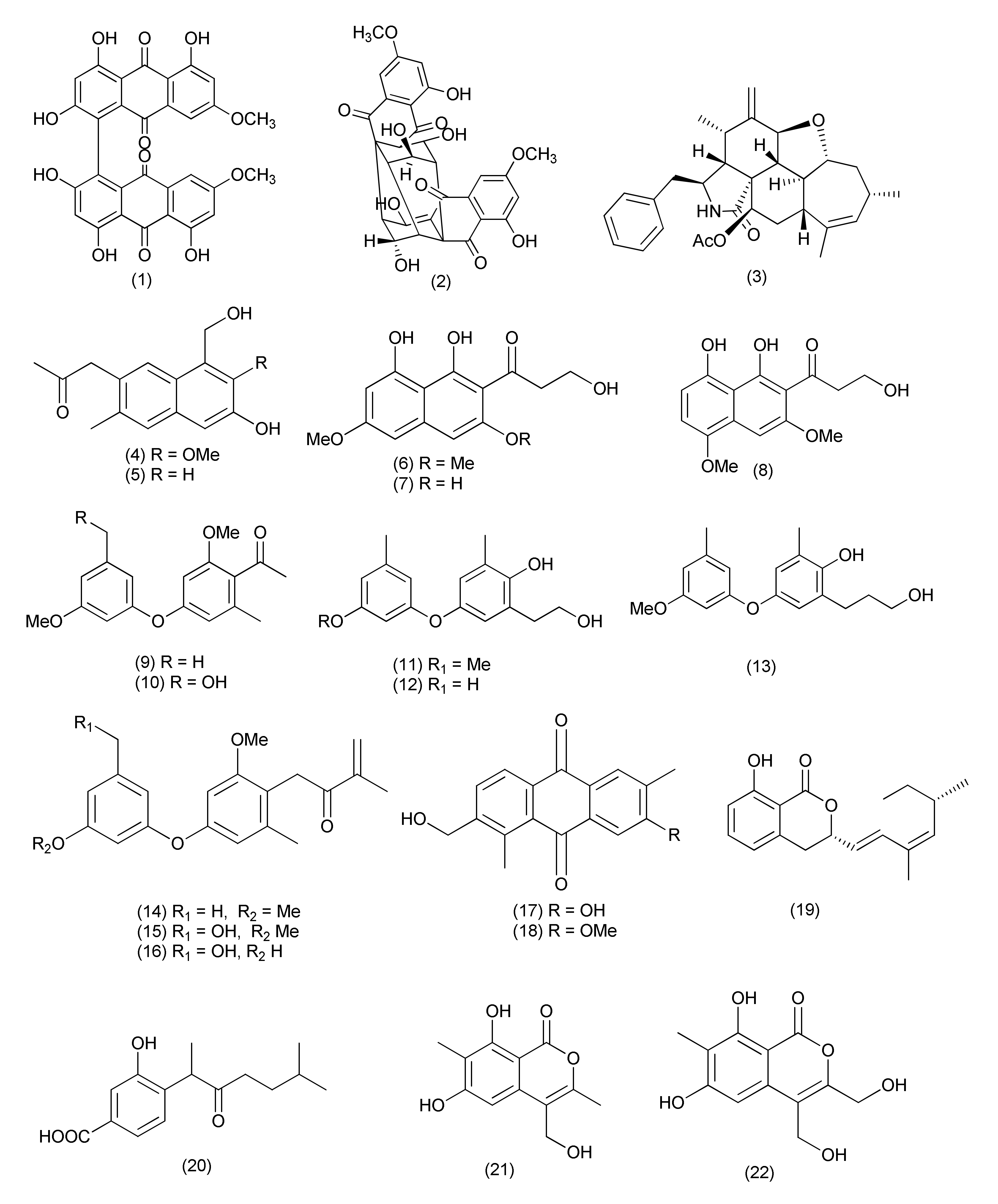
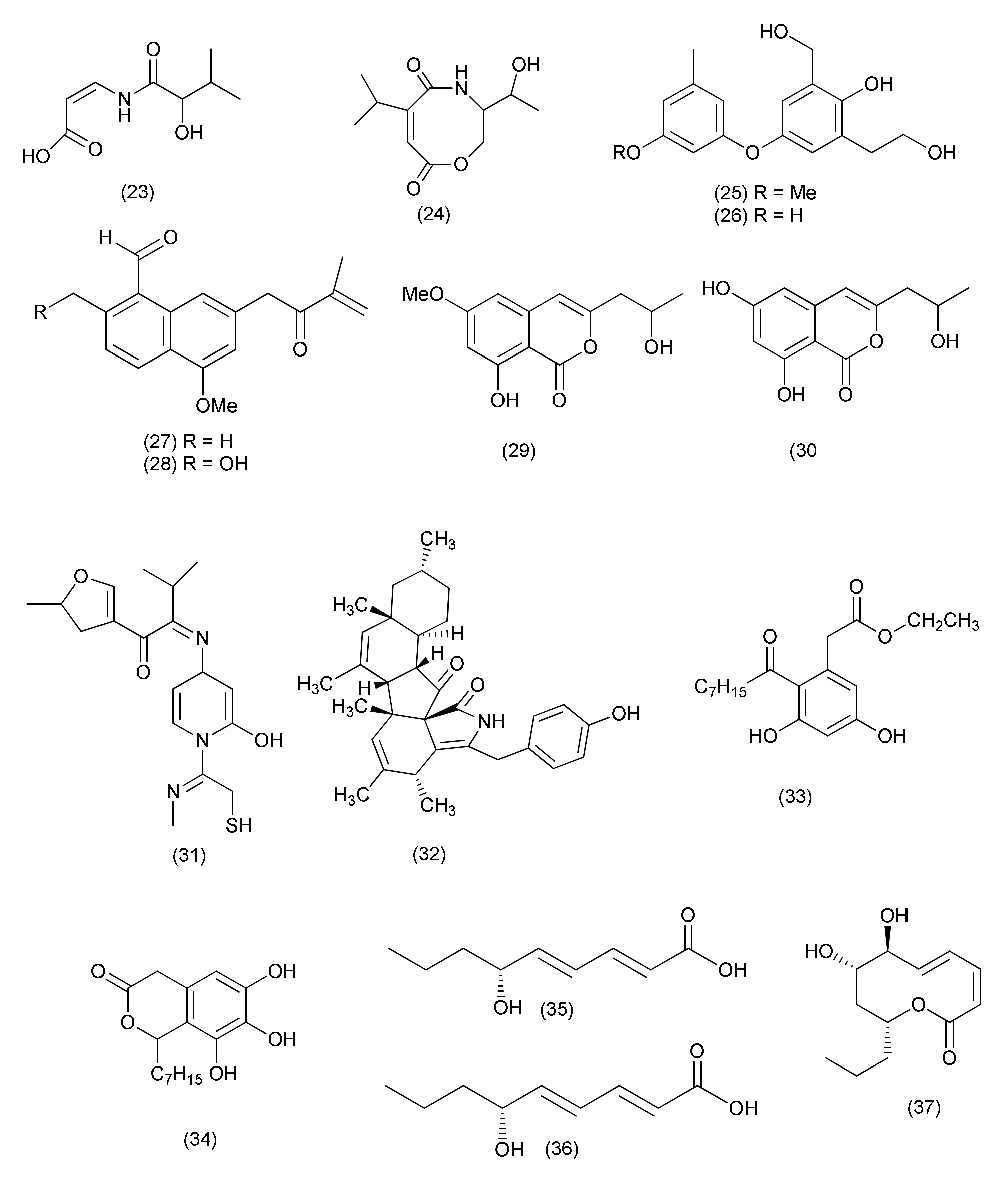
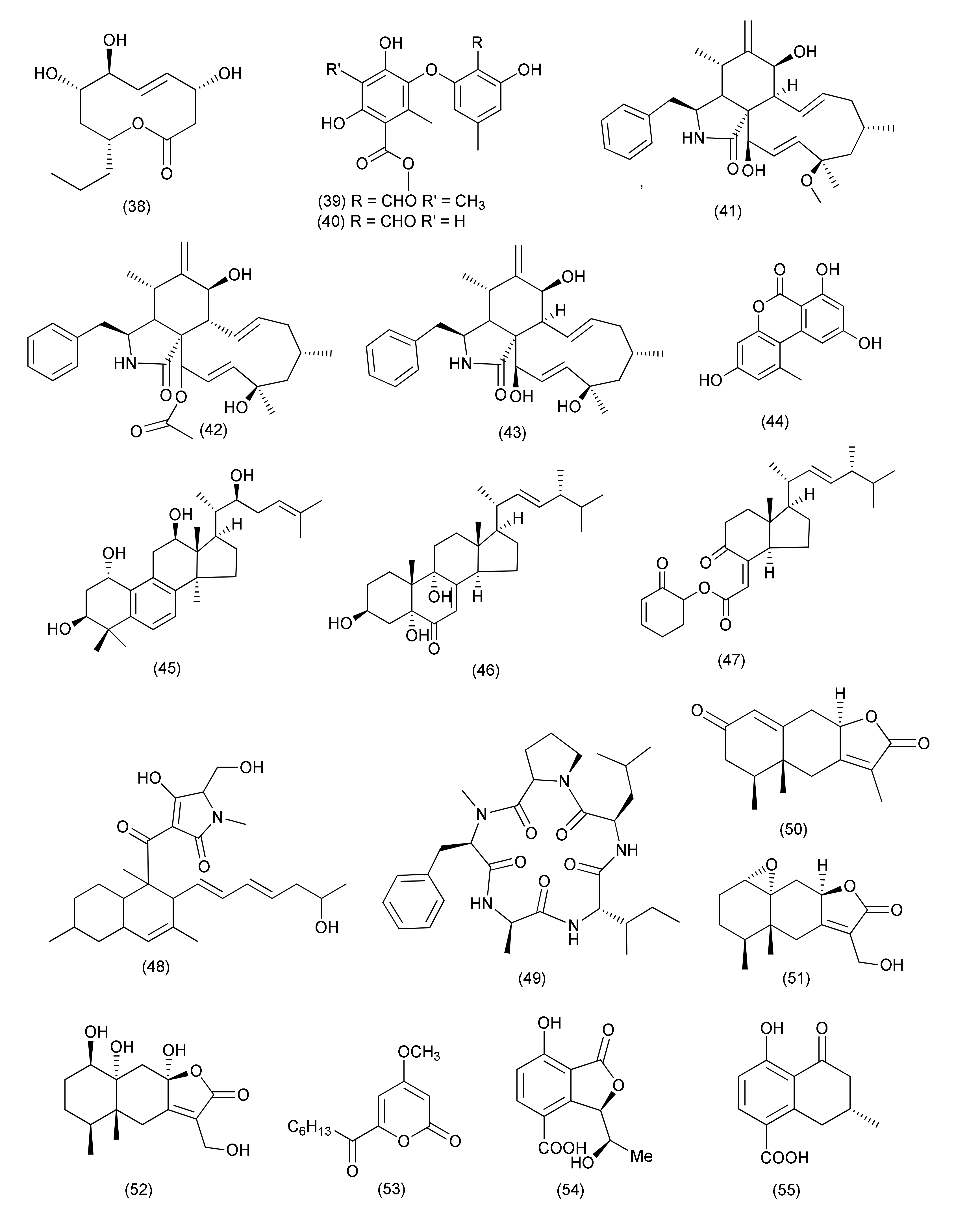
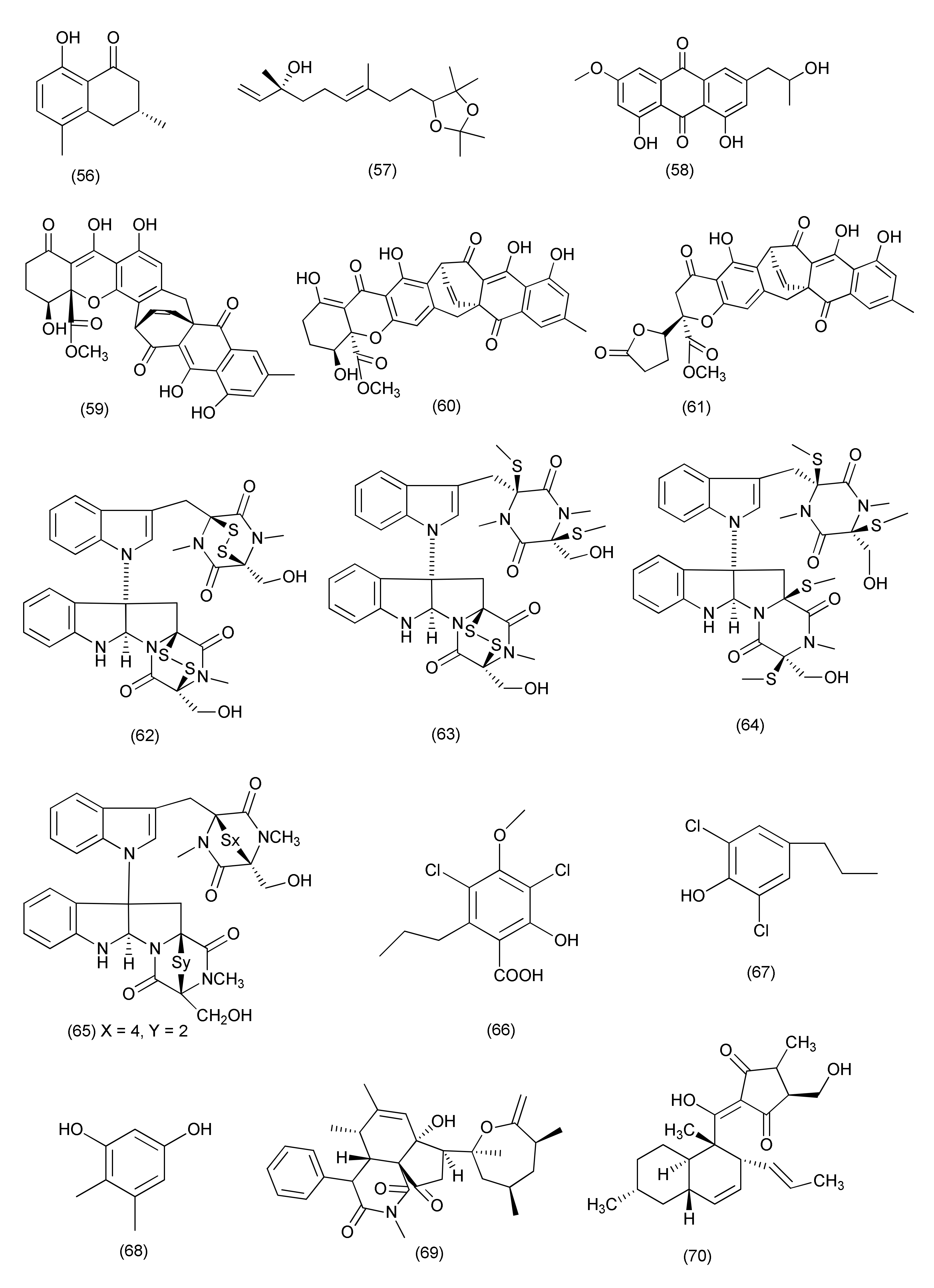
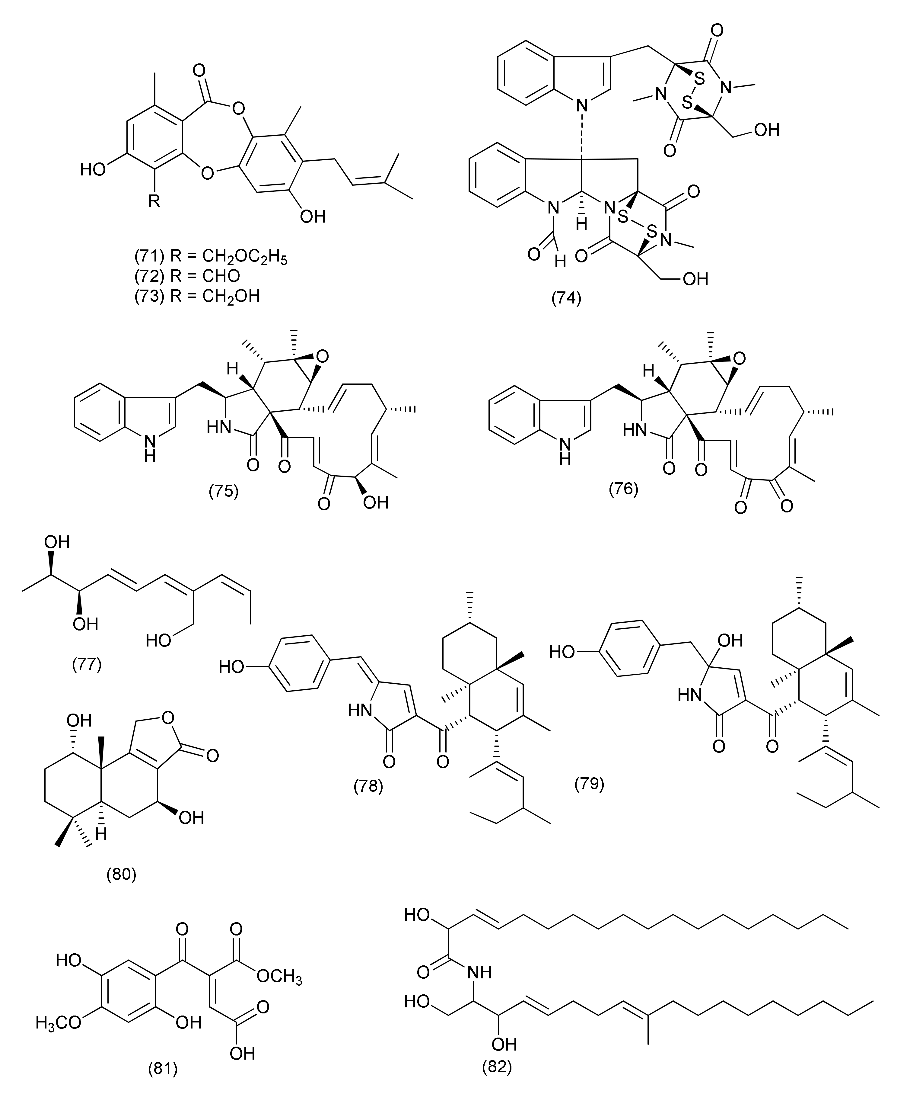
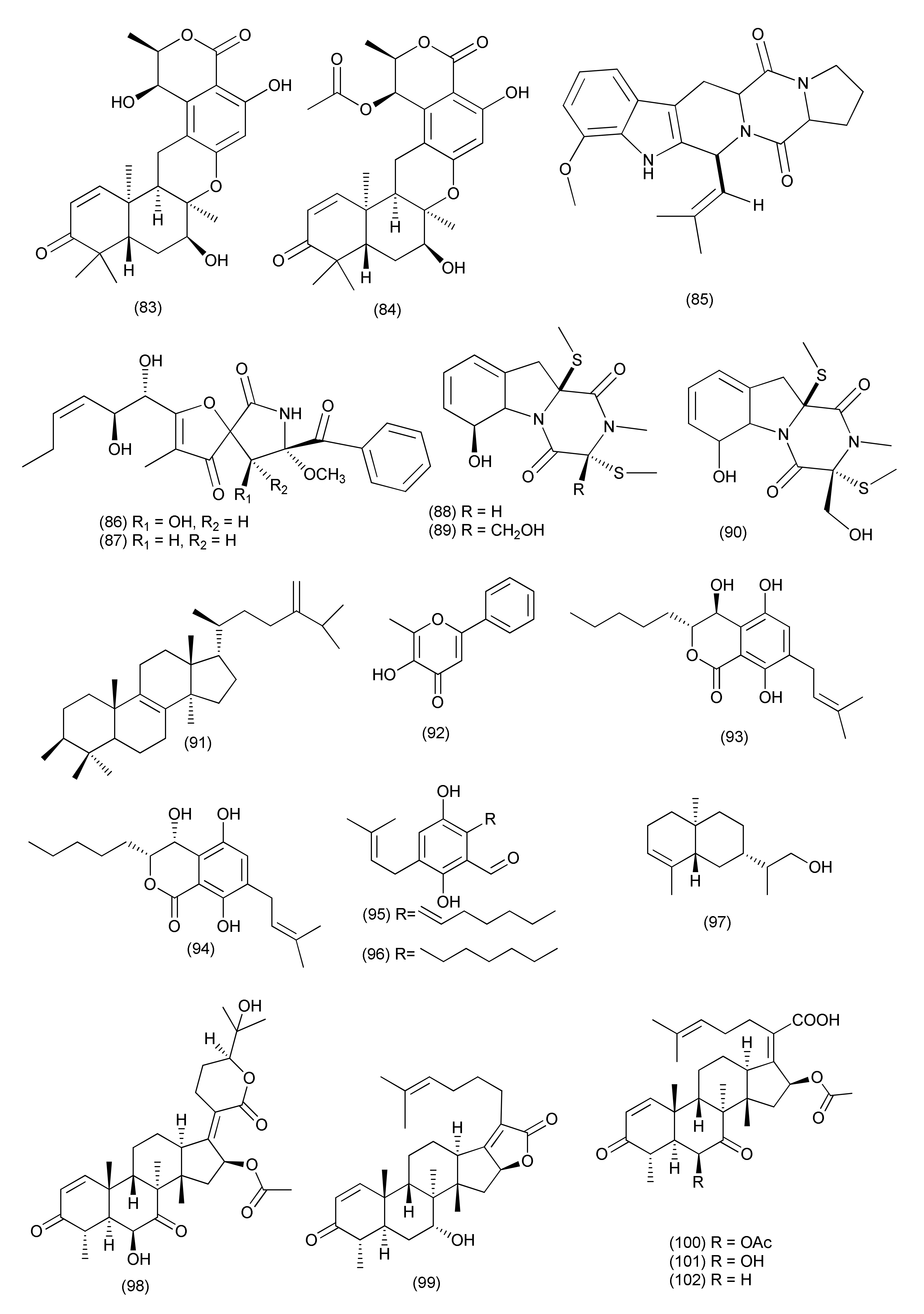
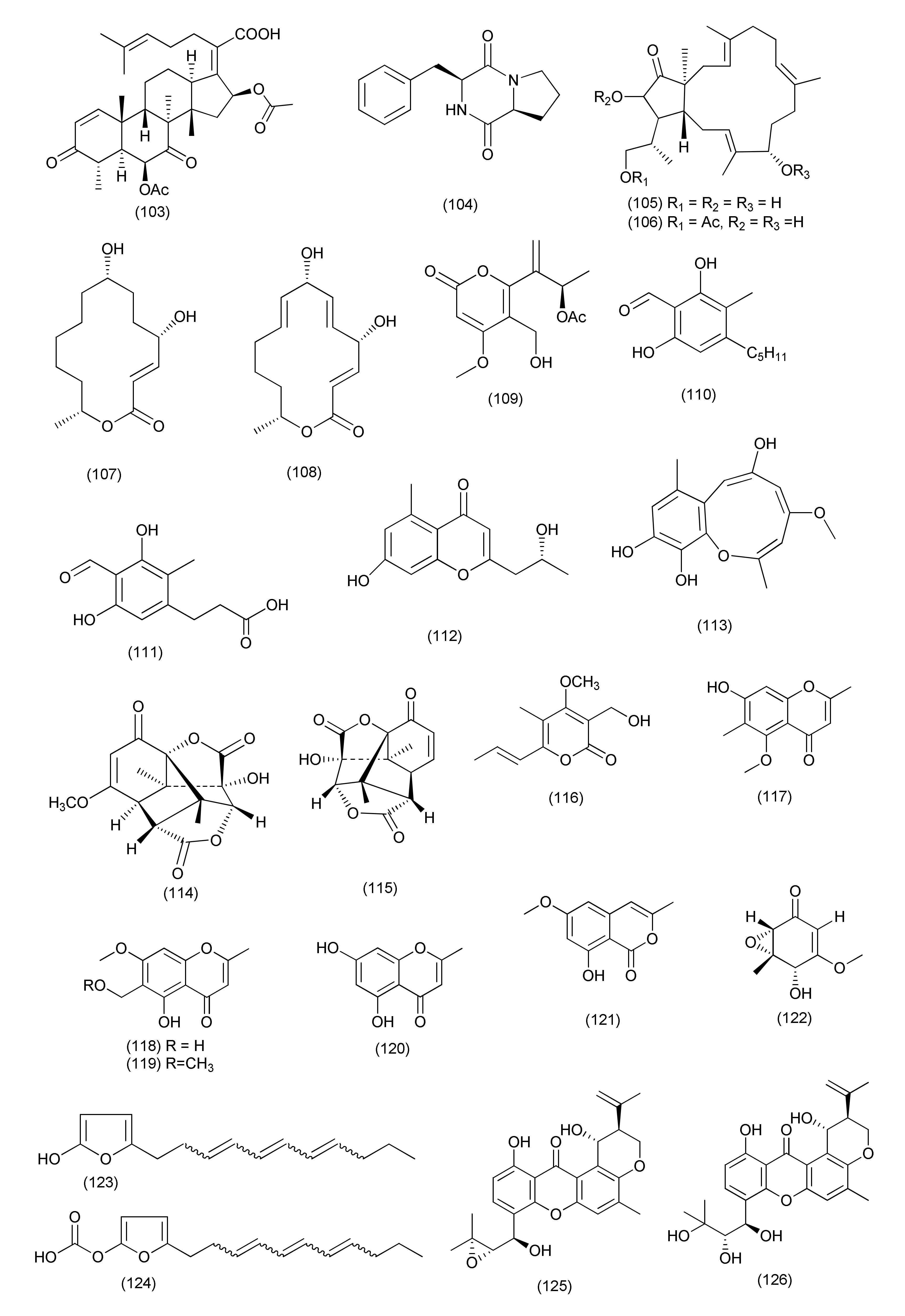
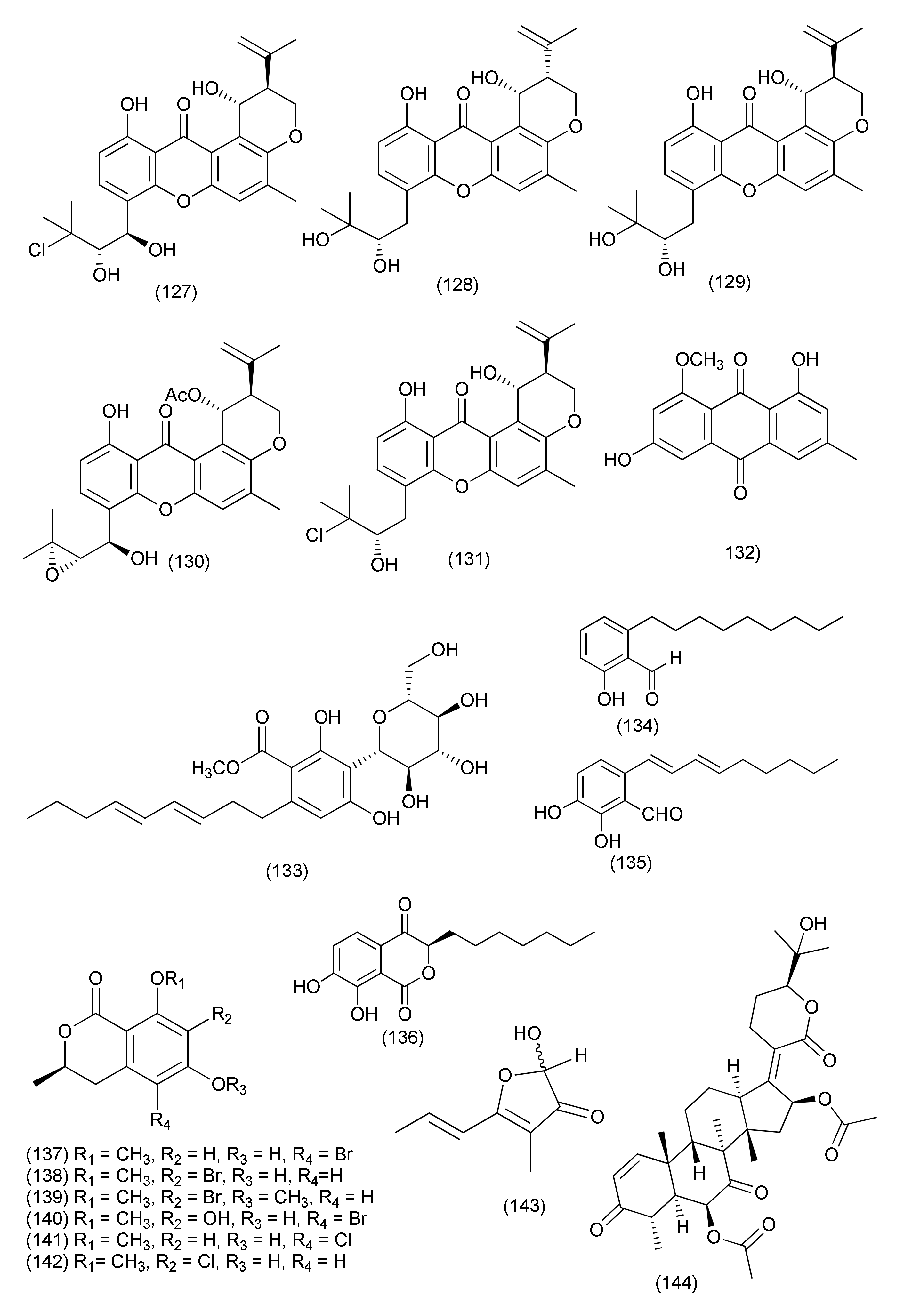
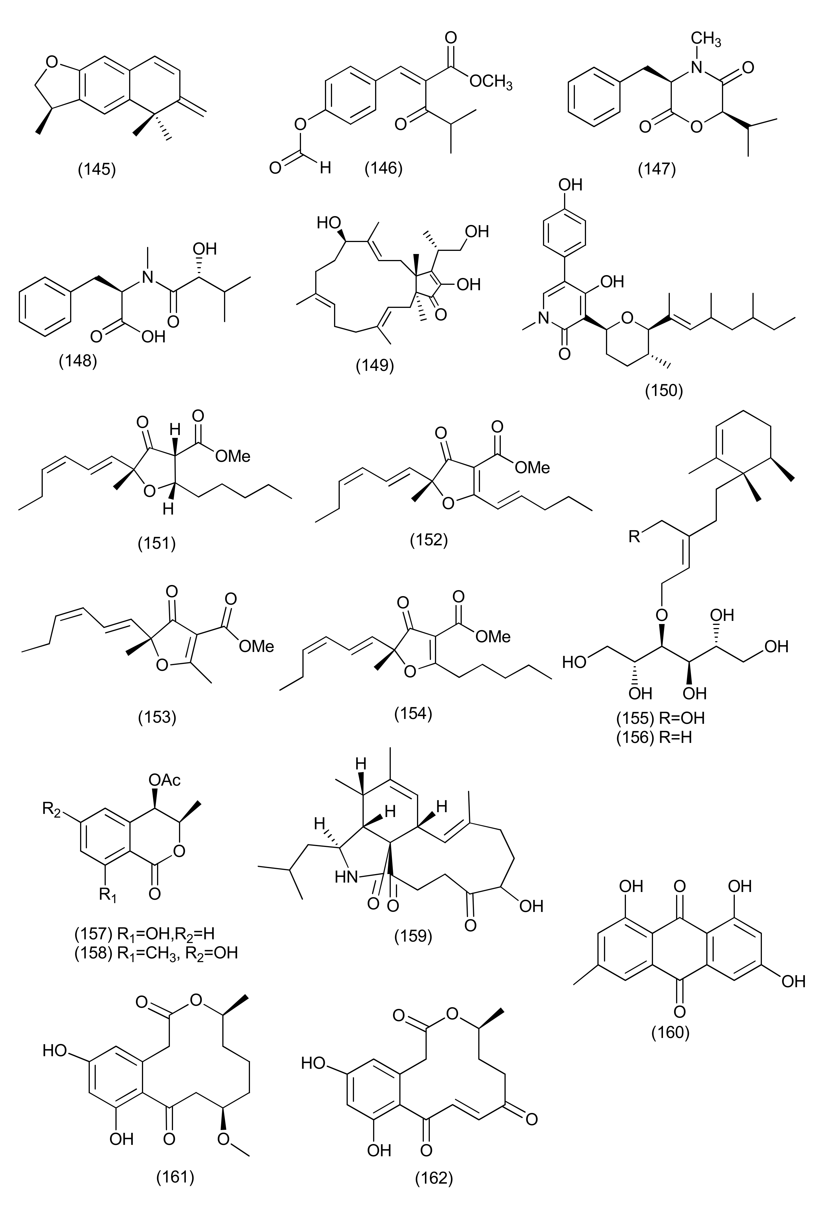
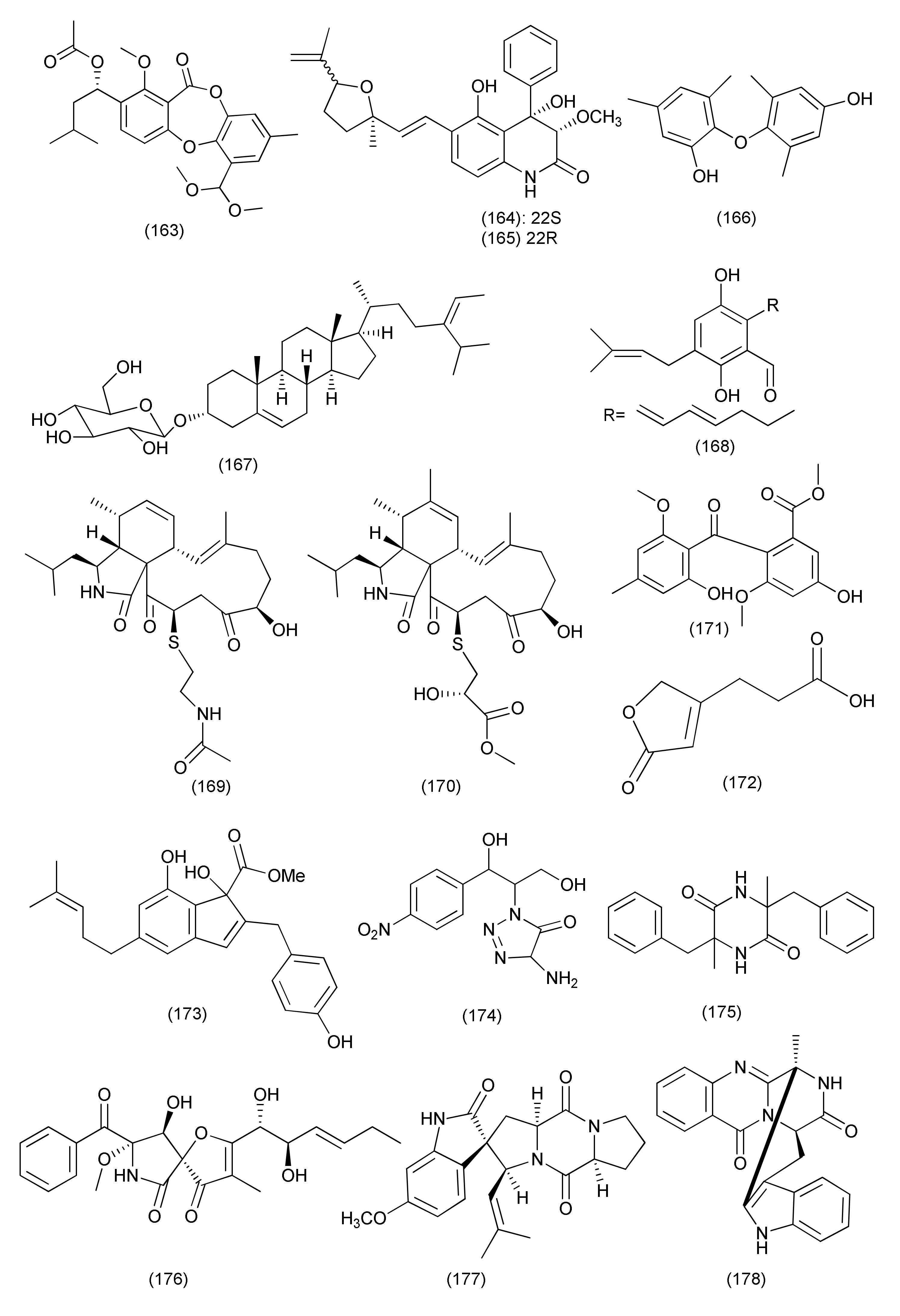
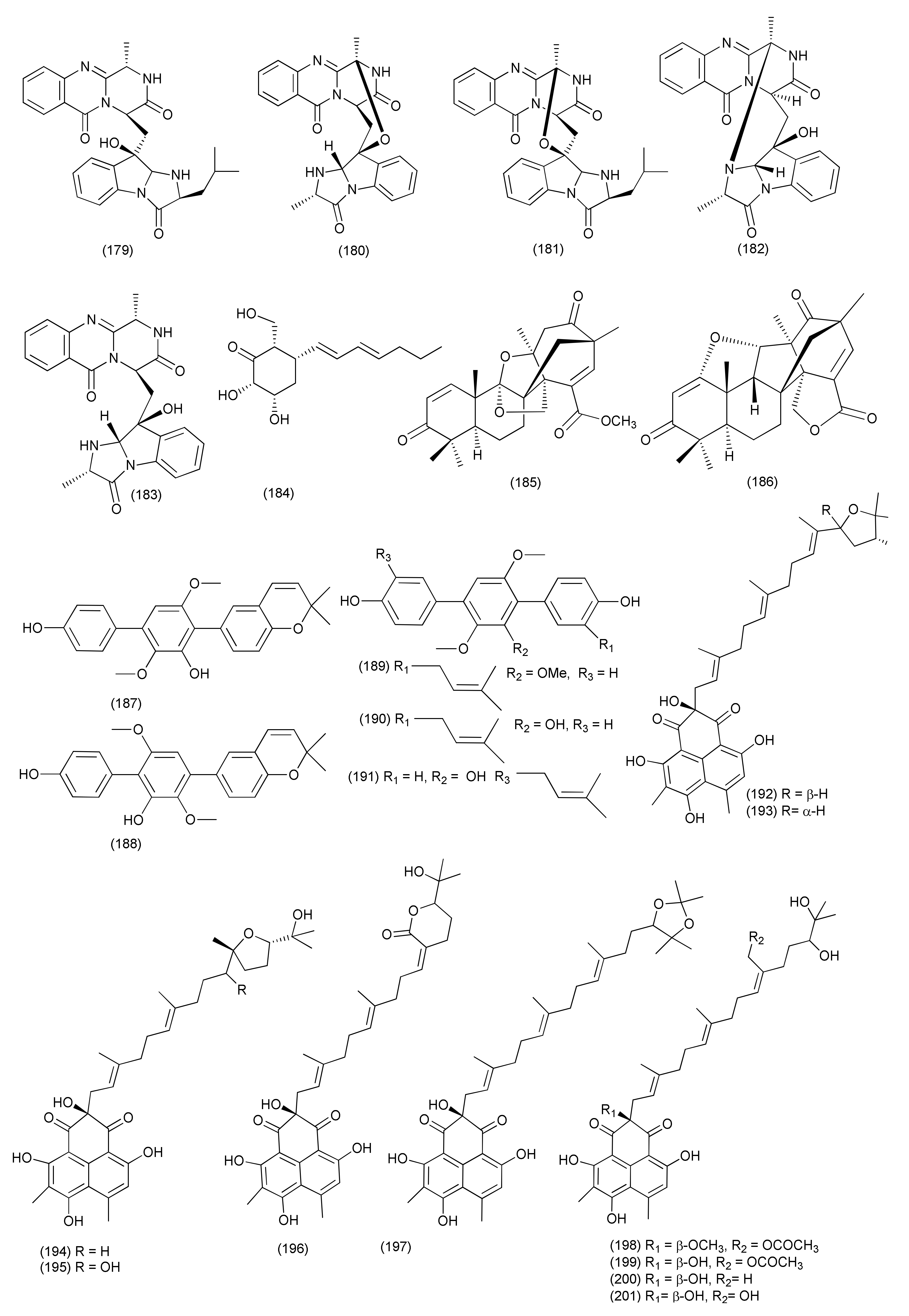
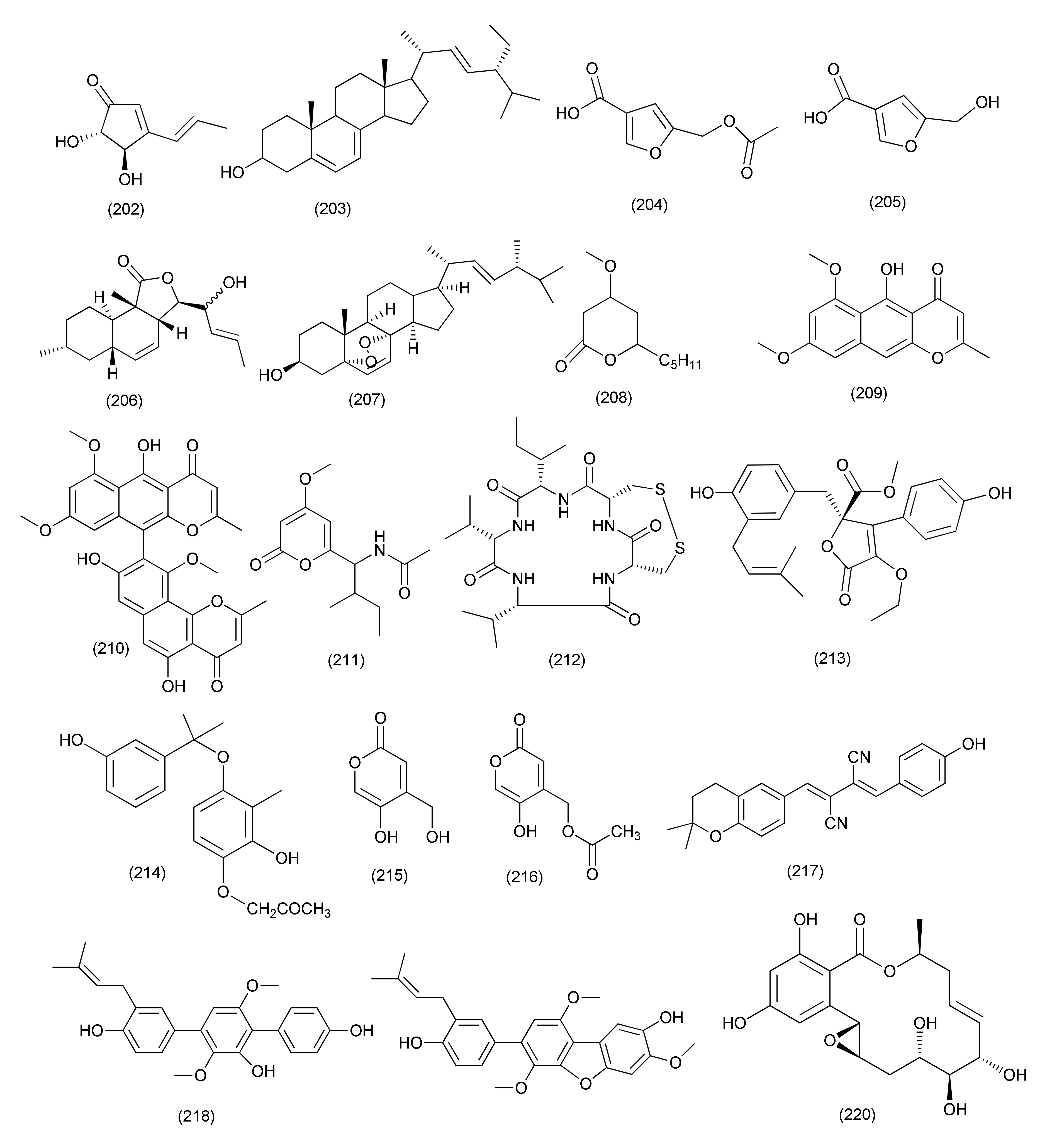
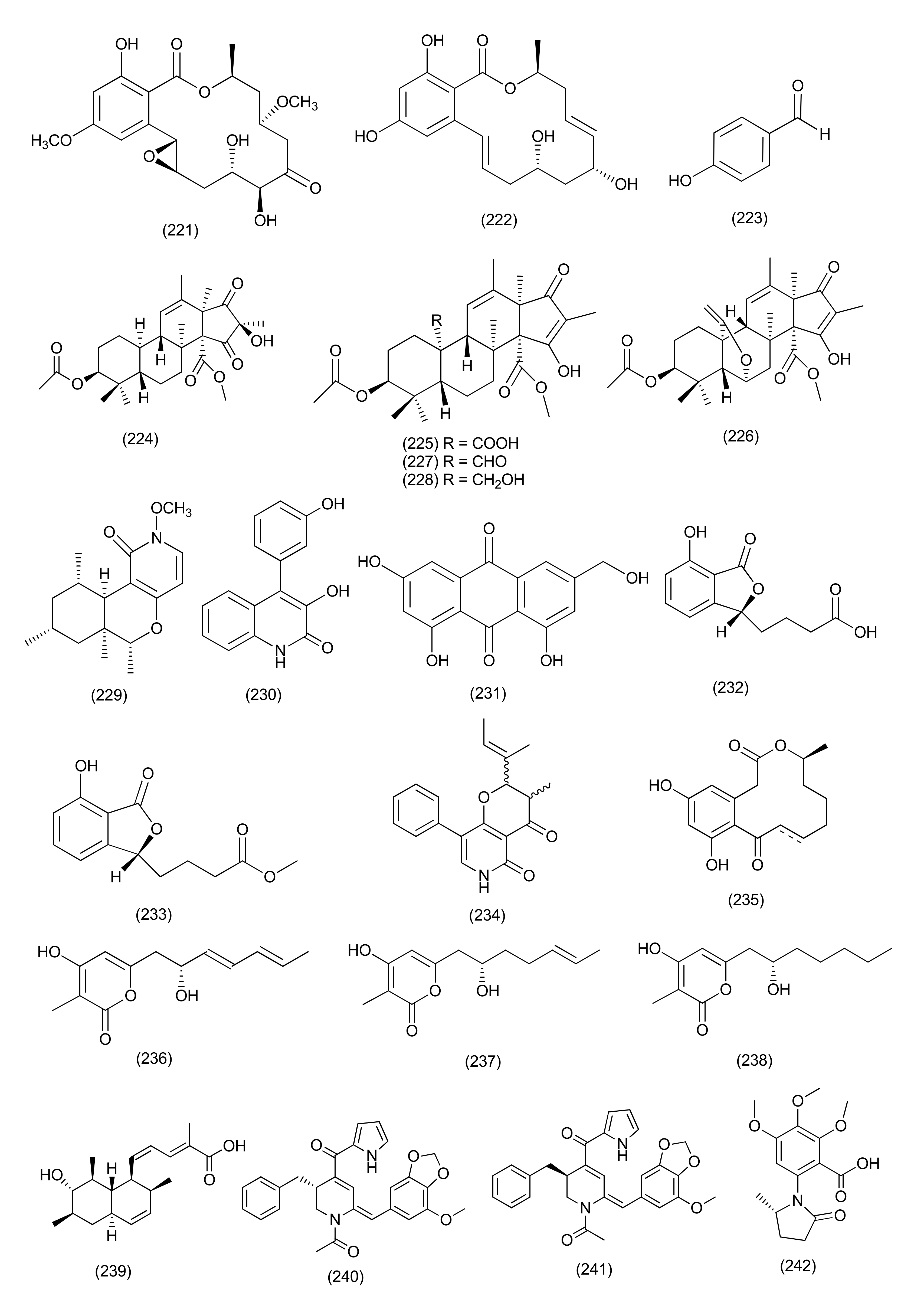
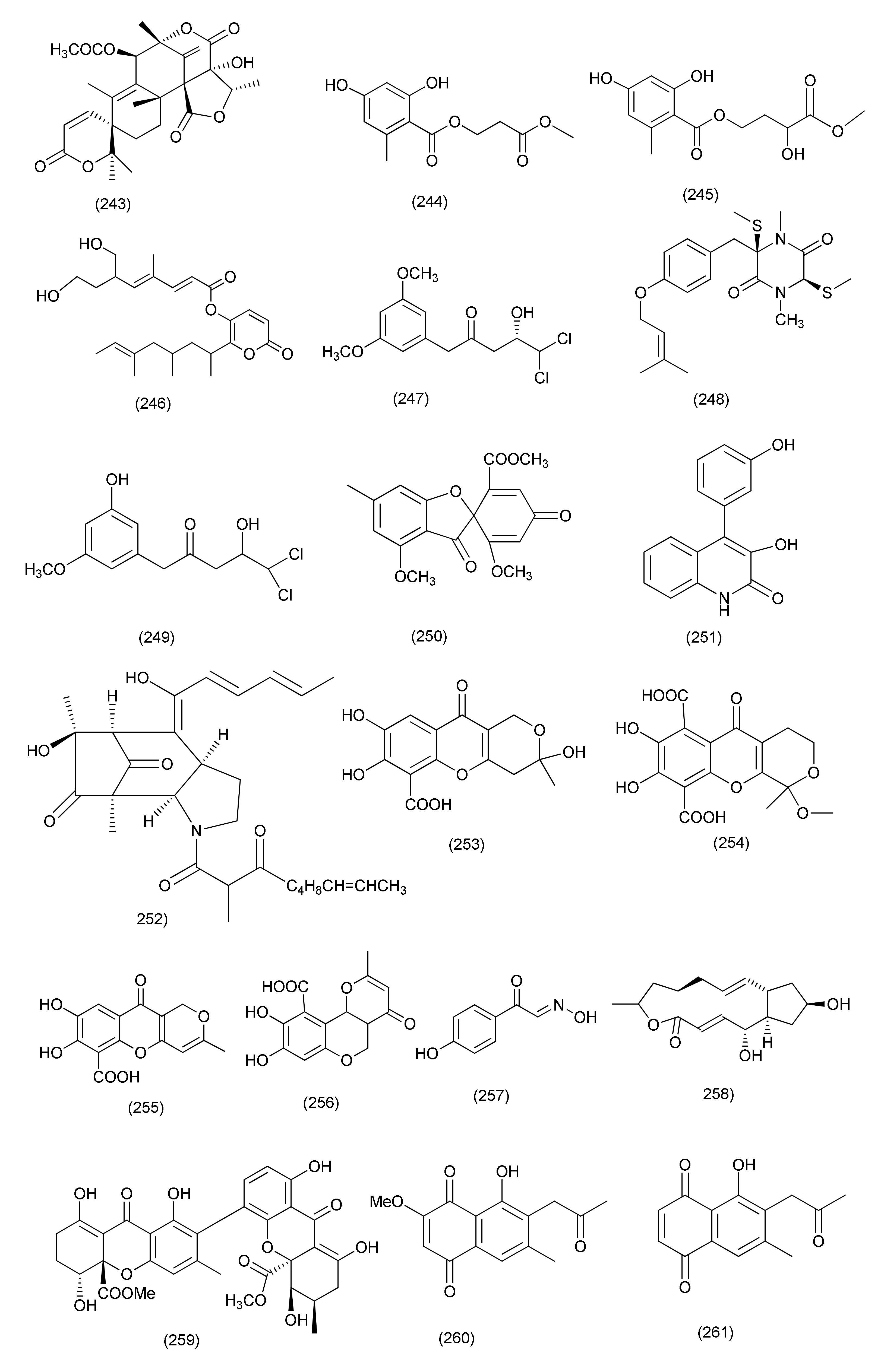
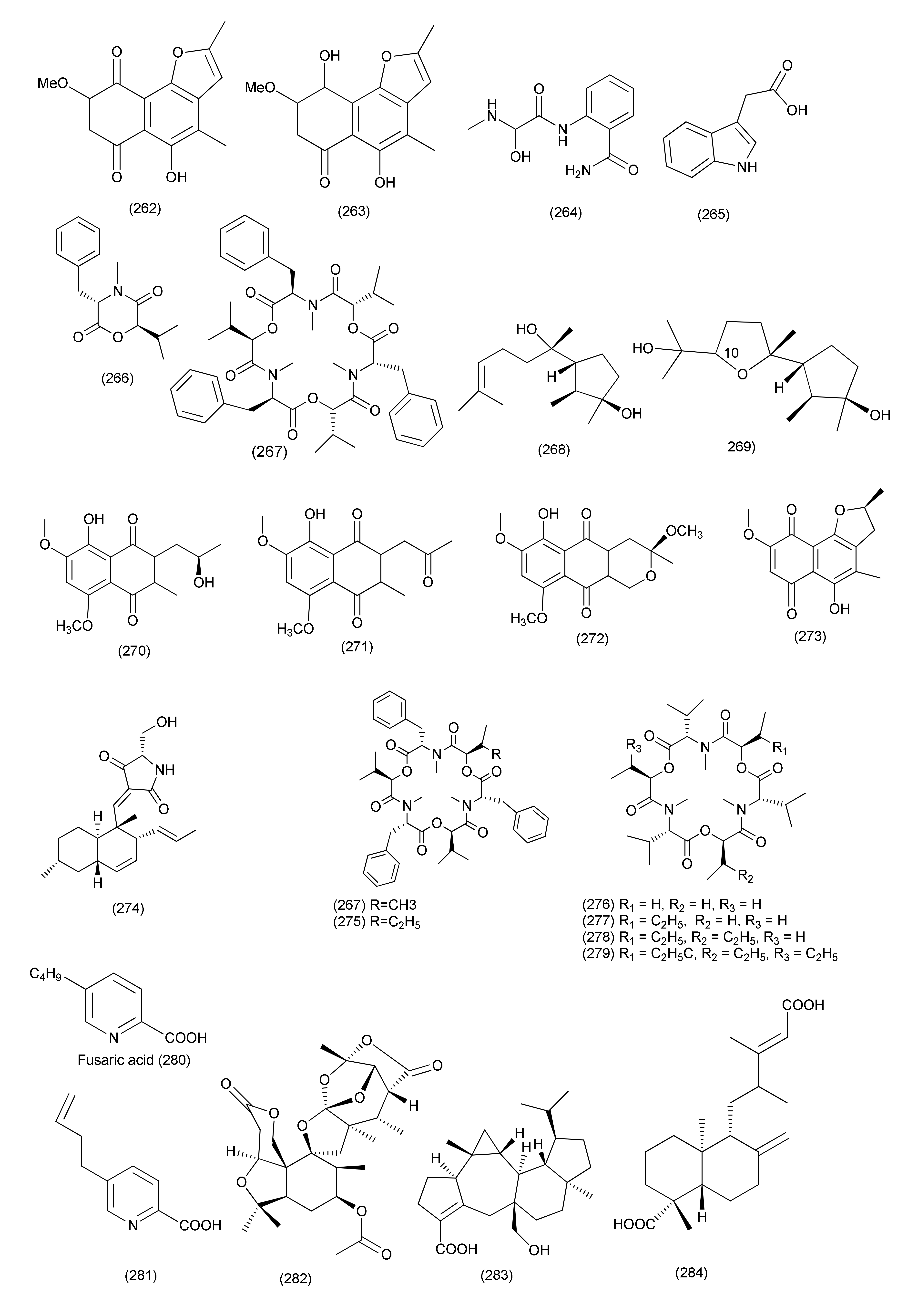
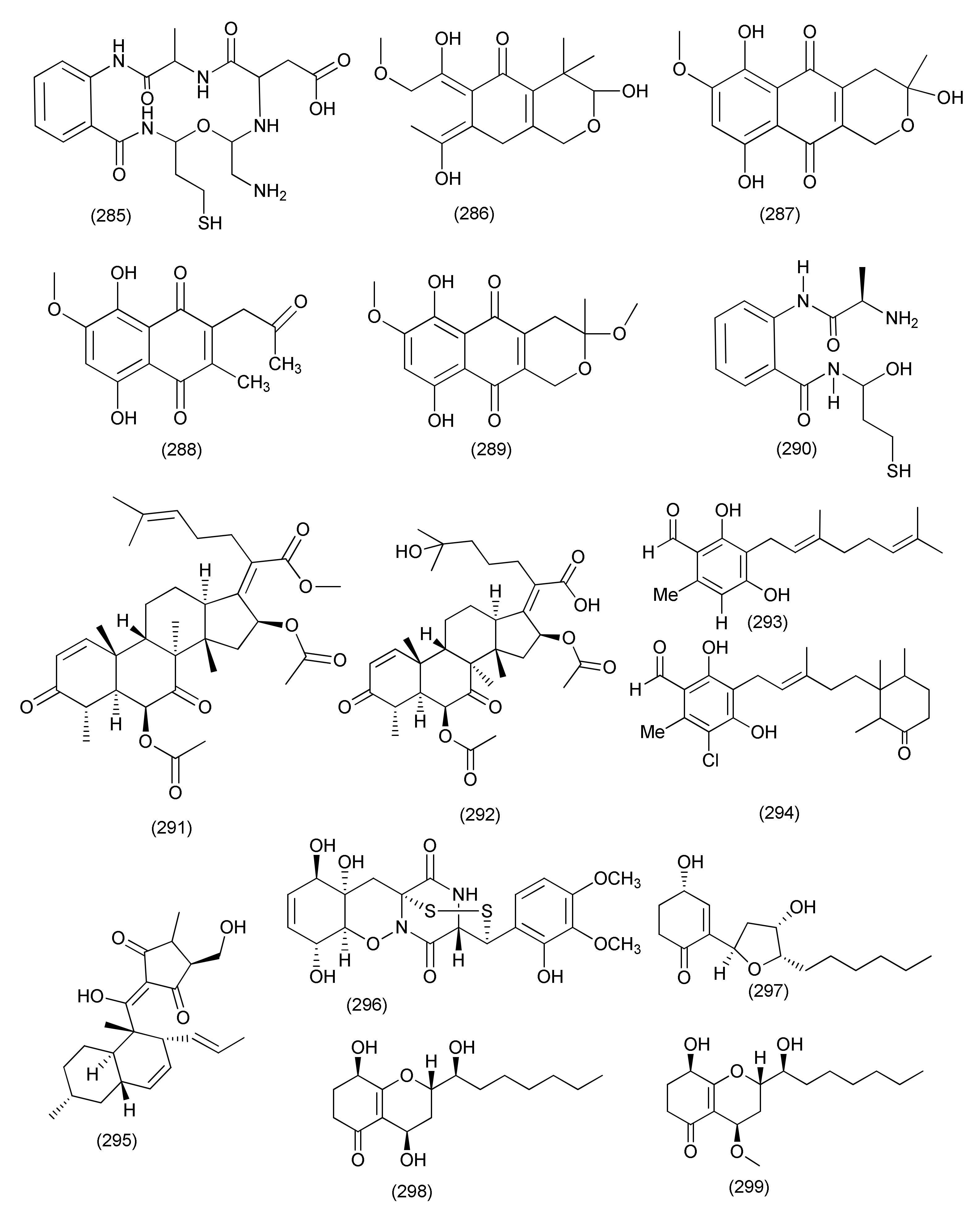
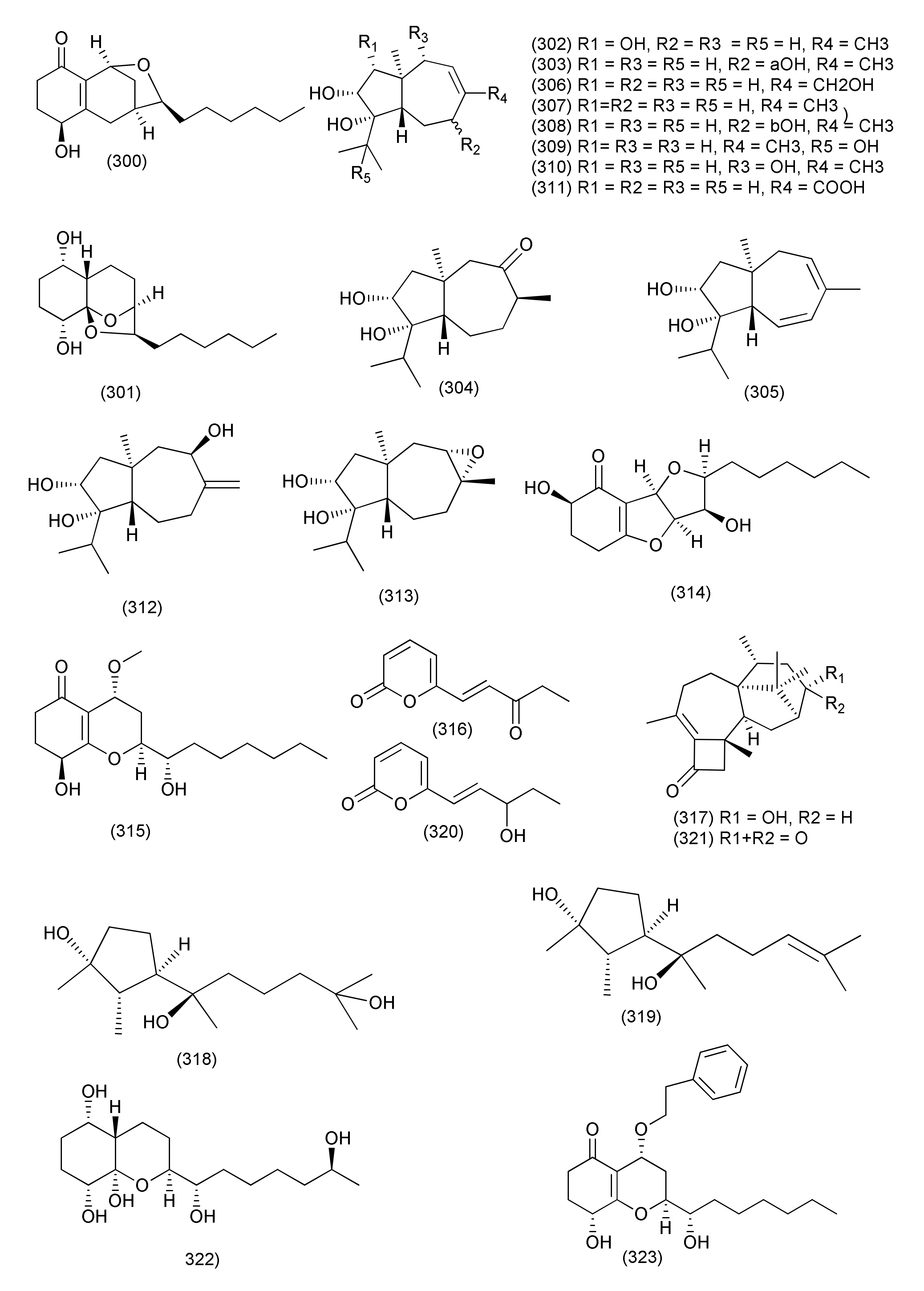
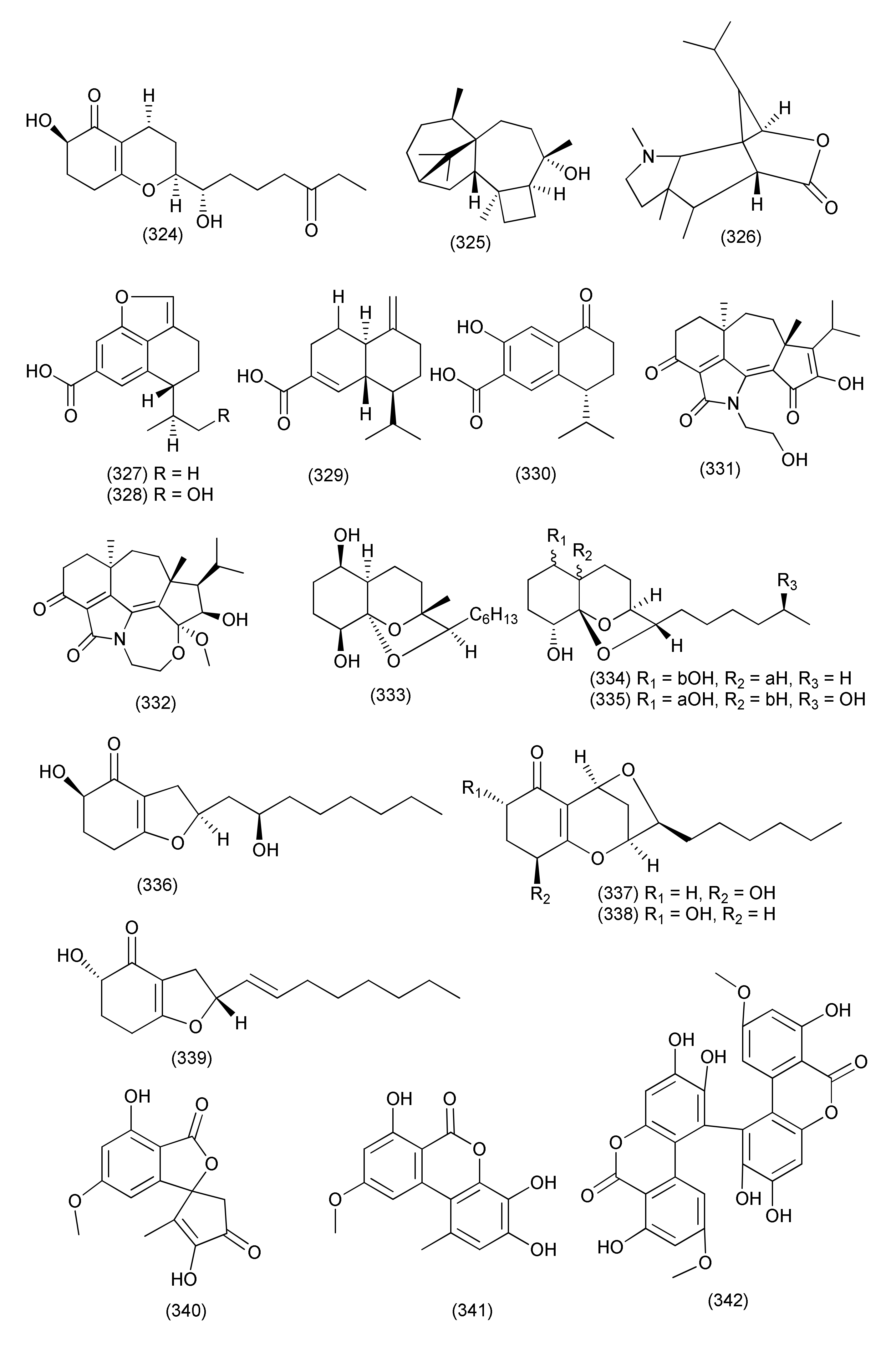
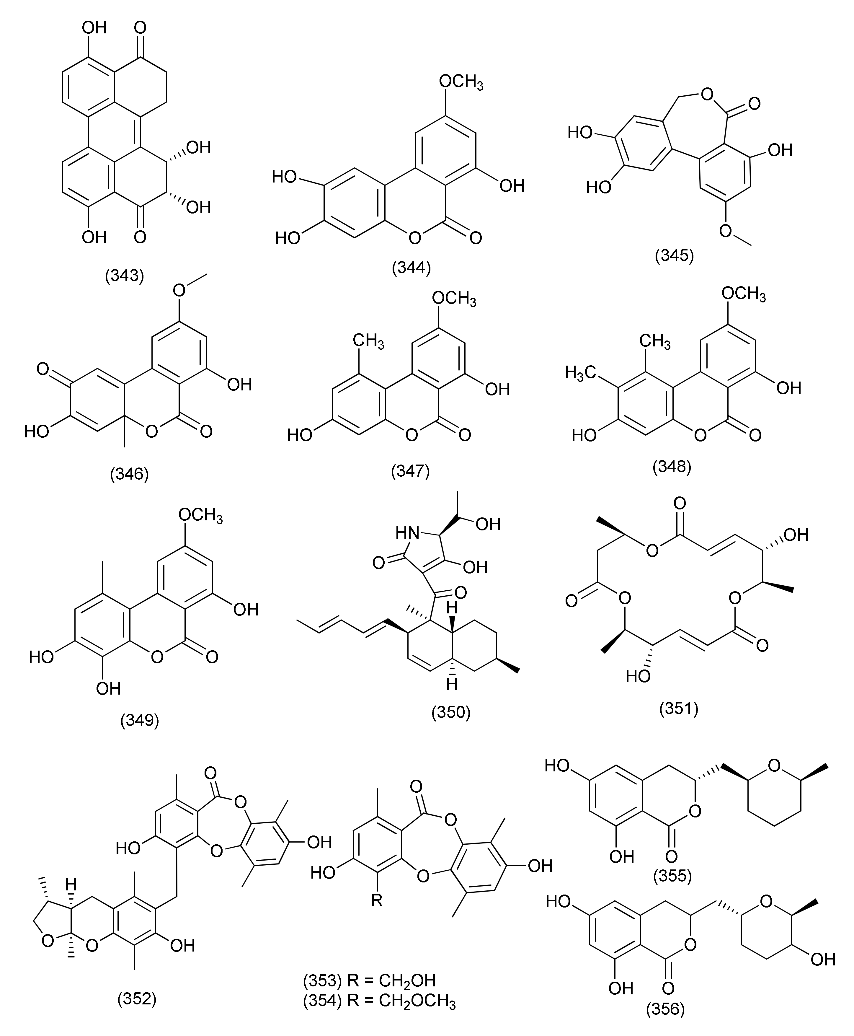
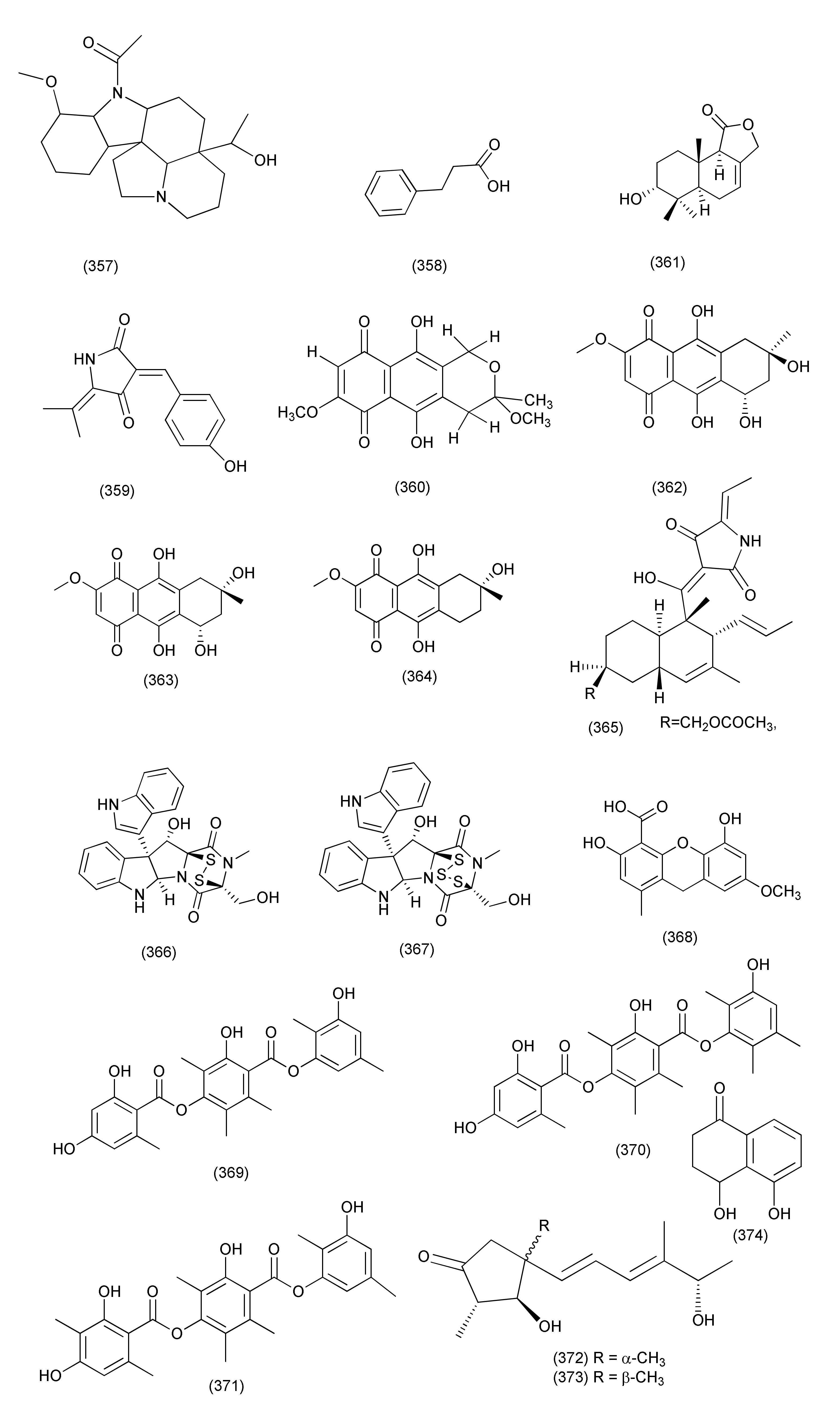
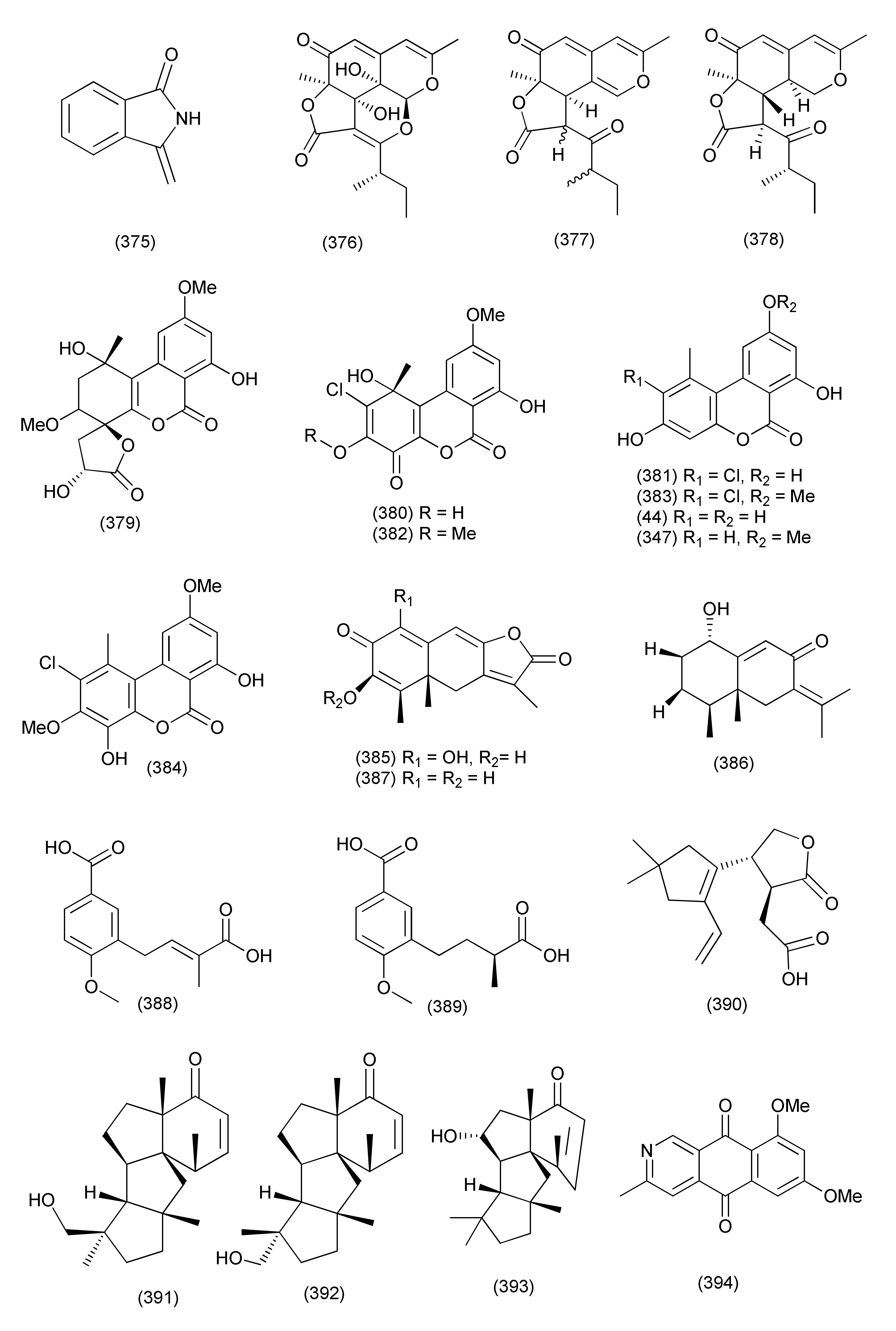
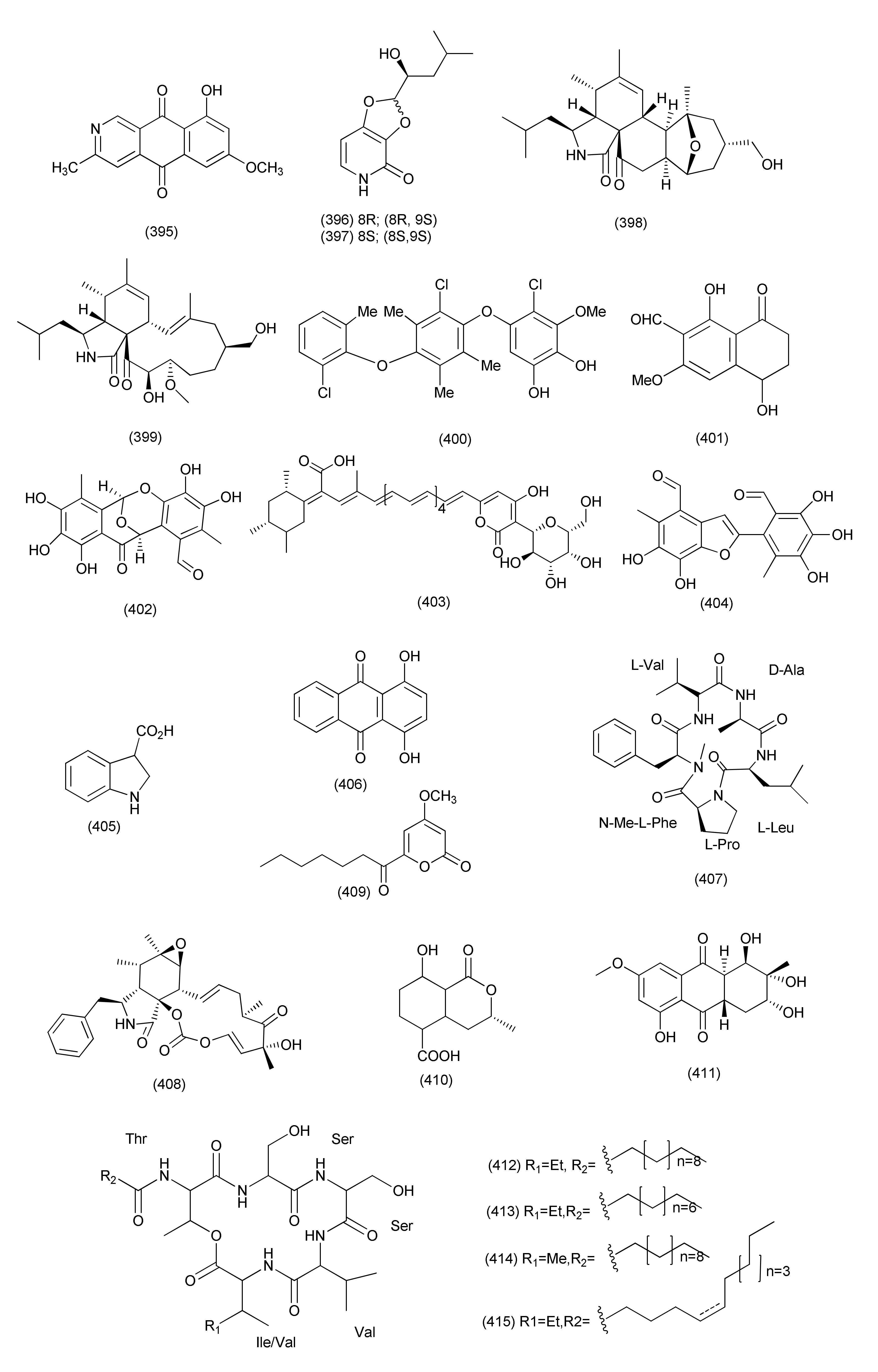
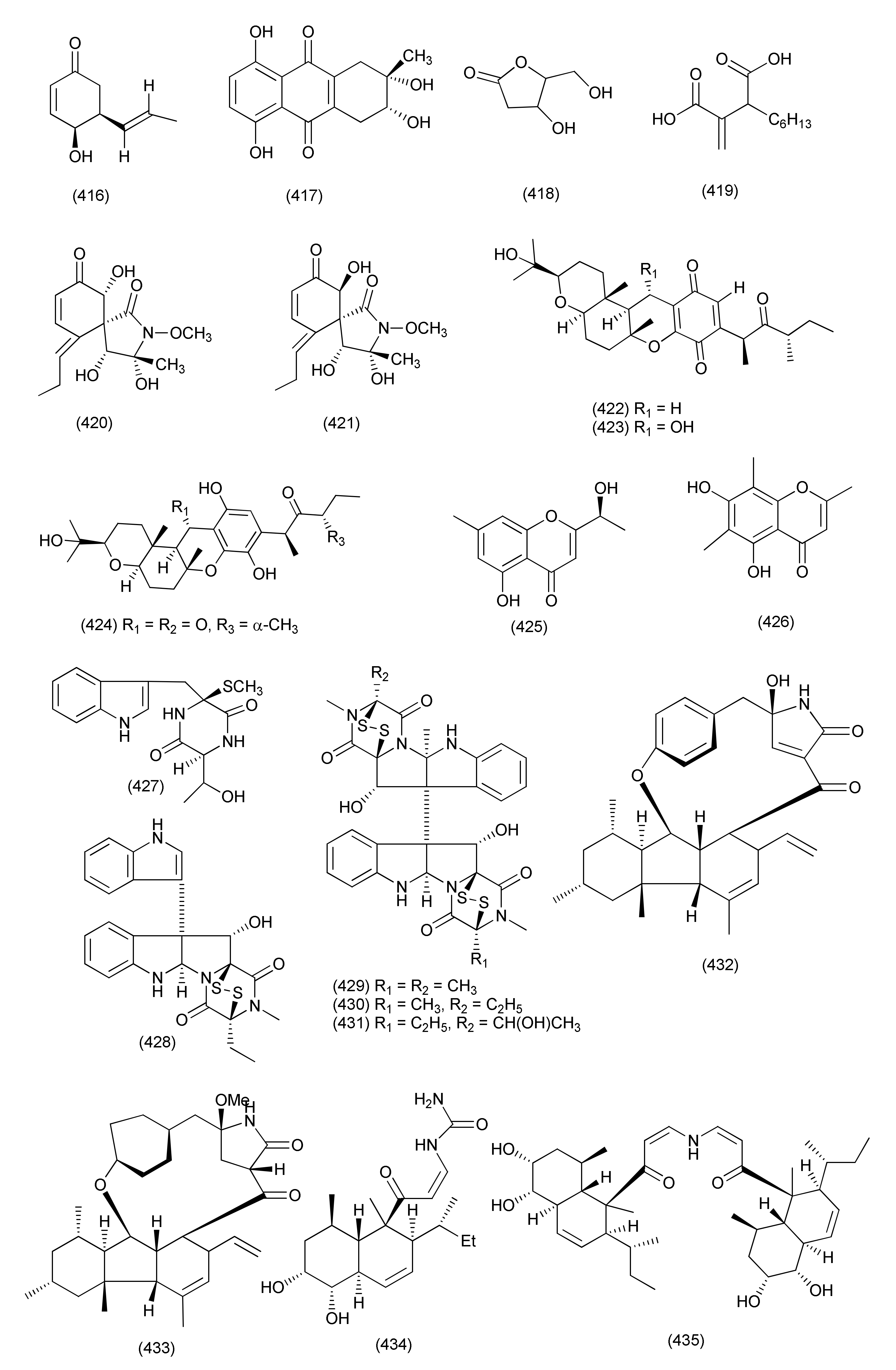
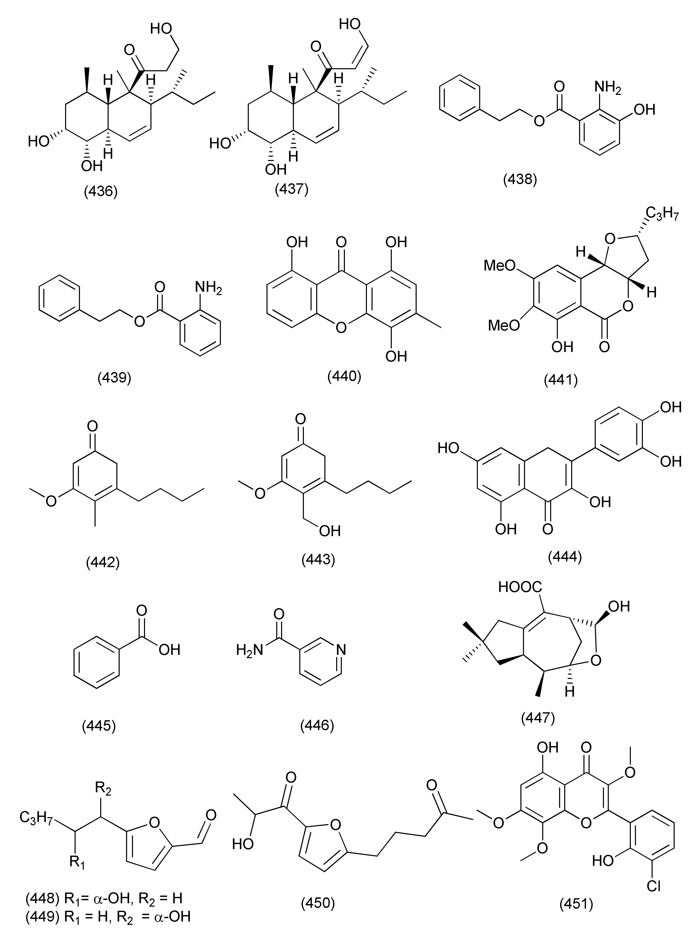
Table 1.
Anti-bacterial metabolites reported from endophytic fungi.
| Sr. No. | Fungus | Source | Locality | Compounds Isolated | Biological Target | Biological Activity (MIC/IC50/ID50) | Reference |
|---|---|---|---|---|---|---|---|
| Ascomycetes | |||||||
| Diaporthe | |||||||
| 1 | Diaporthe sp. | Uncaria gambier | (+)-1,1′-Bislunatin (1) and (+)-2,2′- epicytoskyrin A (2) | Mycobacterium tuberculosis strains H37Rv | MICs 0.422 and 0.844 μM | [18] | |
| 2 | Diaporthe sp. GDG-118 | Sophora tonkinensis | Hechi City, China | 21-Acetoxycytochalasin J3 (3) | Bacillus anthraci and E. coli | inhibited at 12.5 μg/mL concentration | [19] |
| 3 | Phomopsis fukushii. | 1-(3-Hydroxy-1-(hydroxymethyl)-2-methoxy-6-methylnaphthalen-7-yl) propan-2-one (4) and 1-(3-hydroxy-1- (hydroxymethyl)-6-methylnaphthalen-7-yl)propan-2-one (5) | MRSA | Zone of inhibition of 10.2 and 11.3 mm (6 mm strile filterpaper disc were impregnated with 20µL (50 µg) of each compound) | [20] | ||
| 4 | Phomopsis fukushii | Paris polyphylla var. yunnanensis | Kunming, Yunnan, China | 3-Hydroxy-1-(1,8- dihydroxy- 3,6-dimethoxynaphthalen-2-yl)propan-1-one (6), 3-hydroxy-1-(1,3,8-trihydroxy-6-methoxynaphthalen-2-yl)propan-1-one (7) and 3-hydroxy-1-(1,8-dihydroxy3,5-dimethoxynaphthalen-2-yl) propan-1-one (8) | MRSA- ZR11 | MIC, 8, 4, and 4 µg/mL, | [21] |
| 5 | Phomopsis fukushii | Paris polyphylla var. yunnanensis | Kunming, Yunnan, China | 1-[2-Methoxy-4-(3-methoxy-5-methylphenoxy)-6-methylphenyl]-ethanone (9) and 1-[4-(3-(hydroxymethyl)-5-methoxyphenoxy)-2-methoxy-6-methylphenyl]-ethanone (10) | MRSA | Zone of inhibition 13.8 and 14.6 mm | [22] |
| 6 | Phomopsis fukushii | Paris polyphylla var. yunnanensis | Kunming, Yunnan, P. R. China | 4-(3-Methoxy-5-methylphenoxy)-2-(2-hydroxyethyl)-6-methylphenol (11), 4-(3-Hydroxy-5-methylphenoxy)-2-(2-hydroxyethyl)-6-methylphenol (12) and 4-(3-methoxy-5-methylphenoxy)-2-(3-hydroxypropyl) -6-methylphenol (13) | MRSA | Zone of inhibition of 20.2, 17.9 and 15.2 mm (tested at 50µg/6 mm disc) | [23] |
| 7 | Phomopsis fukushii | Paris polyphylla var. yunnanensis | Kunming, Yunnan, China. | 1-(4-(3-Methoxy-5-methylphenoxy)-2-methoxy-6-methylphenyl)-3-methylbut-3-en-2-one (14), 1-(4-(3-(hydroxymethyl)-5-methoxyphenoxy)-2-methoxy-6- methylphenyl)-3-methylbut-3-en-2-one (15), 1-(4-(3-hydroxy-5-(hydroxymethyl)phenoxy)-2-methoxy-6- methylphenyl)-3-methylbut-3-en-2-one (16) | MRSA | Zone of inhibition of 21.8, 16.8 and 15.6 mm, (50 µg/6 mm disc) | [24] |
| 8 | Phomopsis sp. | - | - | 3-Hydroxy-6-hydroxymethyl-2,5-dimethylanthraquinone (17), 6-hydroxymethyl-3-methoxy-2,5-dimethylanthraquinone (18) | MRSA | IZD 14.2 and 14.8 mm | [25] |
| 9 | Diaporthe sp. | Pteroceltis tatarinowii | Mufu Mountain of Nanjing, China. | Diaporone A (19) | B. subtilis | MIC, 66.7 μM, | [26] |
| 10 | Phomopsis prunorum (F4-3). | - | - | (−)-1 and (+)- Phomoterpenes A and B (20) phomoisocoumarins C (21), D (22) | X. citri pv. phaseoli var. fuscans | MIC, 31.2, 62.4, 31.2, and 31.2 μg/mL, | [27] |
| Pseudomonas syringae pv. Lachrymans | MIC, 31.2, 15.6, 31.2 and 15.6 μg/mL | ||||||
| 11 | Diporthe vochysiae LGMF1583 | Vochysia divergens | - | Vochysiamides A (23) | KPC (Klebsiella pneumoniae carbapenemase producing). | MIC, 1.0 μg/mL | [28] |
| Vochysiamides B (24) | KPC, MSSA, MRSA | MIC, 0.08, 1.0, and 1.0 µg/mL | |||||
| 12 | Phomopsis asparagi | Paris polyphylla var. yunnanensis | Kunming, Yunnan, China | 4-(3-Methoxy-5-methylphenoxy)-2-(2-hydroxyethyl)- 6-(hydroxymethyl)phenol (25), 4-(3-Hydroxy-5-methylphenoxy)-2-(2-hydroxyethyl)-6-(hydroxymethyl)phenol(26) | MRSA | Zone of inhibition of 10.8 and 11.4 mm | [29] |
| 13 | Phomopsis sp. | Paris polyphylla var. yunnanensis | ShiZhong, Yunnan, China | 5-Methoxy-2-methyl-7-(3-methyl-2-oxobut-3-enyl)-1-naphthaldehyde (27), 2-(hydroxymethyl)-5-methoxy-7-(3-methyl-2-oxobut-3-enyl)-1-naphthaldehyde (28) | MRSA | Zone of inhibition of 14.5 and 15.2 mm | [30] |
| 14 | Diaporthe terebinthifolii LGMF907 | Schinus terebinthifolius | Curitiba, Paraná, Brazil | Diaporthin (29) | E. coli, Micrococcus luteus, MRSA, and S. aureus | Zone of inhibition 1.73, 2.47, 9.50, and 9.0 mm tested at 100 μg/disk. | [31] |
| Orthosporin (30) | Zone of inhibition of 1.03, 1.53C, 9.0, and 9.33 mm | ||||||
| 15 | Phomopsis/Diaporthe sp. GJJM 16 | Vitex negundo | Azhiyar, Pollachi, Tamilnadu, India | (2Z)-2-(1,4-dihydro-2-hydroxy-1-((E)-2-mercapto-1 (methylimino)ethyl) pyrimidine-4-ylimino)-1-(4,5-dihydro-5-methylfuran-3-yl)-3-methylbutane-1-one (31) | S. aureus, and P. aeroginosa | MIC of 1.25 μg/mL against each organism | [32] |
| 16 | Phomopsis sp. PSU-H188 | Hevea brasiliensis | Trang Province, Thailand. | Diaporthalasin (32) | S. aureus ATCC25923, MRSA | MIC, 4 μg/mL each | [33] |
| Cytosporone B (33) | MIC, 32 and 16 μg/mL | ||||||
| Cytosporone D (34) | MIC, 64 and 32 μg/mL | ||||||
| 17 | Diaporthe terebinthifolii GG3F6 | Glycyrrhiza glabra | Jammu, J & K, India | Diapolic acid A (35), B (36) xylarolide (37) phomolide G (38) | Yersinia enterocolitica | IC50, 78.4, 73.4, 72.1 and 69.2 μM | [34] |
| 18 | Diaporthe sp. F2934 | leaves of Siparuna gesnerioides | Chagres National Park, a protected area of Panama | Phomosine A (39) | S. aureus (ATCC 25923), Streptococcus oralis (ATTC 35037), Enterococcus faecalis (ATCC 19433), Enterococcus cloacae (ATCC 13047), Bordetella bronchiseptica (CECT 440), | Zone of Inhibition 12, 9, 10, 11, 10 and 10 mm at 4 µg/mL concentration | [35] |
| Phomosine C (40) | Zone of Inhibition 9, 6, 8, 8, 8 and 9 mm at 4 µg/mL concentration | ||||||
| 19 | Phomopsis sp., | Garcinia kola nuts | bought at Mokolo local market in Yaounde (Cameroon) | 18-Methoxycytochalasin J (41), cytochalasins H (42) and J (43), alternariol (44) | Shigella flexneri | MIC, 128 μg/mL each | [36] |
| 18-Methoxycytochalasin J (41), cytochalasins H (42) | S. aureus ATCC 25923 | MIC, 128 and 256 μg/mL | |||||
| 20 | Diaporthe sp. LG23 | Mahonia fortunei | Shanghai, China | 19-nor-Lanosta-5(10),6,8,24-tetraene-1α,3β,12β,22S-tetraol (45) | S. aureus, E. coli, Bacillus subtilis, P. aeruginosa, Streptococcus pyogenes | MIC, 5.0, 5.0, 2.0, 2.0 and 0.1 µg/mL | [37] |
| 3β,5α,9α-Trihydroxy-(22E,24R)-ergosta-7,22-dien-6-one (46), and chaxine C (47) | B. subtilis | MIC, 5.0 µg/mL each | |||||
| 21 | Diaporthales sp. E6927E | Ficus sphenophyllum | Ecuadorean dry forest near the Napo River, USA | Pyrrolocin A (48) | S. aureus and E. faecalis | MICs 4 and 5 µg/mL | [38] |
| Xylaria | |||||||
| 22 | Xylaria ellisii | Blueberry (Vaccinium angustifolium) | Ellisiiamide (49) | Escherichia coli | MIC, 100 μg/mL | [39] | |
| 23 | Xylaria sp. GDG-102 | S. tonkinensis | Hechi, Guangxi province, China | Xylareremophil (50) | Micrococcus luteus and Proteus vulgaris | MIC 25 μg/mL each | [40] |
| Mairetolides B (51) | M. luteus | MIC, 50 μg/mL | |||||
| Mairetolide G (52) | P. vulgaris M. luteus | MIC 25 and 50 μg/mL | |||||
| Xylareremophil (50), mairetolides B (51), and G (52) | Micrococcus lysodeikticus and Bacillus subtilis | MIC 100 μg/mL | |||||
| 24 | Xylaria sp. (GDG-102) | Leaves of S. tonkinensis | 6-Heptanoyl-4-methoxy-2H-pyran-2-one (53) | E. coli as well as S. aureus | MIC, 50 μg/mL | [41] | |
| 25 | Xylaria sp. GDG-102 | S. tonkinensis | Hechi, Guangxi province, China | Xylarphthalide A (54) | B. subtilis and E. coli, | MIC, 12.5 μg/mL each | [42] |
| B. megaterium, S. aureus, S. dysenteriae and S. paratyphi | MIC, 25 μg/mL each | ||||||
| (−)-5-Carboxymellein (55) | B. Subtilis | MIC, 12.5 μg/mL | |||||
| B. anthracis, B. megaterium, S. aureus, E. coli, S. dysenteriae and S. paratyphi B | MIC, 25 μg/mL | ||||||
| (−)-5-Methylmellein (56) | B. subtilis and S. aureus | MIC, 12.5 μg/mL | |||||
| B. megaterium, E. coli and S. dysenteriae | 25 μg/mL | ||||||
| 26 | Xylaria sp., | Taxus mairei. | 3,7-Dimethyl-9-(-2,2,5,5-tetramethyl-1,3-dioxolan-4-yl) nona-1,6-dien-3-ol (57) | B. subtilis ATCC 9372, B. pumilus 7061 and S. aureus ATCC 25923 | 48.1, 31.6 and 47.1% inhibition. | [43] | |
| Nalgiovensin (58) | S. aureus ATCC 25923, B. subtilis ATCC 9372, B. pumilus ATCC 7061 and E. coli ATCC 25922 | 42.1, 36.8, 47.1 and 41.2% inhibition. | |||||
| Chaetomium | |||||||
| 27 | C. globosum 7s-1, | Rhapis cochinchinensis | Xanthoquinodin B9 (59), xanthoquinodin A1 (60), xanthoquinodin A3 (61) | B. cereus | MICs of 0.87, 0.44 and 0.22 μM, | [45] | |
| Xanthoquinodin B9 (59), xanthoquinodin A1 (60), xanthoquinodin A3 (61) | S. aureus and MRSA | MIC values ranging from 0.87 to 1.75 μM | |||||
| 3-Epipolythiodioxopiperazines, chetomin (62), chaetocochin C (63) and dethio-tetra(methylthio)chetomin (64) | B. cereus ATCC 11778, S. aureus ATCC 6538, and MRSA | MIC values ranging from 0.02 pM to 10.81 μM. | |||||
| Chetomin (62) | B. cereus, S. aureus and MRSA | MICs, 0.35 μM, 10.74 and 0.02 pM | |||||
| Compounds 59–64 | E. coli ATCC 25922, P. aeruginosa ATCC 27853, and Salmonella typhimurium ATCC 13311 | MICs of 45.06 to >223.72 μM | |||||
| Epipolythiodioxopiperazines (62–64) | Mycobacterium tuberculosis | MICs, 0.55, 4.06 and 8.11 μM, | |||||
| 28 | Chaetomium sp. SYP-F7950 | Panax notoginseng | Wenshan, Yunnan, China | Chaetocochin C (63), chetomin A (65), and chetomin (62) | S. aureus, B. subtilis, Enterococcus faecium | MIC values ranging from 0.12 to 19.3 μg/mL | [46] |
| 29 | Chaetomium sp. HQ-1, | Astragalus chinensis | Tai’an, Shandong Province, China | Differanisole A (66) | L. monocytogenes S. aureus and MRSA, | MIC, 16, 128, 128 μg/mL | [47] |
| 2,6-Dichloro-4-propylphenol (67), 4,5-dimethylresorcinol (68) | L. monocytogenes | MICs of 64 and 32 μg/mL, | |||||
| 30 | Chaetomium nigricolor F5, | Mahonia fortune | Qingdao, People’s Republic of China | Chamiside A (69) | S. aureus | MIC of 25 μg/mL | [48] |
| 31 | C. globosum | Salvia miltiorrhiza | Shenyang, Liaoning province, China | Equisetin (70) | Multidrug-resistant E. faecalis, E. faecium, S. aureus, and S. epidermidis | MIC values of 3.13, 6.25, 3.13, and 6.25 μg/mL | [49] |
| 32 | Chaetomium sp. Eef-10, | Eucalyptus exserta | Guangdong Province, China | Mollicellins H (71) | S. aureus ATCC29213, S. aureus N50, MRSA, | IC50, 5.14, and 6.21 μg/mL | [50] |
| Mollicellin O (72) | S. aureus ATCC29213 and S. aureus N50 | IC50, 79.44 and 76.35 μg/mL | |||||
| Mollicellin I (73) | IC50, 70.14 and 63.15 μg/mL | ||||||
| 33 | Chaetomium sp. M336 | Huperzia serrata | Xichou County, Yunnan Province, China | 6-Formamidochetomin (74) | E. coli, S. aureus, S. typhimurium ATCC 6539 and E. faecalis | MIC, 0.78 μg/mL | [51] |
| 34 | Chaetomium globosum | Nymphaea nouchali | Udugampola in the Gampaha District, Sri Lanka | Chaetoglobosin A (75) | B. subtilis, S. aureus, and MRSA | MIC, 16, 32 and 32 μg/mL | [52] |
| Chaetoglobosin B (76) | >64 μg/mL | ||||||
| Talaromyces | |||||||
| 35 | Talaromyces pinophilus XL-1193 | Salvia miltiorrhiza | Shenyang, Liaoning province, China | Pinophol A (77) | Bacterium paratyphosum B | MIC, 50μg/mL | [53] |
| 36 | Talaromyces purpureogenus XL-25 | Panax notoginseng | Shijiazhuang, Hebei Province, China | Talaroconvolutin A (78) | B. subtilis Micrococcus lysodeikticus, Vibrio parahaemolyticus | MIC value of 1.56 μM | [54] |
| Talaroconvolutin B (79) | MIC = 0.73 and 0.18 μM | ||||||
| 37 | Talaromyces purpureogenus | Panax notoginseng | (1S,5S,7S,10S)-dihydroxyconfertifolin (80) | E. coli | MIC, 25 μM | [55] | |
| 38 | Talaromyces funiculosus -Salicorn 58. | Talafun (81) | E. coli, S. aureus | MIC, 18 and 93 μM | [56] | ||
| N-(2′-hydroxy-3′-octadecenoyl)-9-methyl-4,8-sphingadienin (82) | Mycobacterium smegmatis, S. aureus, Micrococcus tetragenus, and E. coli | MIC, 85, 90, 24, and 68, 93 μM | |||||
| Chrodrimanin A (83) | S. aureus, M. tetragenus, Mycobacterium phlei, and E. coli | MIC, 67, 28, 47, and 26 μM | |||||
| Chrodrimanin B (84) | E.coli | MIC, 43 μM. | |||||
| 39 | Talaromyces sp. LGT-2 | Tripterygium wilfordii. | Alkaloids 85–90 | E. coli, P. aeruginosa, S. aureus, Bnfillus licheniformis, and Streptococcus pneumoniae | MICs in the range of 0.125 to 1.0 50 μg/mL | [57] | |
| 40 | Rhytidhysteron sp. BZM-9 | Leptospermum brachyandrum | Euphorbol (91) | MRSA | MIC, 62.5 ug/mL | [58] | |
| 41 | Stagonosporopsis oculihominis | Dendrobium huoshanense. | Stagonosporopsin C (92) | Staphylococcus aureus subsp. aureus ATCC29213 | MIC50, 41.3 μM | [59] | |
| 42 | Eutypella scoparia SCBG-8. | Leptospermum brachyandrum | SCBG, Chinese Academy of Sciences, China | Eutyscoparols H (93), I (94), tetrahydroauroglaucin (95), flavoglaucin (96) | Staphylococcus aureus and MRSA | MICs in the range of 1.25 to 6.25 μg/mL | [60] |
| 43 | Eutypella scoparia SCBG-8 | Leptospermum brachyandrum | SCBG, Chinese Academy of Sciences, Guangzhou 510650, China | Eutyscoparin G (97) | S. aureus and MRSA | MIC values of 6.3 μg/mL | [61] |
| 44 | Sarocladium oryzae DX-THL3, | Oryza rufipogon Griff. | Sarocladilactone A (98), sarocladilactone B (99), helvolic acid (100), helvolinic acid (101), 6- desacetoxy-helvolic acid (102), 1,2-dihydrohelvolic acid (103) | S. aureus | MIC values of 64, 4, 8, 1, 4 and 16 μg/mL | [62] | |
| Compound 101 | B. subtilis | MIC, 64 μg/mL | |||||
| Compounds 99, 101, 103 | E. coli | MIC 64 μg/mL each | |||||
| 45 | Paraphaeosphaeria sporulosa | Fragaria x ananassa | Caserta province, Southern Italy | Cyclo(L-Pro-L-Phe) (104) | Salmonella strains, S1 and S2 | MIC 71.3 and 78.6 μg/mL | [63] |
| 46 | Aplosporella javeedii | Orychophragmus violaceus | Beijing, China | Terpestacin (105), fusaproliferin (106), mutolide (108) | M. tuberculosis H37Rv | MICs of 100 μM | [64] |
| 6,7,9,10-Tetrahydromutolide (107) | S. aureus, | MICs of 100 μM | |||||
| 47 | Pleosporales sp. Sigrf05 | roots of Siraitia grosvenorii | Guangxi Province of China | Pleospyrone E (109) | B. subtilis, Agrobacterium tumefaciens, Ralstonia solanacearum, and Xanthomonas vesicatoria | MIC 100.0µM each | [65] |
| 48 | Aplosporella javeedii | Orychophragmus violaceus | Beijing, China | Aplojaveediin A (110) | Staphylococcus aureus strain ATCC 29213, S. aureus strain ATCC 700699 and Bacillus subtilis (ATCC 169) | MICs 50, 50 and 25 μM, | [66] |
| Aplojaveediin F (111) | S. aureus ATCC 29213 and ATCC 700699 | MICs of 25 and 50 μM | |||||
| 49 | Paecilomyces variotii | Lawsonia Alba | University of Karachi, Pakistan | Lawsozaheer (112) | S. aureus (NCTC 6571) | 84.26% inhibition at 150 μg/mL | [67] |
| 50 | Preussia isomera OSMAC strategy | Panax notoginseng | Wenshan, Yunnan Province, China | Setosol (113) | Multidrug-resistant E. faecium, methicinllin-resistant S. aureus and multidrug-resistant E. faecalis | MIC 25 μg/mL | [68] |
| Preussia isomera. XL-1326, | Panax notoginseng | (+)- and (−)-Preuisolactone A (114, 115) | Micrococcus luteus and B. megaterium | MIC, 10.2 and 163.4 μM | [69] | ||
| 51 | Neurospora udagawae | Quercus macranthera | Kaleybar region in northwestern Iran | Udagawanones A (116) | S. aureus | MIC, 66 μg/mL | [70] |
| 52 | Xylomelasma sp. Samif07 | Salvia miltiorrhiza Bunge | 2,6-Dimethyl-5-methoxy-7-hydroxychromone (117), 6-hydroxymethyleugenin (118), 6-methoxymethyleugenin (119), isoeugenitol (120), diaporthin (29), 8-hydroxy-6-methoxy-3-methylisocoumarin (121) | Bacillus subtilis, Staphylococcus haemolyticus, A. tumefaciens, Erwinia carotovora, and Xanthomonas vesicatoria | MIC values at the range of 25 ~ 100 μg/mL | [71] | |
| 2,6-Dimethyl-5-methoxy-7-hydroxychromone (117), diaporthin (29) | B. subtilis, E. carotovora | MIC, 50 and 100 μg/mL | |||||
| 6-Hydroxymethyleugenin (118), 6-methoxymethyleugenin (119), isoeugenitol (120), diaporthin (29) | S. haemolyticus and E. carotovora | MIC, 75 μg/mL each | |||||
| 8-Hydroxy-6-methoxy-3-methylisocoumarin (121) | B. subtilis, A. tumefaciens, and X. vesicatoria, | MICs 25, 75, and 25 μg/mL, | |||||
| 53 | Amphirosellinia nigrosporaJS-1675 | Pteris cretica | (4S,5S,6S)-5,6-epoxy-4-hydroxy-3-methoxy-5-methylcyclohex-2-en-1-one (122) | Acidovorax avenae subsp. cattlyae, Agrobacterium konjaci, A. tumefaciens, Burkholderia glumae, Clavibacter michiganensis subsp. michiganensis, Pectobacterium carotovorum subsp. carotovorum, Pectobacterium chrysanthemi, Ralstonia solanacearum, Xanthomonas arboricola pv. pruni, Xanthomonas axonopodis pv. Citri, Xanthomonas euvesicatoria, Xanthomonas oryzae pv. oryzae | MICs ranging between 31.2 and 500 µg/ml | [72] | |
| 54 | Emericella sp. XL029 | Panax notoginseng | 5-(Undeca-3′,5′,7′-trien-1′-yl)furan-2-ol (123) and 5-(undeca-3′,5′,7′-trien-1′-yl)furan-2-carbonate (124) | B. subtilis, B. cereus, S. aureus, B. paratyphosum B, S. typhi, P. aeruginosa, E. coli, and E. aerogenes | MIC values ranging from 6.3 to 50 μg/mL | [73] | |
| 56 | Emericella sp. XL029 | Panax notoginseng | Shijiazhuang, Hebei Province, China | 14-Hydroxytajixanthone (125), 14- hydroxytajixanthone hydrate (126), 14- hydroxy-15-chlorotajixanthone hydrate (127), 14-methoxytajixanthone-25-acetate (130), questin (132), and carnemycin B (133) | M. luteus, S. aureus, B. megaterium, B. anthracis, and B. paratyphosum B | MIC, in the range of of 12.5 and 25μg/mL | [74] |
| Epitajixanthone hydrate (128) | M. luteus, S. aureus, B. megaterium, and B. paratyphosum B | MIC 25 μg/mL | |||||
| Tajixanthone hydrate (129), 15-chlorotajixanthone hydrate (131) | S. aureus, B. megaterium, and B. paratyphosum B | MICs 25 and 12.5 μg/mL, | |||||
| 14-Hydroxytajixanthone (125) Epitajixanthone hydrate (128), carnemycin B (133) | drug resistant S. aureus | MIC 50 μg/mL | |||||
| Compounds 125–133 | P. aeruginosa, E. coli, and E. aerogenes | MIC 50 μg/mL | |||||
| 57 | Byssochlamys spectabilis | Edgeworthia chrysantha | Hangzhou Bay, Hangzhou, Zhejiang Province, China | Bysspectin C (134) | E. coli ATCC 25922 and S. aureus ATCC 25923 | MIC, 32 and 64 µg/mL | [75] |
| 58 | Poculum pseudosydowianum (TNS-F-57853), | Quercus crispula var. crispula | Yoshiwa, Hatsukaichi, Hiroshima prefecture, Japan | Sydowianumols A (135), and B (136) | MRSA | MIC90 values of 12.5 μg/mL | [76] |
| 59 | Lachnum palmae exposure to a HDAC inhibitor SAHA | Przewalskia tangutica | Linzhou Country of the Tibet Autonomous Region, China | Palmaerones A-B, E-G (137, 138, 140, 141, 142) | B. subtilis | MICs, 35, 30, 10, 50, and 55 μg/mL | [77] |
| Palmaerones A-C, E (137, 138, 139, 140) | S. aureus | MICs 65, 55, 60, and 55, μg/mL | |||||
| 60 | Nemania serpens | Vitis vinifera | Canada’s Niagara region | Nemanifuranone A (143) | E. coli | MIC 200 μg/mL | [78] |
| S. aureus, B. subtilis and M. luteus | >75% inhibition at a concentration of 100–200 μg/mL | ||||||
| Triterpenoid 144 | S. cerevisiae | (>25% inhibition) against at 200 μg/mL | |||||
| M. luteus | (>75% inhibition) of at a concentration of 100 μg/mL | ||||||
| 61 | Paraconiothyrium variabile | Cephalotaxus harringtonia | Variabilone (145) | B. subtilis | IC50 of 2.13 μg/mL after 24 h (0.36 μg/mL for kanamycin) | [79] | |
| 62 | Pyronema sp. (A2-1 & D1-2) | Taxus mairei | Shennongjia National Nature Reserve, Hubei province, China. | Methyl 2-{(E)-2-[4-(formyloxy)phenyl] ethenyl}-4-methyl-3-oxopentanoate (146), (3R,6R)-4-methyl-6-(1-methylethyl)-3-phenylmethyl-perhydro-1,4-oxazine-2,5-dione (147), (3R,6R)-N-methyl-N-(1-hydroxy-2-methylpropyl)-phenylalanine (148), siccanol (149), fusaproliferin (106), and sambutoxin (150) | Mycobacterium marinum ATCCBAA-535, | IC50 of 64, 59, 57, 84, 43 and 32 μM, (positive control rifampin IC50 of 2.1 μM) | [80] |
| 63 | Pulvinula sp. 11120 | Cupressus arizonica | Tucson, AZ, USA | Pulvinulin A (151), graminin C (152), cis-gregatin B (153), and graminin B (154) | E. coli | 12, 18, 16 and14 mm zone of inhibition at 100 μg/mL | [81] |
| 64 | Stelliosphaera formicum | Duroia hirsuta | Yasuni’ National Park off the Napo River in Ecuador | Stelliosphaerols A (155) and B (156) | S. aureus | MIC values of 250 μg/mL | [82] |
| 65 | Unidentified Ascomycete | Melilotus dentatus | cis-4-Acetoxyoxymellein (157) | E. coli and B. megaterium | Zone of inhibition of 10 and 10 mm (Partial inhibition) at a concentration of 0.05 mg | [83] | |
| 8-Deoxy-6-hydroxy-cis-4-acetoxyoxymellein (158) | E. coli and B. megaterium | Zone of inhibition of 9 and 9 mm (Partial inhibition) at a concentration of 0.05 mg | |||||
| Anamorphic Ascomycetes | |||||||
| Aspergillus | |||||||
| 66 | Aspergillus sp. FT1307 | Heliotropium sp. | Aspochalasin P (159), alatinone (160), β-11-methoxy curvularine (161), 12-keto-10,11-dehydrocurvularine (162) | S. aureus ATCC12600, B. subtilis ATCC6633 and MRSA ATCC43300 | MIC in the range of 40 to 80 μg/mL | [84] | |
| 67 | Aspergillus cristatus | Pinellia ternata | Aspergillone A (163) | B. subtilis and S. aureus | MIC50, 8.5 and 32.2 μg/mL | [85] | |
| 68 | Aspergillus versicolor strain Eich.5.2.2 | Eichhornia crassipes | El-Kanater El-Khayriah in Egypt | 22S-Aniduquinolone A (164), 22R-aniduquinolone A (165) | S. aureus (ATCC700699) | MIC, 0.4 μg/mL | [86] |
| 69 | Aspergillus versicolor | roots of Pulicaria crispa | Saudi Arabia | Aspergillether B (166) | S. aureus, B. cereus, and E. coli | MICs, 4.3, 3.7, and 3.9 μg/mL | [87] |
| 70 | Aspergillus ochraceus SX-C7 eus SX-C7 | Setaginella stauntoniana | 3-O-β-D-Glucopyranosyl stigmasta-5(6),24(28)-diene (167) | Bacillus subtilis | MIC, 2 μg/mL | [88] | |
| 71 | Aspergillus amstelodami (MK215708) | Ammi majus | Egypt | Dihydroauroglaucin (168) | E. coli, Streptococcus mutans, S. aureus | MIC, 1.95, 1.95 and 3.9 μg/mL | [89] |
| S. aureus, E. coli, Streptococcus mutans, P. aeruginosa | Minimum biofilm inhibitory concentration (MBIC) = 7.81, 7.81, 15.63 and 31.25 μg/mL | ||||||
| 72 | Aspergillus micronesiensis | Phyllanthus glaucus | LuShan Mountain, Jiangxi Province, China | Cyschalasins A (169) and B (170) | MRSA | MIC50, 17.5 and 10.6 μg/mL: MIC90, 28.4 and 14.7 μg/mL | [90] |
| 73 | A. niger | Acanthus montanus | Kala Mountain neighborhood of Yaoundé, Africa | Methylsulochrin (171) | S. aureus, Enterobacter cloacae and Enterobacter aerogenes | MIC, 15.6, 7.8 and 7.8 μg/mL | [91] |
| 74 | Aspergillus tubingensis | stem of Decaisnea insignis | Qinling Mountain, Shaanxi Province, China | 3-(5-Oxo-2,5-dihydrofuran-3-yl) propanoic acid (172) | Streptococcus lactis | MIC value of 32 μg/mL | [92] |
| 75 | Aspergillus flavipes Y-62 | Suaeda glauca | Zhoushan coast, Zhejiang province, East China | Methyl 2-(4-hydroxybenzyl)-1,7-dihydroxy-6-(3-methylbut-2-enyl)-1H-indene-1-carboxylate (173) | MRSA | MIC, 128 μg/mL | [93] |
| K. pneumoniae and P. aeruginosa | MIC, of 32 μg/mL each | ||||||
| 76 | Aspergillus sp. | Rhizome of Zingiber cassumunar | 4-Amino-1-(1,3-dihydroxy-1-(4-nitrophenyl)propan-2-yl)-1H-1,2,3-triazole-5(4H)one (174) | Xanthomonas oryzae, Bacillus subtilis and E. coli | Zone of inhibition 37, 30 and 27 mm | [5] | |
| 3,6-Dibenzyl-3,6-dimethylpiperazine-2,5-dione (175) | E. coli and X. oryzae | Zone of inhibition 21 and 16 mm. | |||||
| 77 | Aspergillus fumigatus | Edgeworthia chrysantha | Hangzhou Bay (Hangzhou, China) | Pseurotin A (176), spirotryprostatin A (177) | S. aureus | MIC of 0.39 µg/mL each | [94] |
| Spirotryprostatin A (177) | E. coli | MIC, 0.39 µg/mL | |||||
| 78 | Aspergillus sp., | Astragalus membranaceus | Fumiquinazoline J (178), fumiquinazoline C (180), fumiquinazoline H (181), fumiquinazoline D (182) | B. subtilis, S. aureus, E. coli and P. aeruginosa | MICs in the range of 0.5–8 μg/mL | [95] | |
| Fumiquinazoline I (179), fumiquinazoline B (183) | MICs in the range of 4–16 μg/mL | ||||||
| 79 | Aspergillus fumigatiaffnis | Tribulus terestris | (−)-Palitantin (184) | E. faecalis UW 2689 and Streptococcus pneumoniae | MIC, 64μg/mL | [96] | |
| 80 | Aspergillus sp. TJ23 | Hypericum perforatum (St John’ Wort) | Shennongjia areas of Hubei Province, China | Aspermerodione (185) | MRSA | MIC, 32 μg/mL/potential inhibitor of PBP2a | [97] |
| Andiconin C (186) | marginal antimicrobial activity (>100μg/mL) | ||||||
| 81 | Aspergillus sp. YXf3 | Ginkgo biloba | Prenylterphenyllin D (187), prenylterphenyllin E (188), 2′-O-Methylprenylterphenyllin (189), prenylterphenyllin (190) | X. oryzae pv. oryzicola Swings and E. amylovora | MIC, 20 μg/mL each | [98] | |
| Prenylterphenyllin B (191) | E. amylovora | MIC, 10 μg/mL | |||||
| 82 | Aspergillus sp. | Pinellia ternata | Nanjing, Jiangsu Province, China | Aspergillussanone D (192) | P. aeruginosa, and S. aureus | MIC50, 38.47 and 29.91 μg/mL | [99] |
| Aspergillussanone E (193) | E. coli | MIC50, 7.83 μg/mL | |||||
| Aspergillussanone F (194) | P. aeruginosa, and S. aureus | MIC50, 26.56, 3.93 and 16.48 μg/mL | |||||
| Aspergillussanone G (195) | P. aeruginosa, and S. aureus, | MIC50, 24.46 and 34.66 μg/mL | |||||
| Aspergillussanone H (196) | P. aeruginosa, and E. coli, | MIC50, 8.59 and 5.87 μg/mL | |||||
| Aspergillussanone I (197) | P. aeruginosa, | MIC50, 12.0 μg/mL | |||||
| Aspergillussanone J (198) | P. aeruginosa, E. coli and S. aureus | MIC50, 28.50, 5.34 and 29.87 μg/mL | |||||
| Aspergillussanone K (199) | P. aeruginosa, and S. aureus, | MIC50, 6.55 and 21.02 μg/mL | |||||
| Aspergillussanone L (200) | P. aeruginosa, S. aureus, and B. subtilis | MIC50, 1.87, 2.77, and 4.80 μg/mL, | |||||
| Compound 201 | P. aeruginosa, and E. coli, | MIC50, 19.07 and 1.88 μg/mL | |||||
| 83 | Aspergillus terreus JAS-2 | Achyranthus aspera | Varanasi, India | Terrein (202) | E. faecalis | IC50, 20 μg/mL | [100] |
| S. aureus and Aeromonas hydrophila | 20 μg/mL | ||||||
| 84 | Aspergillus terreus | roots of Carthamus lanatus | Al-Azhar University campus in Cairo, Egypt | (22E,24R)-Stigmasta-5,7,22-trien-3-β-ol (203) | MRSA | IC50, 2.29 µM | [101] |
| 85 | Aspergillus flavus | Cephalotaxus fortunei | Taibai Mountains, Shaanxi Province, China | 5-Hydroxymethylfuran-3-carboxylic acid (204), 5-acetoxymethylfuran-3-carboxylic acid (205) | S. aureus | MIC, 31.3 and 15.6 μg/mL | [102] |
| 86 | Aspergillus allahabadii BCC45335 | root of Cinnamomum subavenium | Khao Yai National Park, Nakhon Ratchasima Province, Thailand | Allahabadolactone B (206), (22E)-5α,8α-epidioxyergosta-6,22-dien-3β-ol (207) | B. cereus | IC50, 12.50 and 3.13 µg/mL. | [103] |
| 87 | Aspergillus tubingensis | Lycium ruthenicum | 6-Isovaleryl-4-methoxypyran-2-one (208), asperpyrone A (210), campyrone A (211) | E. coli, Pseudomonas aeruginosa, Streptococcus lactis and S. aureus | MIC values ranging from 62.5 to 500 μg/mL | [104] | |
| Rubrofusarin B (209) | E. coli | MIC, 1.95 μg/mL | |||||
| 88 | Aspergillus tamarii FR02 | roots of Ficus carica | Qinling Mountain in China’s Shaanxi province | Malformin E (212) | B. subtilis, S. aureus, P. aeruginosa, and E. coli | MIC, 0.91, 0.45, 1.82, and 0.91 μM | [105] |
| 89 | Aspergillus terreus | Roots of Carthamus lanatus | Al-Azhar University campus, Egypt | (22E,24R)-Stigmasta-5,7,22-trien-3-β-ol (203) | MRSA | IC50, 0.96μg/mL | [106] |
| Aspernolide F (213) | IC50 6.39μg/mL | ||||||
| 90 | Aspergillus sp. (SbD5) | Leaves of Andrographis paniculata | Indralaya, Ogan Ilir, South Sumatra. | 1-(3,8-Dihydroxy-4,6,6-trimethyl-6H-benzochromen-2-yloxy)propane-2-one (214), 5-hydroxy-4-(hydroxymethyl)-2H-pyran-2-one (215), (5-hydroxy-2-oxo-2H-pyran-4-yl)methyl acetate (216) | S. aureus, E. coli, S. dysenteriae and Salmonella typhi | Zone of inhibition diameters ranging from 8.1 to 12.1 mm at a concentration 500 μg/mL. | [107] |
| 91 | Aspergillus sp. IFB-YXS | Ginkgo biloba | Xanthoascin (217) | X. oryzae pv. oryzicola, Swings, E.amylovora, P. syringae pv. Lachrymans and C. michiganense subsp. sepedonicus | MICs, 20, 10, 5.0 and 0.31 µg/mL | [108] | |
| Prenylterphenyllin B (218) | X. oryzae pv.oryzicola Swings, E.amylovora, P. syringae pv. Lachrymans, | MICs of 20 µg/mL each | |||||
| Prenylcandidusin (219) | X. oryzae pv.oryzae Swings X. oryzae pv. oryzicola Swings | MIC values of 10 and 20 µg/mL | |||||
| Penicillium | |||||||
| 92 | Penicillium ochrochloron SWUKD4.1850 | Kadsura angustifolia | 4-O-Desmethylaigialomycin B (220), penochrochlactones C (221) and D (222) | Staphylococcus aureus, Bacillus subtilis, Escherichia coli, and Pseudomonas aeruginosa | MIC values between 9.7 and 32.0 μg/mL | [109] | |
| 93 | Penicillium brefeldianum | Syzygium zeylanicum | p-Hydroxybenzaldehyde (223), | S. typhi, E. coli, and B. subtilis | MIC values of 64 g/mL | [110] | |
| 94 | Penicillium vulpinum GDGJ-91 | Sophorae tonkinensis | Baise, Guangxi Province, China | 10-Demethylated andrastone A (224), andrastin A (227) | Bacillus megaterium | MIC value of 6.25 μg/mL | [111] |
| Citreohybridone E (225), citreohybridonol (226), citreohybridone B (228) | B. megaterium | MIC values of 25, 12.5 and 25 μg/mL | |||||
| Citreohybridonol (226) | B. paratyphosus B, E. coli and S. aureus | MIC, 6.25, 25 and 25 μg/mL | |||||
| 10-Demethylated andrastone A (224), citreohybridone E (225), andrastin A (227), andrastin B (228) | B. paratyphosus B | MIC, 12.5 or 25 μg/mL. | |||||
| 95 | Penicillium nothofagi P-6, | Abies beshanzuensis | Baishanzu Mountain in Lishui, Zhejiang Province of China | Chromenopyridin A (229), viridicatol (230) | S. aureus ATCC29213 | MIC, 62.5 and 15.6 μg/mL | [112] |
| 96 | Penicillium restrictum (strain G85) | Silybum marianum | Horizon Herbs, LLC (Williams, OR, USA). | ω-Hydroxyemodin (231) | Clinical isolates of MRSA | Quorum-sensing inhibition in both in vitro and in vivo models | [113] |
| 97 | Penicillium vulpinum | S. tonkinensis | Baise, Guangxi Province, China | (−)-3-Carboxypropyl-7-hydroxyphthalimide (232) | Shigella dysenteriae and Enterobacter areogenes | MIC, 12.5 μg/mL each | [114] |
| B. subtilis | MIC, 25 μg/mL | ||||||
| B. megaterium and Micrococcus lysodeikticus | MIC, 50 μg/mL | ||||||
| (−)-3-Carboxypropyl-7-hydroxyphthalide methyl ester (233) | E. areogenes | MIC, 12.5 μg/mL | |||||
| B. subtilis, B. megaterium and M. lysodeikticus | MIC, 100 μg/mL. | ||||||
| 98 | Penicillium sumatrense GZWMJZ-313 | Leaf of Garcinia multiflora | Libo, Guizhou Province of China | Citridone E (234), (–)-dehydrocurvularin (235) | S. aureus, P. aeruginosa, Clostridium perfringens, and E. coli | MIC values ranging from 32 to 64 μg/mL | [115] |
| 99 | Penicillium ochrochloronthe | Roots of Taxus media | Qingfeng Mountain, Chongqing, China | 3,4,6-Trisubstituted α-pyrone derivatives, namely 6-(2′R-hydroxy-3′E,5′E-diene-1′-heptyl)-4-hydroxy-3-methyl-2H-pyran-2-one (236), 6-(2′S-hydroxy-5′E-ene-1′-heptyl)-4-hydroxy-3-methyl-2H-pyran2-one (237), 6-(2′S-hydroxy-1′-heptyl)-4 -hydroxy-3-methyl-2H-pyran-2-one (238), trichodermic acid (239) | B. subtilis, Micrococcus luteus, S. aureus, B. megaterium, Salmonella enterica, Proteusbacillm vulgaris, Salmonella typhi, P. aeruginosa, E. coli and Enterobacter aerogenes | MIC values ranging from 25 to 50 μg/mL | [116] |
| 100 | Penicillium janthinellum SYPF 7899 | Panax notoginseng | Wenshan region, Yunnan province, China | Brasiliamide J-a (240), brasiliamide J-b (241) | B. subtilis and S. aureus | MIC, 15 and 18 μg/mL, | [117] |
| Peniciolidone (242), austin (243) | B. subtilis | MIC, 35 and 50 μg/mL | |||||
| S. aureus | MIC 39, and 60 μg/mL | ||||||
| 101 | Penicillium cataractum SYPF 7131 | Ginkgo biloba | Penicimenolidyu A (244), penicimenolidyu B (245) and rasfonin (246) | S. aureus | MIC 65, 59 and 10 μg/mL | [118] | |
| 102 | Penicillium sp., | Tubers of Pinellia ternata | suburb of Nanjing, Jiangsu, China. | 3′-Methoxycitreovirone (247), citreovirone (249) | E. coli and S. aureus | MIC = 62.6 and 76.6 μg/mL | [119] |
| Helvolic acid (100) | S. aureus, P. aeruginosa, B. subtilis and E. coli | MIC = 5.8, 4.6, 42.2 and 75.0 μg/mL | |||||
| cis-bis-(Methylthio)-silvatin (248), trypacidin A (250) | S. aureus | MIC values of 43.4 and 76.0 μg/mL | |||||
| Trypacidin A (250) | B. subtilis | MIC = 54.1 μg/mL | |||||
| 103 | Penicillium sp. R22 | Nerium indicum | Qinling Mountain, Shaanxi Province, China | Viridicatol (251) | S. aureus | MIC value of 15.6 μg/mL | [120] |
| 104 | Penicillium sp. (NO. 24) | Tapiscia sinensis | Shennongjia National Forest Park China | Penicitroamide (252) | Erwinia carotovora subsp. Carotovora | MIC50 at 45 μg/mL | [121] |
| 105 | Penicillium sp. CAM64 | Leaves of Garcinia nobilis | Mount Etinde, Southwest region Cameroon | Penialidin A (253) | Vibrio cholerae SG24 (1), V. cholerae CO6, V. cholerae NB2, V. cholerae PC2, S. flexneri SDINT, | MIC, 8–32 μg/mL | [122] |
| Penialidin B (254) | MIC, 4–32 μg/mL | ||||||
| Penialidin C (255) | MIC, 0.50, 16, 8, 0.50 and 8 μg/mL | ||||||
| Citromycetin (256), brefelfin A (258) | MIC, 64–128 μg/mL | ||||||
| p-Hydroxyphenylglyoxalaldoxime (257) | MIC, 32–64 μg/mL | ||||||
| 106 | Purpureocillium lilacinum | roots of Rauvolfia macrophylla | Mount Kalla in the Center Region of Cameroon | Purpureone (259) | B. cereus, L. monocytogenes, E. coli ATCC 8739, K. pneumoniae ATCC 1296, P. stuartii ATCC 29916, P. aeruginosa ATCC PA01 | Zone of inhibition of 10.6, 12.3, 13.0, 8.7, 12.3, and 10.0, mm against (10 μL/6 mm Filter paper disks). | [123] |
| Fusarium | |||||||
| Neocosmospora sp. MFLUCC 17-0253 | Rhizophora apiculate. | Mixture of 2-methoxy-6-methyl-7-acetonyl-8-hydroxy-1,4-naphthalenedione (260), and 5,8-dihydroxy-7-acetonyl-1,4-naphthalenedione (261) | Acidovorax citrulli | MIC value of 0.0075 mg/mL | [124] | ||
| Anhydrojavanicin (262) | 0.004 mg/mL | ||||||
| Fusarnaphthoquinone (263) | 0.025 mg/mL | ||||||
| 107 | Fusarium sp. | Mentha longifolia | Al Madinah Al Munawwarah, Saudi Arabia. | Fusaribenzamide A (264) | S. aureus and E. coli | MICs, 62.8 and 56.4 μg/disc | [125] |
| 108 | F. proliferatum AF-04 | Green Chinese onion | 5-O-Methylsolaniol (270), 5-O-methyljavanicin (271), methyl ether fusarubin (272), anhydrojavanicin (273) | B. megaterium | MICs 25 μg/mL each. | [126] | |
| 5-O-Methylsolaniol (270), 5-O-methyljavanicin (271), methyl ether fusarubin (272) | B. subtilis | MICs, 50 μg/mL each. | |||||
| Indol-3-acetic acid (265), beauvericin (267), epicyclonerodiol oxide (269) | B. megaterium | MICs 50 μg/mL each | |||||
| Cyclonerodiol (268) | B. megaterium | MIC 12.50 μg/mL. | |||||
| epi-Cyclonerodiol oxide (269), methyl ether fusarubin (272) | E. coli | MIC 50 μg/mL | |||||
| 5-O-Methylsolaniol (270), 5-O-methyljavanicin (271), anhydrojavanicin (273) | E. coli | MIC 25 μg/mL | |||||
| epi-Cyclonerodiol oxide (269), 1,4-naphthoquinones, 5-O-methylsolaniol (270), 5-O-methyljavanicin (271), methyl ether fusarubin (272) | Clostridium perfringens | MICs 50, 50, 12.5 and 50 μg/mL | |||||
| Beauvericin (267), fusaproliferin (106), 5-O-methylsolaniol (270), 5-O-methyljavanicin (271), methyl ether fusarubin (272), anhydrojavanicin (273) | MRSA | MIC value of 50, 50, 12.5, 12.5, 12.5, and 25 μg/mL respectively. | |||||
| 5-O-Methyljavanicin (271), methyl ether fusarubin (272), anhydrojavanicin (273) | RN4220 | MIC value of 50 μg/mL each. | |||||
| Methyl ether fusarubin (272), anhydrojavanicin (273) | NewmanWT | MIC value of 50 μg/mL each. | |||||
| Bassiatin (266) | NewmanWT | MIC, 50 μg/mL | |||||
| 109 | Fusarium sp. TP-G1 | Dendrobium officinable | Chongqing Academy of Chinese Materia Medica in China | Trichosetin (274), beauvericin (267), beauvericin A (275), enniatin H (277), enniatin I (278), enniatin MK1688 (279) | S. aureus and MRSA | IC50 values in the range of 2–32 μg/mL | [127] |
| Enniatin B (276) | S. aureus and MRSA | IC50, 128 μg/mL each | |||||
| Fusaric acid (280), dehydrofusaric acid (281) | Acinetobacter baumannii | MIC, 64 and 128 μg/mL | |||||
| Fusarium sp. YD-2 | Santalum album | Dongguan, Guangdong Province, China | Fusariumin A (282) | S. aureus and P. aeruginosa | MIC, 6.3 μg/mL | [128] | |
| Asperterpenoid A (283) | Salmonella enteritidis and Micrococcus luteus | MIC, 25.2 and 6.3 μg/mL | |||||
| Agathic acid (284) | B. cereus and M. luteus | MIC, 12.5 and 25.4 μg/mL | |||||
| 110 | Fusarium chlamydosporium | Leaves of Anvillea garcinii | Al-Azhar University campus, Egypt | Fusarithioamide B (285) | E. coli, B. cereus, and S. aureus | MIC value of 3.7, 2.5 and 3.1 µg/mL | [129] |
| 111 | Fusarium solani A2 | Glycyrrhiza glabra | Kashmir Himalayas of Jammu and Kashmir State, India | 3,6,9-Trihydroxy-7-methoxy-4,4-dimethyl-3,4-dihydro-1H-benzo[g]-isochromene-5,10-dione (286), fusarubin (287), 3-O-methylfusarubin (288), javanicin (289) | S. aureus (MTCC 96), K. pneumonia (MTCC 109), S. pyogenes (MTCC 442), B. subtilis (MTCC 121), B. cereus (IIIM 25), Micrococcus luteus (MTCC 2470) and E. coli (MTCC 730) | MIC values in the range of <1 to 256 μg/mL. | [130] |
| Fusarubin (287) | Mycobacterium tuberculosis strain H37Rv | MIC, 8 μg/mL, | |||||
| 3,6,9-Trihydroxy-7-methoxy-4,4-dimethyl-3,4-dihydro-1H-benzo[g]-isochromene-5,10-dione (286), 3-O-methylfusarubin (288), javanicin (289) | MIC values of 256, 64, 32 μg/mL | ||||||
| 112 | Fusarium chlamydosporium | Anvillea garcinii | Al-Azhar University, Saudi Arabia | Fusarithioamide A (290) | B. cereus, S. aureus, and E. coli | MICs values of 3.1, 4.4, and 6.9 μg/mL | [131] |
| 113 | Fusarium sp. | Rhoeo spathacea | Pondok Cabe, Banten, Indonesia. | Javanicin (289) | M. tuberculosis and M. phlei | MIC 25 and 50 μg/mL | [132] |
| 114 | Fusarium sp. | Ficus carica | Qinling Mountain, Shaanxi Province, China | Helvolic acid Me ester (291) | B. subtilis, S. aureus, E. coli and P. aeruginosa | MIC, 6.25, 12.5, 6.25, and 3.13 μg/mL | [133] |
| Helvolic acid (100) | MICs 6.25, 6.25, 6.25, and 3.13 μg/mL | ||||||
| hydrohelvolic acid (292) | MICs 6.25, 12.5, 6.25, and 3.13 μg/mL | ||||||
| 115 | Fusarium sp. | - | - | Colletorin B (293), 4,5-dihydroascochlorin (294) | B. megaterium | 5 and 10 mm zone of inhibition at 10 μg/mL concentration of | [134] |
| 116 | Fusarium sp. | Opuntia dillenii | South-Eastern arid zone of Sri Lanka | Equisetin (295) | B. subtilis | MIC, 8 μg/mL | [135] |
| S. aureus and MRSA. | MIC, 16 μg/mL | ||||||
| 117 | Trichoderma harzianum | Zingiber officinale | Banyumas, Central Java, Indonesia | Pretrichodermamide A (296) | M. tuberculosis | MIC, 25 μg/mL (50 μM) | [136] |
| 118 | Trichoderma koningiopsis YIM PH30002 | Panax notoginseng | Koninginin W (297), koninginin D (298), 7-O- and koninginin A (301) | B. subtilis | MIC of 128 μg/mL. | [137] | |
| Koninginin W (297), 7-O-methylkoninginin D (299) | S. typhimurium | MIC, 64 and 128 μg/mL; | |||||
| Koninginin W (297), koninginin (300) | E. coli | MIC of 128 μg/mL. | |||||
| 119 | Trichoderma virens QA-8 | Artemisia argyi | Trichocarotins I–M (302–306), CAF-603 (307), 7β-hydroxy CAF-603 (308), trichocarotins E–H (309–312), and trichocarane A (313) | E. coli EMBLC-1, | MIC values ranging from 0.5 to 32 µg/mL MIC = 0.5 µg/mL | [138] | |
| 7β-Hydroxy CAF-603 (308) | Micrococcus luteus QDIO-3 | ||||||
| 120 | Trichoderma koningiopsis QA-3 | Artemisia argyi. | Trichodermaketone E (314), trichopyranone A (316), 3-hydroxyharziandione (317) and 10,11-dihydro-11-hydroxycyclonerodiol (318), harziandione (321) | E. coli | MIC values ranging from 0.5 to 64 μg/mL | [139] | |
| Trichopyranone A (316), 3-hydroxyharziandione (317), 10,11-dihydro-11-hydroxycyclonerodiol (318), cyclonerodiol (319), 6-(3-hydroxypent-1-en-1-yl)-2H-pyran-2-one (320), harziandione (321) | M. luteus | MIC values ranging from 1 to 16 μg/mL | |||||
| Trichodermaketone E (314), 4-epi-7-O-methylkoninginin D (315), 3-hydroxyharziandione (317), 10,11-dihydro-11-hydroxycyclonerodiol (318), cyclonerodiol (319), 6-(3-hydroxypent-1-en-1-yl)-2H-pyran-2-one (320), harziandione (321) | P. aeruginosa | with MIC values ranging from 4 to 16 μg/mL | |||||
| Trichodermaketone E (314), 10,11-dihydro-11-hydroxycyclonerodiol (318), cyclonerodiol (319), 6-(3-hydroxypent-1-en-1-yl)-2H-pyran-2-one (320), harziandione (321) | V. parahaemolyticus | MIC values ranging from 4 to 16 μg/mL. | |||||
| 3-Hydroxyharziandione (317) | E. coli | MIC value of 0.5 µg/mL | |||||
| 6-(3-Hydroxypent-1-en-1-yl)-2H-pyran-2-one (320) | M. luteus | MIC value of 1 µg/mL | |||||
| 121 | Trichoderma koningiopsis QA-3 | Artemisia argyi | Qichun of the Hubei Province, China | 15-Hydroxy-1,4,5,6-tetra-epi-koninginin G (322) | Vibrio alginolyticus | MIC, 1 μg/mL | [140] |
| Koninginin U (323), 14-ketokoninginin B (324) | Vibrio harveyi and Edwardsiella tarda | MICs 4 and 2 µg/mL | |||||
| 122 | Trichoderma atroviride B7 | Colquhounia coccinea var. mollis | Kunming Botanical Garden, Yunnan, China | Harzianol I (325) | S. aureus, B. subtilis, and M. luteus | EC50 7.7, 7.7, and 9.9 μg/mL | [141] |
| 123 | Trichoderma longibrachiatum MD33 | Dendrobium nobile | Jinshishi, Chishui, China | Dendrobine (326) | Bacillus mycoides, B. subtilis, and Staphylococcus | Zone of inhibition of 9, 12 and 8 mm | [142] |
| 124 | Trichoderma virens QA-8, | Artemisia argyi | Qichun of Hubei Province in central China | Trichocadinins B-D and G (327–330) | E. coli EMBLC-1, Aeromonas hydrophilia QDIO-1, Edwardsiella tarda QDIO-2, E. ictarda QDIO-10, Micrococcus luteus QDIO-3, P. aeruginosa QDIO-4, Vibrio alginolyticus QDIO-5, V. anguillarum QDIO-6, V. harveyi QDIO-7, V. parahemolyticus QDIO-8, and V. vulnificus QDIO-9 | MIC in the range of 8–64 μg/mL | [143] |
| Trichocadinin G (330) | Ed. tarda and V. anguillarum | MIC values of 1 and 2 μg/mL | |||||
| 125 | Trichoderma koningiopsis A729 | Morinda officinalis | Koninginols A-B (331–332) | B. subtilis | MIC values of 10 and 2 μg/mL | [144] | |
| 126 | Trichoderma koningiopsis QA-3 | Artemisia argyi | Qichun | Ent-koninginin A (333) | V. vulnificus | MIC, 4 μg/mL | [145] |
| Ent-koninginin A (333), trichoketide A (339) | E. coli, E. tarda, V. anguillarum, and V. parahemolyticus | MICs ranging from 8 to 64 μg/mL | |||||
| Ent-koninginin A (333), 1,6-di-epi-koninginin A (334), 15-hydroxykoninginin A (335), 10-deacetylkoningiopisin D (336), koninginin T (337), koninginin L (338), trichoketide A (339) | E. coli | MIC, 64 μg/mL each | |||||
| E. tarda, V. alginolyticus, and V. anguillarum | MIC values ranging from 4 to 64 μg/mL | ||||||
| Alternaria | |||||||
| 127 | Alternaria alternata ZHJG5 | Cercis chinensis | Isotalaroflavone (340), 4-hydroxyalternariol-9-methyl ether (341), verrulactone A (342) | Xanthomonas oryzae pv. Oryzae, Xanthomonas oryzae pv. oryzicola and Ralstonia solanacearum (Rs) | MIC ranging from 0.5 to 64 μg/mL. | [146] | |
| 128 | Alternaria sp. PfuH1 | Pogostemon cablin (Pacholi). | Alternariol (44), altertoxin VII (343), altenuisol (344) | S. agalactiae | MIC, 9.3, 17.3 and 85.3 μg/mL | [147] | |
| Altenuisol (344) | E. coli | MIC, 128 μg/mL | |||||
| 129 | Alternaria alternata ZHJG5 | Cercis chinensis | Alternariol (44), altenuisol (344), alterlactone (345), Dehydroaltenusin (346) | FabH of Xanthomonas oryzae pv. oryzae (Xoo) | IC50 values from 29.5 to 74.1 μM | [148] | |
| Xanthomonas oryzae pv. Oryzae | MIC values from 4 to 64 μg/mL. | ||||||
| Alternariol (44), alterlactone (345) | Rice bacterial leaf blight | a protective efficiency of 66.2 and 82.5% at the concentration of 200 μg/mL | |||||
| 130 | Alternaria alternata MGTMMP031 | Vitex negundo | Madurai, Tamil Nadu, India | Alternariol Me ether (347) | B. cereus, Klebsiella pneumoniae | MIC, 30 µM/L | [149] |
| E. coli, Salmonella typhi, Proteus mirabilis, S. aureus and S. epidermidis | MIC, 35 µM/L | ||||||
| 131 | Alternaria alternata | Grewia asiatica | 3,7-Dihydroxy-9-methoxy-2-methyl-6H-benzo[c]chromen-6-one (348) | S. aureus (ATCC 29213), VRE, and MRSA | MIC, 32, 32 and 8 μg/mL | [150] | |
| Alternariol (44) | S. aureus (ATCC 29213), VRE, and MRSA | MIC, 128, 128, and 64 μg/mL | |||||
| 132 | Alternaria sp. Samif01 | Salvia miltiorrhiza | Beijing Medicinal Plant Garden, Beijing, China | Altenuisol (344), 4-hydroxyalternariol-9-methyl ether (349) and alternariol (44) | A. tumefaciens, B. subtilis, Pseudomonas lachrymans, Ralstonia solanacearum, Staphylococcus hemolyticus and Xanthomonas vesicatorya | MIC values in the range of 86.7–364.7 μM | [151] |
| 133 | Alternaria sp. Samif01 | Salvia miltiorrhiza | Beijing, China | Alternariol 9-Me ether (347) | Bacillus subtilis ATCC 11562 and Staphylococcus haemolyticus ATCC 29970, A. tumefaciens ATCC 11158, Pseudomonas lachrymans ATCC 11921, Ralstonia solanacearum ATCC 11696, and Xanthomonas vesicatoria ATCC 11633 | IC50 values varying from 16.00 to 38.27 g/mL | [152] |
| 134 | Alternaria sp. and Pyrenochaeta sp., | Hydrastis canadensis | William Burch in Hendersonville, North Carolina | Altersetin (350), macrosphelide A (351) | S. aureus | MIC, 0.23, and 75 μg/mL | [153] |
| 135 | Simplicillium lanosoniveum | Hevea brasiliensis | Songkhla Province, Thailand | Simplicildones K (352) | S. aureus ATCC25923, MRSA | MIC, 128μg/mL | [154] |
| Botryorhodine C (353), simplicildones A (354) | S. aureus ATCC25923, MRSA | MIC, 32 μg/mL each | |||||
| 136 | Simplicillium sp. PSU-H41 | Hevea brasiliensis | Songkhla Province, Thailand | Botryorhodine C (353), simplicildone A (354) | S. aureus | MIC, 32 μg/mL each | [155] |
| Botryorhodine C (353) | MRSA | MIC, 32 μg/mL | |||||
| Cladosporium | |||||||
| 137 | Cladosporium cladosporioides | Zygophyllum mandavillei | Al-Ahsa, Saudi Arabia | Isocladosporin (355), 5′- hydroxyasperentin (356), 1-acetyl-17-methoxyaspidospermidin-20-ol (357), and 3-phenylpropionic acid (358) | Xanthomonas oryzae and Pseudomonas syringae | MIC values in the range of 7.81 to 125 µg/mL | [156] |
| 138 | Cladosporium sphaerospermum WBS017 | Fritillaria unibracteata var. wabuensis | Western Sichuan Plateau of China | Cladosin L (359) | S. aureus ATCC 29213 and S. aureus ATCC 700699 | MICs, 50 and 25 mM, | [157] |
| 139 | Cladosporium sp. | Rauwolfia serpentina | Me ether of fusarubin (360) | S. aureus, E. coli, P. aeruginosa and B. megaterium | Zone of inhibition of 27, 25, 24 and 22 mm (40μg/disk) | [158] | |
| Pestalotiopsis | |||||||
| 140 | Pestalotiopsis sp. M-23 | Leucosceptrum canum | Kunming Botanical Garden, China | 11-Dehydro-3a-hydroxyisodrimeninol (361) | B. subtilis | IC50, 280.27 µM | [159] |
| 141 | Pestalotiopsis sp. | Melaleuca quinquenervia | Toohey Forest, Queensland, Australia | (1S,3R)-austrocortirubin (362), (1S,3S)-austrocortirubin (363), 1-deoxyaustrocortirubin (364) | Gram-pos. | 100 μM | [160] |
| 142 | Neopestalotiopsis sp. | Neopestalotins B (365) | B. subtilis, S. aureus, S. pneumoniae | MIC, 10, 20, and 20 μg/mL | [161] | ||
| Phoma | |||||||
| 143 | Phoma cucurbitacearum | Glycyrrhiza glabra | Jammu (J&K). | Thiodiketopiperazine derivatives (366) and (367) | S. aureus and Streptococcus pyogenes | IC50, 10 μM | [162] |
| 144 | Phoma sp. JS752 | Phragmites communis | Seochun, South Korea | Barceloneic acid C (368) | Listeria monocytogenes and Staphylococcus pseudintermedius | MIC, 1.02 μg/mL each | [163] |
| 145 | Setophoma sp., | Psidium guajava fruits | Thielavins T (369), U (370) and V (371) | S. aureus ATCC 25923 | MIC, 6.25, 50, and 25 μg/mL | [164] | |
| Colletotrichum | |||||||
| 146 | Colletotrichum gloeosporioides B12 | Illigera rhodantha | Qionghai City, Hainan Province, China | Colletolides A (372) and B (373), and 3-methyleneisoindolinon (374) | Xanthomonas oryzae pv. oryzae, | MIC, 128 μg/mL each | [165] |
| Sclerone (375) | X. oryzae pv. oryzae | MIC, 64 μg/mL | |||||
| 147 | Colletotrichum sp. BS4 | Buxus sinica | Guangzhou, Guangdong Province, China | Colletotrichones A (376) | E. coli and B. subtilis | MIC, 1.0 and 0.1 μg/mL | [166] |
| Colletotrichone B (377) | S. aureus (DSM 799) | MIC, 5.0 μg/mL | |||||
| Colletotrichone C (378) | E. coli | MIC, 5.0 μg/mL | |||||
| Minor Taxa of Anamorphic Ascomycetes | |||||||
| 148 | Rhizopycnis vagum Nitaf22 (synonym Acrocalymma vagum) | Nicotiana tabacum | Agricultural University Beijing China | Rhizopycnolide A (379) | A. tumefaciens, B. subtilis, and P. lachrymans | MICs 100, 75, and 100 μg/mL | [167] |
| Rhizopycnin C (380), penicilliumolide D (384), alternariol (44) | A. tumefaciens, B. subtilis, Pseudomonas lachrymans, Ralstonia solanacearum, Staphylococcus hemolyticus, and Xanthomonas vesicatoria, | MICs in the range 25–100 μg/mL | |||||
| Rhizopycnin D (381) | A. tumefaciens, B. subtilis, and R. solanacearum, | MIC 50 μg/mL each, | |||||
| X. vesicatoria | MIC, 75 μg/mL. | ||||||
| Palmariol B (383), Alternariol 9-methyl ether (347) | A. tumefaciens, B. subtilis, P. lachrymans, R. solanacearum, and X. vesicatoria, | IC50 values in the range 16.7−34.3 μg/mL | |||||
| TMC-264 (382) | B. subtilis | MIC 50 μg/mL | |||||
| 149 | Rhizopycnis vagum Nitaf22 (synonym Acrocalymma vagum) | Nicotiana tabacum | China Agricultural University, Beijing | Rhizoperemophilane K (385), 1α-hydroxyhydroisofukinon (386), 2-oxo-3-hydroxyeremophila-1(10),3,7(11), 8-tetraen-8,12-olide (387) | A. tumefaciens, B. subtilis, P. lachrymans, R. solanacearum, S. haemolyticus, and X. vesicatoria, | MIC, 32~128 μg/mL | [168] |
| 150 | Rhizopycnis vagum Nitaf22 (synonym Acrocalymma vagum) | Nicotiana tabacum | China Agricultural University (CAU), Beijing 100101, China | Rhizopycnis acid A (388) | A. tumefaciens, B. subtilis, P. lachrymans, R. solanacearum, S. hemolyticus and X. vesicatoria | MICs, 20.82, 16.11, 23.48, 29.46, 21.11, and 24.31 µg/mL | [169] |
| Rhizopycnis acid B (389) | MICs, 70.89, 81.28, 21.23, 43.40, 67.61, and 34.86 µg/mL | ||||||
| 151 | Leptosphaeria sp. XL026 | Panax notoginseng | Shijiazhuang, Hebei province, China | Leptosphin B (390), conidiogenone C (391), conidiogenone D (392), conidiogenone G (393) | B. cereus | MICs 12.5–6.25 μg/mL | [170] |
| Conidiogenone D (392) | P. aeruginosa | MIC, 12.5 μg/mL | |||||
| 152 | Lophiostoma sp. Eef-7 | Eucalyptus exserta. | Scorpinone (394), 5-deoxybostrycoidin (395) | Ralstonia solanacearum | Zone of inhibition of 9.86 and 9.58 mm at 64 µg concentration | [171] | |
| Lophiostoma sp. Sigrf10 | Siraitia grosvenorii | Guangxi Province of China | (8R,9S)-dihydroisoflavipucine (396), (8S,9S)-dihydroisoflavipucine (397) | B. subtilis, A. tumefaciens, Ralstonia solanacearum, and Xanthomonas vesicatoria | IC50 in the range of 35.68–44.85 µM | [172] | |
| 153 | Cytospora chrysosperma | Hippophae rhamnoides | Cytochrysin A (398) | Enterococcus faecium | MIC, 25 μg/mL | [173] | |
| Cytochrysin C (399) | MRSA | MIC, 25 μg/mL | |||||
| 154 | Microsphaeropsis sp. Seimatosporium sp. | Salsola oppositifolia | Gomera, Spain | Microsphaerol (400) | B. megaterium and E. coli, | Zone of inhibition 8 and 9 mm at 0.05 mg concentration | [174] |
| Seimatorone (401) | B. megaterium and E. coli, | Zone of inhibition 3 and 7 (partial) mm at a 0.05 mg concentration | |||||
| 155 | Epicoccum nigrum MK214079 | Salix sp. | Caucasus mountains Lago-Naki, Russia | Epicocconigrone A (402), epipyrone A (403), and epicoccolide B (404) | S. aureus ATCC 29213 | MIC values ranging from 25 to 50 μM | [175] |
| 156 | Epicoccum nigrum | Entada abyssinica | Balatchi (Mbouda), in the West region of Cameroon | p-Hydroxybenzaldehyde (223) | S. aureus, B. cereus, P. aeruginosa, and E. coli | MICs 50, 25, 50, and 25 µg/mL | [176] |
| Beauvericin (267) | S. aureus, B. cereus, and Salmonella typhimurium | MICs 3.12, 12.5, and 12.5 µg/mL | |||||
| Indole-3-carboxylic acid (405) | S. aureus and E. faecalis | MIC values of 6.25 and 50 µg/mL | |||||
| Quinizarin (406) | S. aureus, B. cereus St | MIC values of 50 µg/mL each | |||||
| 157 | Stemphylium lycopersici | S. tonkinensis | Xylapeptide B (407) | B. subtilis, S. aureus and E. coli | MIC, 12.5, 25 and 25 μg/mL | [177] | |
| Cytochalasin E (408) | B. subtilis, S. aureus, B. anthracis, S. dysenteriae, and E. coli | MIC 12.5 to 25 μg/mL | |||||
| 6-Heptanoyl-4-methoxy-2H-pyran2-one (409) | S. paratyphi B | MIC, 12.5 μg/mL | |||||
| (–)-5-Carboxymellein (410) | B. subtilis, S. aureus, B. anthracis, S. dysenteriae, S. paratyphi, E. coli and S. paratyphi B | MIC values from 12.5 to 25 μg/mL | |||||
| 158 | Stemphylium globuliferum, | Juncus acutus | Egypt | Dihydroaltersolanol C (411) | S. aureus | MICs of 49.7 μM | [178] |
| 159 | Lecanicillium sp. (BSNB-SG3.7 Strain) | Sandwithia guyanensis | St Elie, France. | Stephensiolides I (412), D (413), G (414), stephensiolide F (415) | MRSA | MICs 4, 32, 16 and 32 μg/mL | [179] |
| 160 | Nigrospora sphaerica | Adiantum philippense | Western Ghats region near Virajpete, India | Phomalactone (416) | E. coli and X. campestris | MIC 3.12 μg/mL | [180] |
| S. typhi, B. subtilis, B. cereus, and K. pneumonia | MIC value of 6.25 μg/mL | ||||||
| S. aureus, S. epidermidis, and C. albicans | MIC of 12.5 μg/mL | ||||||
| 161 | Nigrospora sp. BCC 47789 | Choerospondias axillaris | Khao Yai National Park, Nakhon Ratchasima Province, Thailand | Nigrosporone B (417) | M. tuberculosis, B. cereus and E. faecium | MICs 172.25, 21.53 and 10.78 μM | [181] |
| 162 | Curvularia sorghina BRIP 15900) | Rauwolfia macrophylla | Mount Kalla in Cameroon | 2′-Deoxyribolactone (419), hexylitaconic acid (419) | E. coli, Micrococcus luteus, Pseudomonas agarici and Staphylococcus warneri | MIC ranging between 0.17 μg/mL and 0.58 μg/mL | [182] |
| 163 | Curvularia lunata | Paepalanthus chiquitensis | Serra do Cipó, in Minas Gerais State, Brazil | Triticones E (420), F (421) | E. coli, | MIC 62.5 μg/mL | [183] |
| 164 | Bipolaris sp. L1-2 | Lycium barbarum | Ningxia Province, China | Cochlioquinones B (422), C (423), isocochlioquinones (424) | B. subtilis, C. perfringens, and P. viridiflava | MICs 26 μM | [184] |
| 165 | Bipolaris eleusines | Potatoes | nursery of Yunnan Agricultural University, Kunming, Yunnan China | (S)-5-Hydroxy-2-(1-hydroxyethyl)-7-methylchromone (425), 5,7-dihydroxyl-2,6,8-trimethylchromone (426) | Staphylococcus aureus subsp. Aureus | inhibition rates of 56.3 and 32 %, at the concentration of 128 μg/mL | [185] |
| 166 | Bionectria sp. Y1085, | Huperzia serrata | Xichou County, Yunnan Province, China | Bionectin D (427), bionectin E (428), verticillin A (430), sch 52901 (429), gliocladicillin C (431) | E. coli, S. aureus, and S. typhimurium ATCC 6539, | MIC values ranging from 6.25–25 µg/mL | [186] |
| 167 | Cylindrocarpon sp., | Sapium ellipticum | Haut Plateaux region, Cameroon | Pyrrocidine A (432) | S. aureus, ATCC 25923, S. aueus ATCC 700699, S. aueus ATCC 700699, E. faecalis ATCC 29212, E. faecalis ATCC 51299, E. faecium ATCC 35667, E. faecium ATCC 700221 | MIC values ranging from 0.78 to 25 μM | [187] |
| 19-O-Methylpyrrocidine B (433) | S. aureus ATCC25923 and ATCC700699 | MIC, 50 and 25 μM, | |||||
| 168 | Eupenicillium sp. LG41.9 treated with HDAC inhibitor, nicotinamide (15 mg/100 mL) | Xanthium sibiricum | Taian, Shandong Province, China | Eupenicinicol C (434) | [188] | ||
| Eupenicinicol D (435), | S. aureus | MIC 0.1 μg/mL, | |||||
| Eujavanicol A (436) | E. coli | MIC 5.0 μg/mL | |||||
| Eupenicinicol A (437) | |||||||
| 169 | Dendrothyrium variisporum | Globularia alypum | Ain Touta, Batna 05000, Algeria | 2-Phenylethyl 3-hydroxyanthranilate (438) | B. subtilis and M. luteus | MICs 8.33 and 16.66 μg/mL | [189] |
| 2-Phenylethyl anthranilate (439) | B. subtilis and M. luteus | 66.67 μg/mL each | |||||
| 170 | Exserohilum rostratum | Phanera splendens (Kunth) Vaz | Ravenelin (440) | Bacillus subtilis and Staphylococcus aureus | MICs, 7.5 and 484 μM | [190] | |
| 171 | Exserohilum rostratum | Bauhinia guianensis | Monocerin (441) | P. aeruginosa | MIC, 62.5 µg/mL | [191] | |
| Annularin I (442) | E. coli and B. subtilis | MIC, 62.50 and 31.25 µg/mL | |||||
| Annularin J (443) | E. coli and B. subtilis | MIC, 62.50 µg/mL each | |||||
| Basidiomycete | |||||||
| 172 | Psathyrella candolleana | Ginkgo biloba | Quercetin (444), carboxybenzene (445), and nicotinamide (446) | S. aureus | MIC 0.3906, 0.7812 and 6.25 μg/mL | [192] | |
| 173 | Irpex lacteus DR10-1 | Distylium chinense | Banan district of Chongqing in the TGR area, China | Irpexlacte A (447), irpexlacte B-D (448–450) | P. aeruginosa | MIC values ranging from 23.8 to 35.4 μM | [193] |
| Zygomycetes | |||||||
| 174 | Mucor irregularis | Chlorflavonin (451) | [194] | ||||
4. Methods Used for Activation of Silent Biosynthetic Genes
It has been reported that fungi have various unexpressed gene clusters related to bioactive secondary metabolites, which do not express in mass multiplications of the axenic form [213,214]. The expression of such gene clusters directly or indirectly depends on the surrounding environment of the microorganism. In axenic form, various induction or activation signals are or may be absent for some bioactive molecule production in the culture, which are usually present in natural habitats [215]. Such biosynthetic gene clusters (BGC) are part of the heterochromatin of fungal chromosomes, which do not express at laboratory conditions [216].
To induce such silent biosynthetic gene clusters two major approaches have been reported, including pleiotropic- and pathway-specific approaches, which include various techniques like knocking down, mutation induction [217], co-culture methods [218], heterologous expression [219,220], interspecies crosstalk [221], one strain many compounds (OSMAC) [222] and epigenetic manipulation [223]. Changes in media composition and physical factors like pH, temperature, light, salt concentration, metal and elicitor also support the induction of silent BGC and improve production of secondary metabolites in microbes. The generation of various types of stresses significantly affects the metabolic activities of growing culture and microbes to release compounds for their survival under stress conditions. Changes in physical conditions or stresses impacted gene regulation by upregulating or downregulating the gene expression [126,224]. Nowadays, high throughput elicitor screening technique (HiTES) is also employed to save time in exposing culture against various types of elicitors. In this technique selected culture is grown in 96 well plates with various elicitors in each well and after the incubation period metabolites are identified by mass spectrometry or assay system.
The mutation is one of the other approaches to induce silent biosynthetic gene clusters (BGC). Mutation in RNA polymerase genes and ribosomal proteins changes the transcription and translational process and upregulates the expression of biosynthetic gene clusters. Some of the genes related to biosynthetic gene clusters are silent from decades and overexpression of adpA, a global regulatory gene, induced the expression of silent lucensomycin in Streptomyces cyanogenus S136 [225]. Cloning is another type of molecular technique used to express the silent BGC incompatible strains. In the cloning method, isolation of high-quality DNA, fragmentation, library construction and development of suitable expression vectors for large sequences of BGC is a challenging task and many groups are working on this aspect [226]. In addition to this, use of bioinformatics also helps in direct cloning of silent BGCs and their expression for secondary metabolites production. Development of various bioinformatics tools such as PRISM3, BiG-SCAPE and anti-SMASH etc facilitated the scientist to identify bioactive gene clusters in unknown strains without time consumption used in identification of active BGC sites [227]. The CRISPR-Cas system is also a excellent tool for cloning system or genome editing that provides better expression of silent BGC in comparison to conventional molecular techniques [228]. Similarly, promoter engineering, transcriptional regulation engineering and ribosome engineering also support the activation of silent BGC through molecular approaches [229]. Recent use of Cpf1 nuclease in genome editing was also found to be a suitable tool for induction of silent BGC [230].
4.1. Epigenetic Modification
On the other hand, epigenetic modification played a great role to induce the silent genes related to bioactive molecules, which are actively produced under symbiotic interactions. Epigenetics refers to the study of DNA sequences that do not changes in mutation but change in gene function [231]. The epigenetic regulations such as methylation, demethylation, acetylation, deacetylation and phosphorylation of histones also regulate the transcription of biosynthetic genes of fungi and are helpful in silencing or expression of such genes related to the production of secondary metabolites [232]. The importance of epigenetic regulation in secondary metabolite production by fungi has been shown in a few reports published [231,233,234,235,236]. Modification or alteration in DNA or chromatin changes the expression level of the selected genes, which directly impacted the biosynthesis of the metabolites in the strain.
4.2. The Co-Culture Strategy
The co-culture is another method to induce the silent biosynthetic gene clusters by interspecies cross-talking of microorganisms. In this method, various combinations of inducers with producer microbial strains are screened for the production of novel molecules. In co-culture technique real-time bioactivity screening can also be measured by the growth of pathogen as co-culture [218]. Recently, Kim et al. [237] reviewed the co-culture interactions of fungi with various actinomycetes for induction of silent biosynthetic gene clusters and reported upregulation and production of novel antibiotics and bioactive compounds. Co-culturing of microbes provides the habitat type environment to producers and helps to promote silent BGCs by producing signal molecules. Exchange of chemical signals of growing organisms is helpful in the induction of defense molecules and other silent BGC, and usually results in the production of new natural products or secondary metabolites in the culture [238].
Another concept has also been introduced to elicit the production of silent secondary metabolites by scaffold technique. In this technique, two types of scaffold named cotton and talc powder are introduced in the medium which physically interacts with the grown culture and elicit chemical signaling of the culture and activate the production of silent BGC. The addition of scaffold in the medium supports the grown culture in formation of biofilm and provides a mimic architecture of natural habitat [239,240]. The addition of scaffold in medium affects the morphology of growing culture and sporulation pattern like an agglomeration of spores, oxygen diffusion in comparison to non-scaffold containing medium and then facilitates more metabolites production [241].
4.3. OSMAC
In the OSMAC technique different cultivation approaches are applied to induce silent bioactive gene clusters to promote more production of secondary metabolites including media variations, variation in media composition, co-cultivation with other strains and variations in cultivations strategy [222,242]. Variation in growth conditions also supports the induction of silent biosynthetic gene clusters and the production of novel compounds. Scherlach and Hertweck [243] and Scherlach et al. [244] reported the production of novel aspoquinolone and aspernidine alkaloid compounds from Aspergillus nidulans by variation in growth conditions.
5. Conclusions
Increasing resistance among microbial pathogens against existing antibiotics has been a major concern during the past several decades. Scientists are exploring new sources of novel antibiotics and other bioactive compounds that can curb pathogenic infections and overcome antimicrobial resistance. Endophytic fungi have been reported to secrete a wide spectrum of bioactive compounds to counter pathogens. In the current review, we have reported 453 new bioactive compounds, including volatile compounds, isolated during the period of 2015-21 from various endophytic fungi belonging to the Ascomycetes, Basidiomycetes, and Zygomycetes classes. Newly reported bioactive compounds have shown activity against various pathogenic bacteria and shown scaffold similarity with alkaloids, benzopyranones, chinones, cytochalasins, mullein, peptides, phenols, quinones, flavonoids, steroids, terpenoids, sesquiterpene, tetralones, xanthones, and others. The lowest in vitro activity in terms of minimum inhibitory concentrations (MICs) in the 0.1–1 µg/mL range against various pathogens was reported for the compounds vochysiamides A (23) and B (24), colletotrichone A (376), 15-hydroxy-1,4,5,6-tetra-epi-koninginin G (322), trichocadinin G (330) and eupenicinicol D (435). Compounds like fusarubin (287), chetomin (62), chaetocochin C (63), and dethiotetra(methylthio)chetomin (64), pretrichodermamide A (296), terpestacin (105), fusaproliferin (106), mutolide (108), isoeugenitol (120) and nigrosporone B (417) were reported to have significant in vitro anti-mycobacterial activity and could be developed as potential drugs against resistant mycobacterial infections. The production of such bioactive compounds and their activity is also affected by the surrounding environment and conditions. Various techniques related to induction of silent gene clusters such as epigenetic modifications, co-culture, OSMAC and mutation have been reported
In most of cases only in vitro data against a limited number of bacteria is reported and there is a great need for extensive in vitro studies including their mode of action, kill curve studies, mutation induction frequency, resistance occurrence frequency studies, in vitro cytotoxicity and initial in vivo evaluation followed by formulation studies. Moreover, there is also a need to perform extensive in vitro efficacy testing studies using panels of references strains and clinical strains to establish MIC90 and MIC50 values. Generation of comparative efficacy data with benchmark clinical compounds is very important from a further development perspective. These extensive studies also help to generate data for understanding the scope of work when we consider such potent molecules for semisynthetic work. The exact studies to be performed during screening and further shortlisting of semi-synthetic molecules can be extracted from this initial extensive work.
Still, more research is required to investigate a new generation of antibiotics which can control the increasing resistance of infectious microorganisms in a sustainable manner. The success of this exploration depends upon screening more and more endophytic fungi and ways of their isolation, fermentation and scale-up.
Author Contributions
Conceptualization: (S.K.D., L.D.), Literature search and compilation: (S.K.D., H.C., S.S.); Writing abstract, introduction, conclusion, proof reading: (S.K.D., G.B.M., S.S., H.C., M.K.G.). Preparation of data tables: (M.K.G., S.K.D.). Generating structures: M.K.G., S.K.D. Overall compilation and coordination: (S.K.D., L.D.). All authors have read and agreed to the published version of the manuscript.
Funding
This research received no external funding.
Institutional Review Board Statement
Not Applicable.
Informed Consent Statement
Not Applicable.
Data Availability Statement
Not Applicable.
Conflicts of Interest
The authors declare no conflict of interest.
References
- Deshmukh, S.K.; Verekar, S.A.; Bhave, S. Endophytic fungi: An untapped source for antibacterials. Front. Microbiol. 2015, 5, 715. [Google Scholar] [CrossRef] [PubMed] [Green Version]
- Jakubczyk, D.; Dussart, F. Selected Fungal Natural Products with Antimicrobial Properties. Molecules 2020, 25, 911. [Google Scholar] [CrossRef] [PubMed] [Green Version]
- Xu, T.C.; Lu, Y.H.; Wang, J.F.; Song, Z.Q.; Hou, Y.G.; Liu, S.S.; Liu, C.S.; Wu, S.H. Bioactive secondary metabolites of the genus Diaporthe and anamorph Phomopsis from terrestrial and marine habitats and endophytes: 2010–2019. Microorganisms 2021, 9, 217. [Google Scholar] [CrossRef] [PubMed]
- Kim, J.W.; Choi, H.G.; Song, J.H.; Kang, K.S.; Shim, S.H. Bioactive secondary metabolites from an endophytic fungus Phoma sp. PF2 derived from Artemisia princeps Pamp. J. Antibiot. 2019, 72, 174–177. [Google Scholar] [CrossRef]
- El-hawary, S.S.; Moawad, A.S.; Bahr, H.S.; Abdelmohsen, U.R.; Mohammed, R. Natural product diversity from the endophytic fungi of the genus Aspergillus. RSC Adv. 2020, 10, 22058–22079. [Google Scholar] [CrossRef]
- Deshmukh, S.K.; Mishra, P.D.; Kulkarni-Almeida, A.; Verekar, S.A.; Sahoo, M.R.; Periyasamy, G.; Goswami, H.; Khanna, A.; Balakrishnan, A.; Vishwakarma, R. Anti-inflammatory and anti-cancer activity of ergoflavin isolated from an endophytic fungus. Chem. Biodivers. 2009, 6, 784–789. [Google Scholar] [CrossRef]
- Martínez-Luis, S.; Cherigo, L.; Arnold, E.; Spadafora, C.; Gerwick, W.H.; Cubilla-Rios, L. Antiparasitic and anticancer constituents of the endophytic fungus Aspergillus sp. strain F1544. Nat. Prod. Commun. 2012, 7, 165–168. [Google Scholar] [CrossRef] [Green Version]
- Deshmukh, S.K.; Verekar, S.A.; Ganguli, B.N. Fungi: An Amazing and Hidden Source of Antimycobacterial compounds. In Fungi: Applications and Management Strategies; Deshmukh, S.K., Misra, J.K., Tiwari, J.P., Papp, T., Eds.; CRC Press: Boca Raton, FL, USA, 2016; pp. 32–60. [Google Scholar]
- Deshmukh, S.K.; Gupta, M.K.; Prakash, V.; Saxena, S. Endophytic Fungi: A Source of Potential Antifungal Compounds. J. Fungi 2018, 4, 77. [Google Scholar] [CrossRef] [Green Version]
- Deshmukh, S.K.; Gupta, M.K.; Prakash, V.; Reddy, M.S. Mangrove-associated fungi a novel source of potential anticancer Compounds. J. Fungi 2018, 4, 101. [Google Scholar] [CrossRef] [Green Version]
- Deshmukh, S.K.; Agrawala, S.; Gupta, M.K.; Patidar, R.K.; Ranjan, N. Recent advances in the discovery of antiviral metabolites from fungi. Curr. Pharm. Biotechnol. 2022, 23, 495–537. [Google Scholar] [CrossRef]
- Wang, W.X.; Cheng, G.G.; Li, Z.H.; Ai, H.L.; He, J.; Li, J.; Feng, T.; Liu, J.K. Curtachalasins, immunosuppressive agents from the endophytic fungus Xylaria cf. curta. Org. Biomol. Chem. 2019, 17, 7985–7994. [Google Scholar] [CrossRef]
- Bedi, A.; Gupta, M.K.; Conlan, X.A.; Cahill, D.M.; Deshmukh, S.K. Endophytic and marine fungi are potential source of antioxidants. In Fungi Bio-Prospects in Sustainable Agriculture, Environment and Nano-Technology; Sharma, V.K., Shah, M.P., Parmar, S., Kumar, A., Eds.; Elsevier: San Diego, CA, USA, 2021; pp. 23–89. [Google Scholar]
- Toghueo, R.M.K.; Boyom, F.F. Endophytic Penicillium species and their agricultural, biotechnological, and pharmaceutical applications. 3 Biotech 2020, 10, 1–35. [Google Scholar] [CrossRef]
- Toghueo, R.M.K. Bioprospecting endophytic fungi from Fusarium genus as sources of bioactive metabolites. Mycology 2020, 11, 1–21. [Google Scholar] [CrossRef] [Green Version]
- Selvakumar, V.; Panneerselvam, A. Bioactive compounds from endophytic fungi. In Fungi and Their Role in Sustainable Development: Current Perspectives; Gehlot, P., Singh, J., Eds.; Springer: Singapore, 2018; pp. 699–717. [Google Scholar]
- Preethi, K.; Manon Mani, V.; Lavanya, N. Endophytic fungi: A potential source of bioactive compounds for commercial and therapeutic applications. In Endophytes; Patil, R.H., Maheshwari, V.L., Eds.; Springer: Singapore, 2021; pp. 247–272. [Google Scholar]
- Oktavia, L.; Krishna, V.S.; Rekha, E.M.; Fathoni, A.; Sriram, D.; Agusta, A. Anti-mycobacterial activity of two natural Bisanthraquinones:(+)-1, 1′-Bislunatin and (+)-2, 2′-Epicytoskyrin A. In IOP Conference Series: Earth and Environmental Science. IOP Publ. 2020, 591, 12025. [Google Scholar]
- Huang, X.; Zhou, D.; Liang, Y.; Liu, X.; Cao, F.; Qin, Y.; Mo, T.; Xu, Z.; Li, J.; Yang, R. Cytochalasins from endophytic Diaporthe sp. GDG-118. Nat. Prod. Res. 2021, 35, 3396–3403. [Google Scholar] [CrossRef]
- Li, X.M.; Mi, Q.L.; Gao, Q.; Li, J.; Song, C.M.; Zeng, W.L.; Xiang, H.Y.; Liu, X.; Chen, J.H.; Zhang, C.M.; et al. Antibacterial naphthalene derivatives from the fermentation products of the endophytic fungus Phomopsis fukushii. Chem. Nat. Compd. 2021, 57, 293–296. [Google Scholar] [CrossRef]
- Yang, H.Y.; Duan, Y.Q.; Yang, Y.K.; Li, J.; Liu, X.; Ye, L.; Mi, Q.L.; Kong, W.S.; Zhou, M.; Yang, G.Y.; et al. Three new naphthalene derivatives from the endophytic fungus Phomopsis fukushii. Phytochem. Lett. 2017, 22, 266–269. [Google Scholar] [CrossRef]
- Yang, H.Y.; Duan, Y.Q.; Yang, Y.K.; Liu, X.; Ye, L.; Mi, Q.L.; Kong, W.S.; Zhou, M.; Yang, G.Y.; Hu, Q.F.; et al. Two new diphenyl ether derivatives from the fermentation products of an endophytic fungus Phomopsis fukushii. Chem. Nat. Compd. 2019, 55, 428–431. [Google Scholar] [CrossRef]
- Gao, Y.H.; Zheng, R.; Li, J.; Kong, W.S.; Liu, X.; Ye, L.; Mi, Q.L.; Kong, W.S.; Zhou, M.; Yang, G.Y.; et al. Three new diphenyl ether derivatives from the fermentation products of an endophytic fungus Phomopsis fukushii. J. Asian Nat. Prod. Res. 2019, 21, 316–322. [Google Scholar] [CrossRef]
- Li, Z.J.; Yang, H.Y.; Li, J.; Liu, X.; Ye, L.; Kong, W.S.; Tang, S.Y.; Du, G.; Liu, Z.H.; Zhou, M.; et al. Isopentylated diphenyl ether derivatives from the fermentation products of an endophytic fungus Phomopsis fukushii. J. Antibiot. 2018, 71, 359–362. [Google Scholar] [CrossRef]
- Wu, F.; Zhu, Y.N.; Hou, Y.T.; Mi, Q.L.; Chen, J.H.; Zhang, C.M.; Miao, D.; Zhou, M.; Wang, W.G.; Hu, Q.F.; et al. Two new antibacterial anthraquinones from cultures of an endophytic fungus Phomopsis sp. Chem. Nat. Compd. 2021, 57, 823–827. [Google Scholar] [CrossRef]
- Guo, L.; Niu, S.; Chen, S.; Liu, L. Diaporone A, a new antibacterial secondary metabolite from the plant endophytic fungus Diaporthe sp. J. Antibiot. 2020, 73, 116–119. [Google Scholar] [CrossRef] [PubMed]
- Qu, H.R.; Yang, W.W.; Zhang, X.Q.; Lu, Z.H.; Deng, Z.S.; Guo, Z.Y.; Cao, F.; Zou, K.; Proksch, P. Antibacterial bisabolane sesquiterpenoids and isocoumarin derivatives from the endophytic fungus Phomopsis prunorum. Phytochem. Lett. 2020, 37, 1–4. [Google Scholar] [CrossRef]
- Noriler, S.A.; Savi, D.C.; Ponomareva, L.V.; Rodrigues, R.; Rohr, J.; Thorson, J.S.; Glienke, C.; Shaaban, K.A. Vochysiamides A and B: Two new bioactive carboxamides produced by the new species Diaporthe vochysiae. Fitoterapia 2019, 138, 104273. [Google Scholar] [CrossRef] [PubMed]
- Hu, S.S.; Liang, M.J.; Mi, Q.L.; Chen, W.; Ling, J.; Chen, X.; Li, J.; Yang, G.Y.; Hu, Q.F.; Wang, W.G.; et al. Two new diphenyl ether derivatives from the fermentation products of the endophytic fungus Phomopsis asparagi. Chem. Nat. Compd. 2019, 55, 843–846. [Google Scholar] [CrossRef]
- Li, X.M.; Zeng, Y.C.; Chen, J.H.; Yang, Y.K.; Li, J.; Ye, L.; Du, G.; Zhou, M.; Hu, Q.F.; Yang, H.Y.; et al. Two new naphthalene derivatives from the fermentation products of an endophytic fungus Phomopsis sp. Chem. Nat. Compd. 2019, 55, 618–621. [Google Scholar] [CrossRef]
- De Medeiros, A.G.; Savi, D.C.; Mitra, P.; Shaaban, K.A.; Jha, A.K.; Thorson, J.S.; Rohr, J.; Glienke, C. Bioprospecting of Diaporthe terebinthifolii LGMF907 for antimicrobial compounds. Folia Microbiol. 2018, 63, 499–505. [Google Scholar] [CrossRef]
- Jayanthi, G.; Arun Babu, R.; Ramachandran, R.; Karthikeyan, K.; Muthumary, J. Production, isolation and structural elucidation of a novel antimicrobial metabolite from the endophytic fungus, Phomopsis/Diaporthe theae. Int. J. Pharm. Biol. Sci. 2018, 8, 8–26. [Google Scholar]
- Kongprapan, T.; Xu, X.; Rukachaisirikul, V.; Phongpaichit, S.; Sakayaroj, J.; Chen, J.; Shen, X. Cytosporone derivatives from the endophytic fungus Phomopsis sp. PSU-H188. Phytochem. Lett. 2017, 22, 219–223. [Google Scholar] [CrossRef]
- Yedukondalu, N.; Arora, P.; Wadhwa, B.; Malik, F.A.; Vishwakarma, R.A.; Gupta, V.K.; Riyaz-Ul-Hassan, S.; Ali, A. Diapolic acid A-B from an endophytic fungus, Diaporthe terebinthifolii depicting antimicrobial and cytotoxic activity. J. Antibiot. 2017, 70, 212–215. [Google Scholar] [CrossRef]
- Sousa, J.P.B.; Aguilar-Pérez, M.M.; Arnold, A.E.; Rios, N.; Coley, P.D.; Kursar, T.A.; Cubilla-Rios, L. Chemical constituents and their antibacterial activity from the tropical endophytic fungus Diaporthe sp. F2934. J. Appl. Microbiol. 2016, 120, 1501–1508. [Google Scholar] [CrossRef] [Green Version]
- Jouda, J.B.; Mbazoa, C.D.; Douala-Meli, C.; Sarkar, P.; Bag, P.K.; Wandji, J. Antibacterial and cytotoxic cytochalasins from the endophytic fungus Phomopsis sp. harbored in Garcinia kola (Heckel) nut. BMC Complement Altern. Med. 2016, 16, 1–9. [Google Scholar] [CrossRef] [Green Version]
- Li, G.; Kusari, S.; Kusari, P.; Kayser, O.; Spiteller, M. Endophytic Diaporthe sp. LG23 produces a potent antibacterial tetracyclic triterpenoid. J. Nat. Prod. 2015, 78, 2128–2132. [Google Scholar] [CrossRef]
- Patridge, E.V.; Darnell, A.; Kucera, K.; Phillips, G.M.; Bokesch, H.R.; Gustafson, K.R.; Spakowicz, D.J.; Zhou, L.; Hungerford, W.M.; Plummer, M.; et al. Pyrrolocin a, a 3-decalinoyltetramic acid with selective biological activity, isolated from Amazonian cultures of the novel endophyte Diaporthales sp. E6927E. Nat. Prod. Commun. 2015, 10, 1649–1654. [Google Scholar] [CrossRef]
- Ibrahim, A.; Tanney, J.B.; Fei, F.; Seifert, K.A.; Cutler, G.C.; Capretta, A.; Miller, J.D.; Sumarah, M.W. Metabolomic-guided discovery of cyclic nonribosomal peptides from Xylaria ellisii sp. nov., a leaf and stem endophyte of Vaccinium angustifolium. Sci. Rep. 2020, 10, 1–17. [Google Scholar] [CrossRef] [Green Version]
- Liang, Y.; Xu, W.; Liu, C.; Zhou, D.; Liu, X.; Qin, Y.; Cao, F.; Li, J.; Yang, R.; Qin, J. Eremophilane sesquiterpenes from the endophytic fungus Xylaria sp. GDG-102. Nat. Prod. Res. 2019, 33, 1304–1309. [Google Scholar] [CrossRef]
- Zheng, N.; Yao, F.; Liang, X.; Liu, Q.; Xu, W.; Liang, Y.; Liu, X.; Li, J.; Yang, R. A new phthalide from the endophytic fungus Xylaria sp. GDG-102. Nat. Prod. Res. 2018, 32, 755–760. [Google Scholar] [CrossRef]
- Zheng, N.; Liu, Q.; He, D.L.; Liang, Y.; Li, J.; Yang, R.Y. A New compound from the endophytic fungus Xylaria sp. from Sophora tonkinensis. Chem. Nat. Compd. 2018, 54, 447–449. [Google Scholar] [CrossRef]
- Lin, X.; Yu, M.; Lin, T.; Zhang, L. Secondary metabolites of Xylaria sp., an endophytic fungus from Taxus mairei. Nat. Prod. Res. 2016, 30, 2442–2447. [Google Scholar] [CrossRef]
- Zhang, Q.; Li, H.Q.; Zong, S.C.; Gao, J.M.; Zhang, A.L. Chemical and bioactive diversities of the genus Chaetomium secondary metabolites. Mini Rev. Med. Chem. 2012, 12, 127–148. [Google Scholar] [CrossRef]
- Tantapakul, C.; Promgool, T.; Kanokmedhakul, K.; Soytong, K.; Song, J.; Hadsadee, S.; Jungsuttiwong, S.; Kanokmedhakul, S. Bioactive xanthoquinodins and epipolythiodioxopiperazines from Chaetomium globosum 7s-1, an endophytic fungus isolated from Rhapis cochinchinensis (Lour.) Mart. Nat. Prod. Res. 2020, 34, 494–502. [Google Scholar] [CrossRef] [PubMed]
- Peng, F.; Hou, S.Y.; Zhang, T.Y.; Wu, Y.Y.; Zhang, M.Y.; Yan, X.M.; Xia, M.Y.; Zhang, Y.X. Cytotoxic and antimicrobial indole alkaloids from an endophytic fungus Chaetomium sp. SYP-F7950 of Panax notoginseng. RSC Adv. 2019, 9, 28754–28763. [Google Scholar] [CrossRef] [Green Version]
- Liu, P.; Zhang, D.; Shi, R.; Yang, Z.; Zhao, F.; Tian, Y. Antimicrobial potential of endophytic fungi from Astragalus chinensis. 3 Biotech 2019, 9, 1–9. [Google Scholar] [CrossRef] [PubMed]
- Wang, H.H.; Li, G.; Qiao, Y.N.; Sun, Y.; Peng, X.P.; Lou, H.X. Chamiside A, a cytochalasan with a tricyclic core skeleton from the endophytic fungus Chaetomium nigricolor F5. Org. Lett. 2019, 21, 3319–3322. [Google Scholar] [CrossRef]
- Yang, S.X.; Zhao, W.T.; Chen, H.Y.; Zhang, L.; Liu, T.K.; Chen, H.P.; Yang, J.; Yang, X.L. Aureonitols A and B, Two New C13-Polyketides from Chaetomium globosum, an endophytic fungus in Salvia miltiorrhiza. Chem. Biodivers. 2019, 16, e1900364. [Google Scholar] [CrossRef]
- Ouyang, J.; Mao, Z.; Guo, H.; Xie, Y.; Cui, Z.; Sun, J.; Wu, H.; Wen, X.; Wang, J.; Shan, T. Mollicellins O–R, Four new depsidones isolated from the endophytic fungus Chaetomium sp. Eef-10. Molecules 2018, 23, 3218. [Google Scholar] [CrossRef] [Green Version]
- Yu, F.X.; Chen, Y.; Yang, Y.H.; Li, G.H.; Zhao, P.J. A new epipolythiodioxopiperazine with antibacterial and cytotoxic activities from the endophytic fungus Chaetomium sp. M336. Nat. Prod Res. 2018, 32, 689–694. [Google Scholar] [CrossRef]
- Dissanayake, R.K.; Ratnaweera, P.B.; Williams, D.E.; Wijayarathne, C.D.; Wijesundera, R.L.; Andersen, R.J.; de Silva, E.D. Antimicrobial activities of endophytic fungi of the Sri Lankan aquatic plant Nymphaea nouchali and chaetoglobosin A and C, produced by the endophytic fungus Chaetomium globosum. Mycology 2016, 7, 1–8. [Google Scholar] [CrossRef] [Green Version]
- Zhao, W.T.; Shi, X.; Xian, P.J.; Feng, Z.; Yang, J.; Yang, X.L. A new fusicoccane diterpene and a new polyene from the plant endophytic fungus Talaromyces pinophilus and their antimicrobial activities. Nat. Prod. Res. 2021, 35, 124–130. [Google Scholar] [CrossRef]
- Feng, L.X.; Zhang, B.Y.; Zhu, H.J.; Pan, L.; Cao, F. Bioactive metabolites from Talaromyces purpureogenus, an endophytic fungus from Panax notoginseng. Chem. Nat. Compd. 2020, 56, 974–976. [Google Scholar] [CrossRef]
- Bingyang, Z.; Yangyang, M.; Hua, G.; Huajie, Z.; Wan, L. Absolute configuration determination of two drimane sesquiterpenoids from the endophytic fungi Talaromyces Purpureogenus of Panax notoginseng. Chem. J. Chin. Univ.-Chin. 2017, 38, 1046–1051. [Google Scholar]
- Guo, J.; Ran, H.; Zeng, J.; Liu, D.; Xin, Z. Tafuketide, a phylogeny-guided discovery of a new polyketide from Talaromyces funiculosus Salicorn 58. Appl. Microbiol. Biotechnol. 2016, 100, 5323–5338. [Google Scholar] [CrossRef]
- Zhao, Q.H.; Yang, Z.D.; Shu, Z.M.; Wang, Y.G.; Wang, M.G. Secondary metabolites and biological activities of Talaromyces sp. LGT-2, an endophytic fungus from Tripterygium wilfordii. Iran. J. Pharm Res. 2016, 15, 453–457. [Google Scholar]
- Zhang, S.; Chen, D.; Kuang, M.; Peng, W.; Chen, Y.; Tan, J.; Kang, F.; Xu, K.; Zou, Z. Rhytidhylides A and B, two new phthalide derivatives from the endophytic fungus Rhytidhysteron sp. BZM -9. Molecules 2021, 26, 6092. [Google Scholar] [CrossRef]
- Wang, J.T.; Li, H.Y.; Rao, R.; Yue, J.Y.; Wang, G.K.; Yu, Y. (±)-Stagonosporopsin A, stagonosporopsin B and stagonosporopsin C, antibacterial metabolites produced by endophytic fungus Stagonosporopsis oculihominis. Phytochem. Lett. 2021, 45, 157–160. [Google Scholar] [CrossRef]
- Zhang, W.; Lu, X.; Wang, H.; Chen, Y.; Zhang, J.; Zou, Z.; Tan, H. Antibacterial secondary metabolites from the endophytic fungus Eutypella scoparia SCBG-8. Tetrahedron Lett. 2021, 79, 153314. [Google Scholar] [CrossRef]
- Zhang, W.; Lu, X.; Huo, L.; Zhang, S.; Chen, Y.; Zou, Z.; Tan, H. Sesquiterpenes and steroids from an endophytic Eutypella scoparia. J. Nat. Prod. 2021, 84, 1715–1724. [Google Scholar] [CrossRef]
- Zhang, Z.B.; Du, S.Y.; Ji, B.; Ji, C.J.; Xiao, Y.W.; Yan, R.M.; Zhu, D. New Helvolic Acid derivatives with antibacterial activities from Sarocladium oryzae DX-THL3, an endophytic fungus from Dongxiang wild rice (Oryza rufipogon Griff.). Molecules 2021, 26, 1828. [Google Scholar] [CrossRef]
- Carrieri, R.; Borriello, G.; Piccirillo, G.; Lahoz, E.; Sorrentino, R.; Cermola, M.; Bolletti Censi, S.; Grauso, L.; Mangoni, A.; Vinale, F. Antibiotic Activity of a Paraphaeosphaeria sporulosa -Produced diketopiperazine against Salmonella enterica. J. Fungi 2020, 6, 83. [Google Scholar] [CrossRef]
- Gao, Y.; Stuhldreier, F.; Schmitt, L.; Wesselborg, S.; Wang, L.; Müller, W.E.; Kalscheuer, R.; Guo, Z.; Zou, K.; Liu, Z.; et al. Sesterterpenes and macrolide derivatives from the endophytic fungus Aplosporella javeedii. Fitoterapia 2020, 146, 104652. [Google Scholar] [CrossRef]
- Lai, D.; Mao, Z.; Zhou, Z.; Zhao, S.; Xue, M.; Dai, J.; Zhou, L.; Li, D. New chlamydosporol derivatives from the endophytic fungus Pleosporales sp. Sigrf05 and their cytotoxic and antimicrobial activities. Sci. Rep. 2020, 10, 1–9. [Google Scholar] [CrossRef]
- Gao, Y.; Wang, L.; Kalscheuer, R.; Liu, Z.; Proksch, P. Antifungal polyketide derivatives from the endophytic fungus Aplosporella javeedii. Bioorg. Med. Chem. 2020, 28, 115456. [Google Scholar] [CrossRef]
- Abbas, Z.; Siddiqui, B.S.; Shahzad, S.; Sattar, S.; Begum, S.; Batool, A.; Choudhary, M.I. Lawsozaheer, a new chromone produced by an endophytic fungus Paecilomyces variotii isolated from Lawsonia Alba Lam. inhibits the growth of Staphylococcus aureus. Nat. Prod. Res. 2021, 35, 4448–4453. [Google Scholar] [CrossRef]
- Chen, H.L.; Zhao, W.T.; Liu, Q.P.; Chen, H.Y.; Zhao, W.; Yang, D.F.; Yang, X.L. (±)-Preisomide: A new alkaloid featuring a rare naturally occurring tetrahydro-2H-1, 2-oxazin skeleton from an endophytic fungus Preussia isomera by using OSMAC strategy. Fitoterapia 2020, 141, 104475. [Google Scholar] [CrossRef]
- Xu, L.L.; Chen, H.L.; Hai, P.; Gao, Y.; Xie, C.D.; Yang, X.L.; Abe, I. (+)-and (−)-Preuisolactone A: A pair of caged norsesquiterpenoidal enantiomers with a tricyclo.4.4. 01, 6.02, 8. decane carbon skeleton from the endophytic fungus Preussia isomera. Org. Lett. 2019, 21, 1078–1081. [Google Scholar] [CrossRef]
- Macabeo, A.P.G.; Cruz, A.J.C.; Narmani, A.; Arzanlou, M.; Babai-Ahari, A.; Pilapil, L.A.E.; Garcia, K.Y.M.; Huch, V.; Stadler, M. Tetrasubstituted α-pyrone derivatives from the endophytic fungus, Neurospora udagawae. Phytochem. Lett. 2020, 35, 147–151. [Google Scholar] [CrossRef]
- Lai, D.; Li, J.; Zhao, S.; Gu, G.; Gong, X.; Proksch, P.; Zhou, L. Chromone and isocoumarin derivatives from the endophytic fungus Xylomelasma sp. Samif07, and their antibacterial and antioxidant activities. Nat. Prod. Res. 2021, 35, 4616–4620. [Google Scholar] [CrossRef]
- Nguyen, H.T.; Kim, S.; Yu, N.H.; Park, A.R.; Yoon, H.; Bae, C.H.; Yeo, J.H.; Kim, I.S.; Kim, J.C. Antimicrobial activities of an oxygenated cyclohexanone derivative isolated from Amphirosellinia nigrospora JS-1675 against various plant pathogenic bacteria and fungi. J. Appl. Microbiol. 2019, 126, 894–904. [Google Scholar] [CrossRef]
- Wu, X.; Pang, X.J.; Xu, L.L.; Zhao, T.; Long, X.Y.; Zhang, Q.Y.; Qin, H.L.; Yang, D.F.; Yang, X.L. Two new alkylated furan derivatives with antifungal and antibacterial activities from the plant endophytic fungus Emericella sp. XL029. Nat. Prod. Res. 2018, 32, 2625–2631. [Google Scholar] [CrossRef]
- Wu, X.; Fang, L.Z.; Liu, F.L.; Pang, X.J.; Qin, H.L.; Zhao, T.; Xu, L.L.; Yang, D.F.; Yang, X.L. New prenylxanthones, polyketide hemiterpenoid pigments from the endophytic fungus Emericella sp. XL029 and their anti-agricultural pathogenic fungal and antibacterial activities. RSC Adv. 2017, 7, 31115–31122. [Google Scholar] [CrossRef] [Green Version]
- Wu, Y.Z.; Zhang, H.W.; Sun, Z.H.; Dai, J.G.; Hu, Y.C.; Li, R.; Lin, P.C.; Xia, G.Y.; Wang, L.Y.; Qiu, B.L.; et al. Bysspectin A, an unusual octaketide dimer and the precursor derivatives from the endophytic fungus Byssochlamys spectabilis IMM0002 and their biological activities. Eur. J. Med. Chem. 2018, 145, 717–725. [Google Scholar] [CrossRef] [PubMed]
- Kawashima, D.; Hosoya, T.; Tomoda, H.; Kita, M.; Shigemori, H. Sydowianumols A, B, and C, Three new compounds from discomycete Poculum pseudosydowianum. Chem. Pharm. Bull. 2018, 66, 826–829. [Google Scholar] [CrossRef] [PubMed] [Green Version]
- Zhao, M.; Yuan, L.Y.; Guo, D.L.; Ye, Y.; Da-Wa, Z.M.; Wang, X.L.; Ma, F.W.; Chen, L.; Gu, Y.C.; Ding, L.S.; et al. Bioactive halogenated dihydroisocoumarins produced by the endophytic fungus Lachnum palmae isolated from Przewalskia tangutica. Phytochemistry 2018, 148, 97–103. [Google Scholar] [CrossRef] [PubMed]
- Ibrahim, A.; Sørensen, D.; Jenkins, H.A.; Ejim, L.; Capretta, A.; Sumarah, M.W. Epoxynemanione A, nemanifuranones A–F, and nemanilactones A–C, from Nemania serpens, an endophytic fungus isolated from Riesling grapevines. Phytochemistry 2017, 140, 16–26. [Google Scholar] [CrossRef] [PubMed]
- Amand, S.; Vallet, M.; Guedon, L.; Genta-Jouve, G.; Wien, F.; Mann, S.; Dupont, J.; Prado, S.; Nay, B. A reactive eremophilane and its antibacterial 2 (1 H)-naphthalenone rearrangement product, witnesses of a microbial chemical warfare. Org. Lett. 2017, 19, 4038–4041. [Google Scholar] [CrossRef] [PubMed]
- Deng, Z.; Li, C.; Luo, D.; Teng, P.; Guo, Z.; Tu, X.; Zou, K.; Gong, D. A new cinnamic acid derivative from plant-derived endophytic fungus Pyronema sp. Nat. Prod. Res. 2017, 31, 2413–2419. [Google Scholar] [CrossRef] [PubMed]
- Wijeratne, E.K.; Xu, Y.; Arnold, A.E.; Gunatilaka, A.L. Pulvinulin A, graminin C, and cis-gregatin B–new natural furanones from Pulvinula sp. 11120, a fungal endophyte of Cupressus arizonica. Nat. Prod. Commun. 2015, 10, 107–111. [Google Scholar] [CrossRef] [Green Version]
- Forcina, G.C.; Castro, A.; Bokesch, H.R.; Spakowicz, D.J.; Legaspi, M.E.; Kucera, K.; Villota, S.; Narva’ez-Trujillo, A.; McMahon, J.B.; Gustafson, K.R.; et al. Stelliosphaerols A and B, sesquiterpene–polyol conjugates from an ecuadorian fungal endophyte. J. Nat. Prod. 2015, 78, 3005–3010. [Google Scholar] [CrossRef]
- Hussain, H.; Jabeen, F.; Krohn, K.; Al-Harrasi, A.; Ahmad, M.; Mabood, F.; Shah, A.; Badshah, A.; Rehman, N.U.; Green, I.R.; et al. Antimicrobial activity of two mellein derivatives isolated from an endophytic fungus. Med. Chem. Res. 2015, 24, 2111–2114. [Google Scholar] [CrossRef]
- Qader, M.; Zaman, K.H.; Hu, Z.; Wang, C.; Wu, X.; Cao, S. Aspochalasin H1: A New Cyclic Aspochalasin from Hawaiian Plant-Associated Endophytic Fungus Aspergillus sp. T1307. Molecules 2021, 26, 4239. [Google Scholar] [CrossRef]
- Wang, M.L.; Chen, R.; Sun, F.J.; Cao, P.R.; Chen, X.R.; Yang, M.H. Three alkaloids and one polyketide from Aspergillus cristatus harbored in Pinellia ternate tubers. Tetrahedron Lett. 2021, 68, 152914. [Google Scholar] [CrossRef]
- Ebada, S.S.; Ebrahim, W. A new antibacterial quinolone derivative from the endophytic fungus Aspergillus versicolor strain Eich. 5.2.2. S. Afr. J. Bot. 2020, 134, 151–155. [Google Scholar] [CrossRef]
- Mohamed, G.A.; Ibrahim, S.R.; Asfour, H.Z. Antimicrobial metabolites from the endophytic fungus Aspergillus versicolor. Phytochem. Lett. 2020, 35, 152–155. [Google Scholar] [CrossRef]
- Luo, P.; Shao, G.; Zhang, S.Q.; Zhu, L.; Ding, Z.T.; Cai, L. Secondary metabolites of endophytic fungus Aspergillus ochraceus SX-C7 from Selaginella stauntoniana. Zhongcaoyao 2020, 51, 17–23. [Google Scholar]
- Fathallah, N.; Raafat, M.M.; Issa, M.Y.; Abdel-Aziz, M.M.; Bishr, M.; Abdelkawy, M.A.; Salama, O. Bio-guided fractionation of prenylated benzaldehyde derivatives as potent antimicrobial and antibiofilm from Ammi majus L. fruits-associated Aspergillus amstelodami. Molecules 2019, 24, 4118. [Google Scholar] [CrossRef] [Green Version]
- Wu, Z.; Zhang, X.; Anbari, W.H.A.; Zhou, Q.; Zhou, P.; Zhang, M.; Zeng, F.; Chen, C.; Tong, Q.; Wang, J.; et al. Cysteine Residue Containing Merocytochalasans and 17, 18-seco-Aspochalasins from Aspergillus micronesiensis. J. Nat. Prod. 2019, 82, 2653–2658. [Google Scholar] [CrossRef]
- Mawabo, I.K.; Nkenfou, C.; Notedji, A.; Jouda, J.B.; Lunga, P.K.; Eke, P.; Fokou, V.T.; Kuiate, J.R. Antimicrobial activities of two secondary metabolites isolated from Aspergillus niger, endophytic fungus harbouring stems of Acanthus montanus. Issues Biol. Sci. Pharm. Res. 2019, 7, 7–15. [Google Scholar]
- Yang, X.F.; Wang, N.N.; Kang, Y.F.; Ma, Y.M. A new furan derivative from an endophytic Aspergillus tubingensis of Decaisnea insignis (Griff.) Hook. f. & Thomson. Nat. Prod. Res. 2019, 33, 2777–2783. [Google Scholar]
- Akhter, N.; Pan, C.; Liu, Y.; Shi, Y.; Wu, B. Isolation and structure determination of a new indene derivative from endophytic fungus Aspergillus flavipes Y-62. Nat. Prod. Res. 2019, 33, 2939–2944. [Google Scholar] [CrossRef]
- Zhang, H.; Ruan, C.; Bai, X.; Chen, J.; Wang, H. Heterocyclic alkaloids as antimicrobial agents of Aspergillus fumigatus D endophytic on Edgeworthia chrysantha. Chem. Nat. Compd. 2018, 54, 411–414. [Google Scholar] [CrossRef]
- Liu, R.; Li, H.; Yang, J.; An, Z. Quinazolinones isolated from Aspergillus sp., an endophytic fungus of Astragalus membranaceus. Chem. Nat. Compd. 2018, 54, 808–810. [Google Scholar] [CrossRef]
- Ola, A.R.; Tawo, B.D.; Belli, H.L.L.; Proksch, P.; Tommy, D.; Hakim, E.H. A new antibacterial polyketide from the endophytic fungi Aspergillus fumigatiaffinis. Nat. Prod. Commun. 2018, 13, 1573–1574. [Google Scholar] [CrossRef]
- Qiao, Y.; Zhang, X.; He, Y.; Sun, W.; Feng, W.; Liu, J.; Hu, Z.; Xu, Q.; Zhu, H.; Zhang, J.; et al. Aspermerodione, a novel fungal metabolite with an unusual 2, 6-dioxabicyclo.2.2. 1. heptane skeleton, as an inhibitor of penicillin-binding protein 2a. Sci. Rep. 2018, 8, 1–11. [Google Scholar]
- Yan, W.; Li, S.J.; Guo, Z.K.; Zhang, W.J.; Wei, W.; Tan, R.X.; Jiao, R.H. New p-terphenyls from the endophytic fungus Aspergillus sp. YXf3. Bioorg. Med. Chem. Lett. 2017, 27, 51–54. [Google Scholar] [CrossRef]
- Gombodorj, S.; Yang, M.H.; Shang, Z.C.; Liu, R.H.; Li, T.X.; Yin, G.P.; Kong, L.Y. New phenalenone derivatives from Pinellia ternata tubers derived Aspergillus sp. Fitoterapia 2017, 120, 72–78. [Google Scholar] [CrossRef]
- Goutam, J.; Sharma, G.; Tiwari, V.K.; Mishra, A.; Kharwar, R.N.; Ramaraj, V.; Koch, B. Isolation and characterization of “terrein” an antimicrobial and antitumor compound from endophytic fungus Aspergillus terreus (JAS-2) associated from Achyranthus aspera Varanasi, India. Front. Microbiol. 2017, 8, 1334. [Google Scholar] [CrossRef] [Green Version]
- Elkhayat, E.S.; Ibrahim, S.R.; Mohamed, G.A.; Ross, S.A. Terrenolide S, a new antileishmanial butenolide from the endophytic fungus Aspergillus terreus. Nat. Prod. Res. 2016, 30, 814–820. [Google Scholar] [CrossRef]
- Ma, Y.M.; Ma, C.C.; Li, T.; Wang, J. A new furan derivative from an endophytic Aspergillus flavus of Cephalotaxus fortunei. Nat. Prod. Res. 2016, 30, 79–84. [Google Scholar] [CrossRef]
- Sadorn, K.; Saepua, S.; Boonyuen, N.; Laksanacharoen, P.; Rachtawee, P.; Prabpai, S.; Kongsaeree, P.; Pittayakhajonwut, P. Allahabadolactones A and B from the endophytic fungus, Aspergillus allahabadii BCC45335. Tetrahedron 2016, 72, 489–495. [Google Scholar] [CrossRef]
- Ma, Y.M.; Li, T.; Ma, C.C. A new pyrone derivative from an endophytic Aspergillus tubingensis of Lycium ruthenicum. Nat. Prod. Res. 2016, 30, 1499–1503. [Google Scholar] [CrossRef]
- Ma, Y.M.; Liang, X.A.; Zhang, H.C.; Liu, R. Cytotoxic and antibiotic cyclic pentapeptide from an endophytic Aspergillus tamarii of Ficus carica. J. Agric. Food Chem. 2016, 64, 3789–3793. [Google Scholar] [CrossRef] [PubMed]
- Ibrahim, S.R.M.; Elkhayat, E.S.; Mohamed, G.A.; Khedr, A.I.M.; Fouad, M.A.; Kotb, M.H.R.; Ross, S.A. Aspernolides F and G, new butyrolactones from the endophytic fungus Aspergillus terreus. Phytochem. Lett. 2015, 14, 84–90. [Google Scholar] [CrossRef]
- Elfita, E.; Munawar, M.; Muharni, M.; Ivantri, I. Chemical constituen from an endophytic fungus Aspergillus sp. (SbD5) isolated from Sambiloto (Andrographis paniculata Nees). Microbiol. Indones. 2015, 9, 6. [Google Scholar]
- Zhang, W.; Wei, W.; Shi, J.; Chen, C.; Zhao, G.; Jiao, R.; Tan, R. Natural phenolic metabolites from endophytic Aspergillus sp. IFB-YXS with antimicrobial activity. Bioorg. Med. Chem. Lett. 2015, 25, 2698–2701. [Google Scholar] [CrossRef]
- Song, H.C.; Qin, D.; Liu, H.Y.; Dong, J.Y.; You, C.; Wang, Y.M. Resorcylic acid lactones produced by an endophytic Penicillium ochrochloron strain from Kadsura angustifolia. Planta Med. 2021, 87, 225–235. [Google Scholar] [CrossRef]
- Syarifah, S.; Elfita, E.; Widjajanti, H.; Setiawan, A.; Kurniawati, A.R. Diversity of endophytic fungi from the root bark of Syzygium zeylanicum, and the antibacterial activity of fungal extracts, and secondary metabolite. Biodivers. J. 2021, 22, 4572–4582. [Google Scholar] [CrossRef]
- Qin, Y.Y.; Huang, X.S.; Liu, X.B.; Mo, T.X.; Xu, Z.L.; Li, B.C.; Qin, X.Y.; Li, J.; Schӓberle, T.F.; Yang, R.Y. Three new andrastin derivatives from the endophytic fungus Penicillium vulpinum. Nat. Prod. Res. 2020, 1–9. [Google Scholar] [CrossRef]
- Zhu, Y.X.; Peng, C.; Ding, W.; Hu, J.F.; Li, J. Chromenopyridin A, a new N-methoxy-1-pyridone alkaloid from the endophytic fungus Penicillium nothofagi P-6 isolated from the critically endangered conifer Abies beshanzuensis. Nat. Prod. Res. 2020, 1–7. [Google Scholar] [CrossRef]
- Graf, T.N.; Kao, D.; Rivera-Chávez, J.; Gallagher, J.M.; Raja, H.A.; Oberlies, N.H. Drug leads from endophytic fungi: Lessons learned via scaled production. Planta Med. 2020, 86, 988–996. [Google Scholar] [CrossRef]
- Qin, Y.; Liu, X.; Lin, J.; Huang, J.; Jiang, X.; Mo, T.; Xu, Z.; Li, J.; Yang, R. Two new phthalide derivatives from the endophytic fungus Penicillium vulpinum isolated from Sophora tonkinensis. Nat. Prod. Res. 2021, 35, 421–427. [Google Scholar] [CrossRef]
- Xu, Y.; Wang, L.; Zhu, G.; Zuo, M.; Gong, Q.; He, W.; Li, M.; Yuan, C.; Hao, X.; Zhu, W. New phenylpyridone derivatives from the Penicillium sumatrense GZWMJZ-313, a fungal endophyte of Garcinia multiflora. Chin. Chem. Lett. 2019, 30, 431–434. [Google Scholar] [CrossRef]
- Zhao, T.; Xu, L.L.; Zhang, Y.; Lin, Z.H.; Xia, T.; Yang, D.F.; Chen, Y.M.; Yang, X.L. Three new α-pyrone derivatives from the plant endophytic fungus Penicillium ochrochloronthe and their antibacterial, antifungal, and cytotoxic activities. J. Asian Nat. Prod. Res. 2019, 21, 851–858. [Google Scholar] [CrossRef]
- Xie, J.; Wu, Y.Y.; Zhang, T.Y.; Zhang, M.Y.; Peng, F.; Lin, B.; Zhang, Y.X. New antimicrobial compounds produced by endophytic Penicillium janthinellum isolated from Panax notoginseng as potential inhibitors of FtsZ. Fitoterapia 2018, 131, 35–43. [Google Scholar] [CrossRef]
- Wu, Y.Y.; Zhang, T.Y.; Zhang, M.Y.; Cheng, J.; Zhang, Y.X. An endophytic Fungi of Ginkgo biloba L. produces antimicrobial metabolites as potential inhibitors of FtsZ of Staphylococcus aureus. Fitoterapia 2018, 128, 265–271. [Google Scholar] [CrossRef]
- Yang, M.H.; Li, T.X.; Wang, Y.; Liu, R.H.; Luo, J.; Kong, L.Y. Antimicrobial metabolites from the plant endophytic fungus Penicillium sp. Fitoterapia 2017, 116, 72–76. [Google Scholar] [CrossRef]
- Ma, Y.M.; Qiao, K.; Kong, Y.; Li, M.Y.; Guo, L.X.; Miao, Z.; Fan, C. A new isoquinolone alkaloid from an endophytic fungus R22 of Nerium indicum. Nat. Prod. Res. 2017, 31, 951–958. [Google Scholar] [CrossRef]
- Feng, Z.W.; Lv, M.M.; Li, X.S.; Zhang, L.; Liu, C.X.; Guo, Z.Y.; Deng, Z.S.; Zou, K.; Proksch, P. Penicitroamide, an antimicrobial metabolite with high carbonylization from the endophytic fungus Penicillium sp. (No. 24). Molecules 2016, 21, 1438. [Google Scholar] [CrossRef] [Green Version]
- Jouda, J.B.; Mbazoa, C.D.; Sarkar, P.; Bag, P.K.; Wandji, J. Anticancer and antibacterial secondary metabolites from the endophytic fungus Penicillium sp. CAM64 against multi-drug resistant Gram-negative bacteria. Afr. Health Sci. 2016, 16, 734–743. [Google Scholar] [CrossRef]
- Lenta, B.N.; Ngatchou, J.; Frese, M.; Ladoh-Yemeda, F.; Voundi, S.; Nardella, F.; Michalek, C.; Wibberg, D.; Ngouela, S.; Tsamo, E.; et al. Purpureone, an antileishmanial ergochrome from the endophytic fungus Purpureocillium lilacinum. Z. Naturforsch. B. 2016, 71, 1159–1167. [Google Scholar] [CrossRef]
- Klomchit, A.; Calderin, J.D.; Jaidee, W.; Watla-Iad, K.; Brooks, S. Napthoquinones from Neocosmospora sp.—Antibiotic Activity against Acidovorax citrulli, the Causative Agent of Bacterial Fruit Blotch in Watermelon and Melon. J. Fungi 2021, 7, 370. [Google Scholar] [CrossRef]
- Ibrahim, S.R.M.; Mohamed, G.A.; Khayat, M.T.; Al Haidari, R.A.; El-Kholy, A.A.; Zayed, M.F. A new antifungal aminobenzamide derivative from the endophytic fungus Fusarium sp. Pharmacogn. Mag. 2019, 15, 204–207. [Google Scholar] [CrossRef]
- Jiang, C.X.; Li, J.; Zhang, J.M.; Jin, X.J.; Yu, B.; Fang, J.G.; Wu, Q.X. Isolation, identification, and activity evaluation of chemical constituents from soil fungus Fusarium avenaceum SF-1502 and endophytic fungus Fusarium proliferatum AF-04. J. Agric. Food Chem. 2019, 67, 1839–1846. [Google Scholar] [CrossRef]
- Shi, S.; Li, Y.; Ming, Y.; Li, C.; Li, Z.; Chen, J.; Luo, M. Biological activity and chemical composition of the endophytic fungus Fusarium sp. TP-G1 obtained from the root of Dendrobium officinale Kimura et Migo. Rec. Nat. Prod. 2018, 12, 549–556. [Google Scholar] [CrossRef]
- Yan, C.; Liu, W.; Li, J.; Deng, Y.; Chen, S.; Liu, H. Bioactive terpenoids from Santalum album derived endophytic fungus Fusarium sp. YD-2. RSC Adv. 2018, 8, 14823–14828. [Google Scholar] [CrossRef] [Green Version]
- Ibrahim, S.R.; Mohamed, G.A.; Al Haidari, R.A.; Zayed, M.F.; El-Kholy, A.A.; Elkhayat, E.S.; Ross, S.A. Fusarithioamide B, a new benzamide derivative from the endophytic fungus Fusarium chlamydosporium with potent cytotoxic and antimicrobial activities. Bioorg. Med. Chem. 2018, 26, 786–790. [Google Scholar] [CrossRef]
- Shah, A.; Rather, M.A.; Hassan, Q.P.; Aga, M.A.; Mushtaq, S.; Shah, A.M.; Hussain, A.; Baba, S.A.; Ahmad, Z. Discovery of anti-microbial and anti-tubercular molecules from Fusarium solani: An endophyte of Glycyrrhiza glabra. J. Appl. Microbiol. 2017, 122, 1168–1176. [Google Scholar] [CrossRef]
- Ibrahim, S.R.M.; Elkhayat, E.S.; Mohamed, G.A.A.; Fat’hi, S.M.; Ross, S.A. Fusarithioamide A, a new antimicrobial and cytotoxic benzamide derivative from the endophytic fungus Fusarium chlamydosporium. Biochem. Biophys Res. Commun. 2016, 479, 211–216. [Google Scholar] [CrossRef] [PubMed]
- Alvin, A.; Kalaitzis, J.A.; Sasia, B.; Neilan, B.A. Combined genetic and bioactivity-based prioritization leads to the isolation of an endophyte-derived antimycobacterial compound. J. Appl. Microbiol. 2016, 120, 1229–1239. [Google Scholar] [CrossRef] [Green Version]
- Liang, X.A.; Ma, Y.M.; Zhang, H.C.; Liu, R. A new helvolic acid derivative from an endophytic Fusarium sp. of Ficus carica. Nat. Prod. Res. 2016, 30, 2407–2412. [Google Scholar] [CrossRef]
- Hussain, H.; Drogies, K.H.; Al-Harrasi, A.; Hassan, Z.; Shah, A.; Rana, U.A.; Green, I.R.; Draeger, S.; Schulz, B.; Krohn, K. Antimicrobial constituents from endophytic fungus Fusarium sp. Asian Pac. J. Trop. Dis. 2015, 5, 186–189. [Google Scholar] [CrossRef]
- Ratnaweera, P.B.; de Silva, E.D.; Williams, D.E.; Andersen, R.J. Antimicrobial activities of endophytic fungi obtained from the arid zone invasive plant Opuntia dillenii and the isolation of equisetin, from endophytic Fusarium sp. BMC Complement. Altern. Med. 2015, 15, 1–7. [Google Scholar] [CrossRef] [Green Version]
- Harwoko, H.; Daletos, G.; Stuhldreier, F.; Lee, J.; Wesselborg, S.; Feldbrügge, M.; Müller, W.E.; Kalscheuer, R.; Ancheeva, E.; Proksch, P. Dithiodiketopiperazine derivatives from endophytic fungi Trichoderma harzianum and Epicoccum nigrum. Nat. Prod. Res. 2021, 35, 257–265. [Google Scholar] [CrossRef] [PubMed]
- Wang, Y.L.; Hu, B.Y.; Qian, M.A.; Wang, Z.H.; Zou, J.M.; Sang, X.Y.; Li, L.; Luo, X.D.; Zhao, L.X. Koninginin W, a new polyketide from the endophytic fungus Trichoderma koningiopsis YIM PH30002. Chem. Biodivers. 2021, 18, e2100460. [Google Scholar] [CrossRef]
- Shi, X.S.; Song, Y.P.; Meng, L.H.; Yang, S.Q.; Wang, D.J.; Zhou, X.W.; Ji, N.Y.; Wang, B.G.; Li, X.M. Isolation and Characterization of antibacterial carotane sesquiterpenes from Artemisia argyi associated endophytic Trichoderma virens QA-8. Antibiotics 2021, 10, 213. [Google Scholar] [CrossRef]
- Shi, X.S.; Meng, L.H.; Li, X.; Wang, D.J.; Zhou, X.W.; Du, F.Y.; Wang, B.G.; Li, X.M. Polyketides and Terpenoids with Potent Antibacterial Activities from the Artemisia argyi-Derived Fungus Trichoderma koningiopsis QA-3. Chem. Biodivers. 2020, 17, e2000566. [Google Scholar] [CrossRef]
- Shi, X.S.; Li, H.L.; Li, X.M.; Wang, D.J.; Li, X.; Meng, L.H.; Zhou, X.W.; Wang, B.G. Highly oxygenated polyketides produced by Trichoderma koningiopsis QA-3, an endophytic fungus obtained from the fresh roots of the medicinal plant Artemisia argyi. Bioorg. Chem. 2020, 94, 103448. [Google Scholar] [CrossRef]
- Li, W.Y.; Liu, Y.; Lin, Y.T.; Liu, Y.C.; Guo, K.; Li, X.N.; Luo, S.H.; Li, S.H. Antibacterial harziane diterpenoids from a fungal symbiont Trichoderma atroviride isolated from Colquhounia coccinea var. mollis. Phytochemistry 2020, 170, 112198. [Google Scholar] [CrossRef]
- Sarsaiya, S.; Jain, A.; Fan, X.; Jia, Q.; Xu, Q.; Shu, F.; Zhou, Q.; Shi, J.; Chen, J. New insights into detection of Front Microbiol, 11a dendrobine compound from a novel endophytic Trichoderma longibrachiatum strain and its toxicity against phytopathogenic bacteria. Front. Microbiol. 2020, 11, 337. [Google Scholar] [CrossRef]
- Shi, X.S.; Meng, L.H.; Li, X.M.; Li, X.; Wang, D.J.; Li, H.L.; Zhou, X.W.; Wang, B.G. Trichocadinins B–G: Antimicrobial cadinane sesquiterpenes from Trichoderma virens QA-8, an endophytic fungus obtained from the medicinal plant Artemisia argyi. J. Nat. Prod. 2019, 82, 2470–2476. [Google Scholar] [CrossRef]
- Chen, S.; Li, H.; Chen, Y.; Li, S.; Xu, J.; Guo, H.; Liu, Z.; Zhu, S.; Liu, H.; Zhang, W. Three new diterpenes and two new sesquiterpenoids from the endophytic fungus Trichoderma koningiopsis A729. Bioorg. Chem. 2019, 86, 368–374. [Google Scholar] [CrossRef]
- Shi, X.S.; Wang, D.J.; Li, X.M.; Li, H.L.; Meng, L.H.; Li, X.; Pi, Y.; Zhou, X.W.; Wang, B.G. Antimicrobial polyketides from Trichoderma koningiopsis QA-3, an endophytic fungus obtained from the medicinal plant Artemisia argyi. Rsc. Adv. 2017, 7, 51335–51342. [Google Scholar] [CrossRef] [Green Version]
- Zhao, S.; Wang, B.; Tian, K.; Ji, W.; Zhang, T.; Ping, C.; Yan, W.; Ye, Y. Novel metabolites from the Cercis chinensis derived endophytic fungus Alternaria alternata ZHJG5 and their antibacterial activities. Pest Manag. Sci. 2021, 77, 2264–2271. [Google Scholar] [CrossRef] [PubMed]
- Kong, F.D.; Yi, T.F.; Ma, Q.Y.; Xie, Q.Y.; Zhou, L.M.; Chen, J.P.; Dai, H.F.; Wu, Y.G.; Zhao, Y.X. Biphenyl metabolites from the patchouli endophytic fungus Alternaria sp. PfuH1. Fitoterapia 2020, 146, 104708. [Google Scholar] [CrossRef] [PubMed]
- Zhao, S.; Xiao, C.; Wang, J.; Tian, K.; Ji, W.; Yang, T.; Khan, B.; Qian, G.; Yan, W.; Ye, Y. Discovery of natural FabH inhibitors using an immobilized enzyme column and their antibacterial activity against Xanthomonas oryzae pv. oryzae. J. Agric. Food Chem. 2020, 68, 14204–14211. [Google Scholar] [CrossRef]
- Palanichamy, P.; Kannan, S.; Murugan, D.; Alagusundaram, P.; Marudhamuthu, M. Purification, crystallization and anticancer activity evaluation of the compound alternariol methyl ether from endophytic fungi Alternaria alternata. J. Appl. Microbiol. 2019, 127, 1468–1478. [Google Scholar] [CrossRef]
- Deshidi, R.; Devari, S.; Kushwaha, M.; Gupta, A.P.; Sharma, R.; Chib, R.; Khan, I.A.; Jaglan, S.; Shah, B.A. Isolation and quantification of alternariols from endophytic fungus, Alternaria alternata: LC-ESI-MS/MS analysis. ChemistrySelect 2017, 2, 364–368. [Google Scholar] [CrossRef]
- Tian, J.; Fu, L.; Zhang, Z.; Dong, X.; Xu, D.; Mao, Z.; Liu, Y.; Lai, D.; Zhou, L. Dibenzo-α-pyrones from the endophytic fungus Alternaria sp. Samif01: Isolation, structure elucidation, and their antibacterial and antioxidant activities. Nat. Prod. Res. 2017, 31, 387–396. [Google Scholar] [CrossRef]
- Lou, J.; Yu, R.; Wang, X.; Mao, Z.; Fu, L.; Liu, Y.; Zhou, L. Alternariol 9-methyl ether from the endophytic fungus Alternaria sp. Samif01 and its bioactivities. Braz. J. Microbiol. 2016, 47, 96–101. [Google Scholar] [CrossRef] [Green Version]
- Kellogg, J.J.; Todd, D.A.; Egan, J.M.; Raja, H.A.; Oberlies, N.H.; Kvalheim, O.M.; Cech, N.B. Biochemometrics for natural products research: Comparison of data analysis approaches and application to identification of bioactive compounds. J. Nat. Prod. 2016, 79, 376–386. [Google Scholar] [CrossRef] [Green Version]
- Rukachaisirikul, V.; Chinpha, S.; Saetang, P.; Phongpaichit, S.; Jungsuttiwong, S.; Hadsadee, S.; Sakayaroj, J.; Preedanon, S.; Temkitthawon, P.; Ingkaninan, K. Depsidones and a dihydroxanthenone from the endophytic fungi Simplicillium lanosoniveum (JFH Beyma) Zare & W. Gams PSU-H168 and PSU-H261. Fitoterapia 2019, 138, 104286. [Google Scholar]
- Saetang, P.; Rukachaisirikul, V.; Phongpaichit, S.; Preedanon, S.; Sakayaroj, J.; Borwornpinyo, S.; Seemakhan, S.; Muanprasat, C. Depsidones and an α-pyrone derivative from Simpilcillium sp. PSU-H41, an endophytic fungus from Hevea brasiliensis leaf. Phytochemistry 2017, 143, 115–123. [Google Scholar] [CrossRef]
- Yehia, R.S.; Osman, G.H.; Assaggaf, H.; Salem, R.; Mohamed, M.S. Isolation of potential antimicrobial metabolites from endophytic fungus Cladosporium cladosporioides from endemic plant Zygophyllum mandavillei. S. Afr. J. Bot. 2020, 134, 296–302. [Google Scholar] [CrossRef]
- Pan, F.; El-Kashef, D.H.; Kalscheuer, R.; Müller, W.E.; Lee, J.; Feldbrügge, M.; Mándi, A.; Kurtán, T.; Liu, Z.; Wu, W.; et al. Cladosins LO, new hybrid polyketides from the endophytic fungus Cladosporium sphaerospermum WBS017. Eur. J. Med. Chem. 2020, 191, 112159. [Google Scholar] [CrossRef]
- Khan, M.I.H.; Sohrab, M.H.; Rony, S.R.; Tareq, F.S.; Hasan, C.M.; Mazid, M.A. Cytotoxic and antibacterial naphthoquinones from an endophytic fungus, Cladosporium sp. Toxicol. Rep. 2016, 3, 861–865. [Google Scholar] [CrossRef]
- Kuang, C.; Jing, S.X.; Liu, Y.; Luo, S.H.; Li, S.H. Drimane sesquiterpenoids and isochromone derivative from the endophytic fungus Pestalotiopsis sp. M-23. Nat. Prod. Bioprospect. 2016, 6, 155–160. [Google Scholar] [CrossRef] [Green Version]
- Beattie, K.D.; Ellwood, N.; Kumar, R.; Yang, X.; Healy, P.C.; Choomuenwai, V.; Quinn, R.J.; Elliott, A.G.; Huang, J.X.; Chitty, J.L.; et al. Antibacterial and antifungal screening of natural products sourced from Australian fungi and characterisation of pestalactams D–F. Phytochemistry 2016, 124, 79–85. [Google Scholar] [CrossRef]
- Zhao, S.; Chen, S.; Wang, B.; Niu, S.; Wu, W.; Guo, L.; Che, Y. Four new tetramic acid and one new furanone derivatives from the plant endophytic fungus Neopestalotiopsis sp. Fitoterapia 2015, 103, 106–112. [Google Scholar] [CrossRef]
- Arora, P.; Wani, Z.A.; Nalli, Y.; Ali, A.; Riyaz-Ul-Hassan, S. Antimicrobial potential of thiodiketopiperazine derivatives produced by Phoma sp., an endophyte of Glycyrrhiza glabra Linn. Microb. Ecol. 2016, 72, 802–812. [Google Scholar] [CrossRef]
- Xia, X.; Kim, S.; Bang, S.; Lee, H.J.; Liu, C.; Park, C.I.; Shim, S.H. Barceloneic acid C, a new polyketide from an endophytic fungus Phoma sp. JS752 and its antibacterial activities. J. Antibiot. 2015, 68, 139–141. [Google Scholar] [CrossRef]
- De Medeiros, L.S.; Abreu, L.M.; Nielsen, A.; Ingmer, H.; Larsen, T.O.; Nielsen, K.F.; Rodrigues-Filho, E. Dereplication-guided isolation of depsides thielavins S–T and lecanorins D–F from the endophytic fungus Setophoma sp. Phytochemistry 2015, 111, 154–162. [Google Scholar] [CrossRef]
- Li, Y.; Wei, W.; Wang, R.L.; Liu, F.; Wang, Y.K.; Li, R.; Khan, B.; Lin, J.; Yan, W.; Ye, Y.H. Colletolides A and B, two new γ-butyrolactone derivatives from the endophytic fungus Colletotrichum gloeosporioides. Phytochem. Lett. 2019, 33, 90–93. [Google Scholar] [CrossRef]
- Wang, W.X.; Kusari, S.; Laatsch, H.; Golz, C.; Kusari, P.; Strohmann, C.; Kayser, O.; Spiteller, M. Antibacterial azaphilones from an endophytic fungus, Colletotrichum sp. BS4. J. Nat. Prod. 2016, 79, 704–710. [Google Scholar] [CrossRef] [Green Version]
- Wang, A.; Yin, R.; Zhou, Z.; Gu, G.; Dai, J.; Lai, D.; Zhou, L. Eremophilane-type sesquiterpenoids from the endophytic fungus Rhizopycnis vagum and their antibacterial, cytotoxic, and phytotoxic activities. Front. Chem. 2020, 8, 980. [Google Scholar] [CrossRef]
- Wang, A.; Li, P.; Zhang, X.; Han, P.; Lai, D.; Zhou, L. Two new anisic acid derivatives from endophytic fungus Rhizopycnis vagum Nitaf22 and their antibacterial activity. Molecules 2018, 23, 591. [Google Scholar] [CrossRef] [PubMed] [Green Version]
- Lai, D.; Wang, A.; Cao, Y.; Zhou, K.; Mao, Z.; Dong, X.; Tian, J.; Xu, D.; Dai, J.; Peng, Y.; et al. Bioactive dibenzo-α-pyrone derivatives from the endophytic fungus Rhizopycnis vagum Nitaf22. J. Nat. Prod. 2016, 79, 2022–2031. [Google Scholar] [CrossRef] [PubMed]
- Chen, H.Y.; Liu, T.K.; Shi, Q.; Yang, X.L. Sesquiterpenoids and diterpenes with antimicrobial activity from Leptosphaeria sp. XL026, an endophytic fungus in Panax notoginseng. Fitoterapia 2019, 137, 104243. [Google Scholar] [CrossRef]
- Mao, Z.; Zhang, W.; Wu, C.; Feng, H.; Peng, Y.; Shahid, H.; Cui, Z.; Ding, P.; Shan, T. Diversity and antibacterial activity of fungal endophytes from Eucalyptus exserta. BMC Microbiol. 2021, 21, 1–12. [Google Scholar] [CrossRef] [PubMed]
- Mou, Q.L.; Yang, S.X.; Xiang, T.; Liu, W.W.; Yang, J.; Guo, L.P.; Wang, W.J.; Yang, X.L. New cytochalasan alkaloids and cyclobutane dimer from an endophytic fungus Cytospora chrysosperma in Hippophae rhamnoides and their antimicrobial activities. Tetrahedron Lett. 2021, 87, 153207. [Google Scholar] [CrossRef]
- Mao, Z.; Xue, M.; Gu, G.; Wang, W.; Li, D.; Lai, D.; Zhou, L. Lophiostomin A–D: New 3, 4-dihydroisocoumarin derivatives from the endophytic fungus Lophiostoma sp. Sigrf10. RSC Adv. 2020, 10, 6985–6991. [Google Scholar] [CrossRef] [Green Version]
- Hussain, H.; Root, N.; Jabeen, F.; Al-Harrasi, A.; Ahmad, M.; Mabood, F.; Hassan, Z.; Shah, A.; Green, I.R.; Schulz, B.; et al. Microsphaerol and seimatorone: Two new compounds isolated from the endophytic fungi, Microsphaeropsis sp. and Seimatosporium sp. Chem. Biodivers. 2015, 12, 289–294. [Google Scholar] [CrossRef]
- Harwoko, H.; Lee, J.; Hartmann, R.; Mándi, A.; Kurtán, T.; Müller, W.E.; Feldbrügge, M.; Kalscheuer, R.; Ancheeva, E.; Daletos, G.; et al. Azacoccones FH, new flavipin-derived alkaloids from an endophytic fungus Epicoccum nigrum MK214079. Fitoterapia 2020, 146, 104698. [Google Scholar] [CrossRef]
- Dzoyem, J.P.; Melong, R.; Tsamo, A.T.; Maffo, T.; Kapche, D.G.; Ngadjui, B.T.; McGaw, L.J.; Eloff, J.N. Cytotoxicity, antioxidant and antibacterial activity of four compounds produced by an endophytic fungus Epicoccum nigrum associated with Entada abyssinica. Rev. Bras. Farmacogn. 2017, 27, 251–253. [Google Scholar] [CrossRef]
- Xu, Z.L.; Zheng, N.; Cao, S.M.; Li, S.T.; Mo, T.X.; Qin, Y.Y.; Li, J.; Yang, R.Y. Secondary metabolites from the endophytic fungus Stemphylium lycopersici and their antibacterial activities. Chem. Nat. Compd. 2020, 56, 1162–1165. [Google Scholar] [CrossRef]
- Liu, Y.; Marmann, A.; Abdel-Aziz, M.S.; Wang, C.Y.; Müller, W.E.; Lin, W.H.; Mándi, A.; Kurtán, T.; Daletos, G.; Proksch, P. Tetrahydroanthraquinone derivatives from the endophytic fungus Stemphylium globuliferum. Eur. J. Org. Chem. 2015, 2015, 2646–2653. [Google Scholar] [CrossRef]
- Mai, P.Y.; Levasseur, M.; Buisson, D.; Touboul, D.; Eparvier, V. Identification of antimicrobial compounds from Sandwithia guyanensis-associated endophyte using molecular network approach. Plants 2020, 9, 47. [Google Scholar] [CrossRef] [Green Version]
- Ramesha, K.P.; Mohana, N.C.; Nuthan, B.R.; Rakshith, D.; Satish, S. Antimicrobial metabolite profiling of Nigrospora sphaerica from Adiantum philippense L. J. Genet. Eng. Biotechnol. 2020, 18, 1–9. [Google Scholar] [CrossRef]
- Kornsakulkarn, J.; Choowong, W.; Rachtawee, P.; Boonyuen, N.; Kongthong, S.; Isaka, M.; Thongpanchang, C. Bioactive hydroanthraquinones from endophytic fungus Nigrospora sp. BCC 47789. Phytochem. Lett. 2018, 24, 46–50. [Google Scholar] [CrossRef]
- Kaaniche, F.; Hamed, A.; Abdel-Razek, A.S.; Wibberg, D.; Abdissa, N.; El Euch, I.Z.; Allouche, N.; Mellouli, L.; Shaaban, M.; Sewald, N. Bioactive secondary metabolites from new endophytic fungus Curvularia sp. isolated from Rauwolfia macrophylla. PLoS ONE 2019, 14, e0217627. [Google Scholar] [CrossRef] [Green Version]
- Hilario, F.; Polinário, G.; de Amorim, M.R.; de Sousa Batista, V.; do Nascimento Júnior, N.M.; Araújo, A.R.; Bauab, T.M.; Dos Santos, L.C. Spirocyclic lactams and curvulinic acid derivatives from the endophytic fungus Curvularia lunata and their antibacterial and antifungal activities. Fitoterapia 2020, 141, 104466. [Google Scholar] [CrossRef]
- Long, Y.; Tang, T.; Wang, L.Y.; He, B.; Gao, K. Absolute configuration and biological activities of meroterpenoids from an endophytic fungus of Lycium barbarum. J. Nat. Prod. 2019, 82, 2229–2237. [Google Scholar] [CrossRef]
- He, J.; Li, Z.H.; Ai, H.L.; Feng, T.; Liu, J.K. Anti-bacterial chromones from cultures of the endophytic fungus Bipolaris eleusines. Nat. Prod. Res. 2019, 33, 3515–3520. [Google Scholar] [CrossRef]
- Yang, Y.H.; Yang, D.S.; Li, G.H.; Pu, X.J.; Mo, M.H.; Zhao, P.J. Antibacterial diketopiperazines from an endophytic fungus Bionectria sp. Y1085. J. Antibiot. 2019, 72, 752–758. [Google Scholar] [CrossRef]
- Kamdem, R.S.; Pascal, W.; Rehberg, N.; van Geelen, L.; Höfert, S.P.; Knedel, T.O.; Janiak, C.; Sureechatchaiyan, P.; Kassack, M.U.; Lin, W.; et al. Metabolites from the endophytic fungus Cylindrocarpon sp. isolated from tropical plant Sapium ellipticum. Fitoterapia 2018, 128, 175–179. [Google Scholar] [CrossRef]
- Li, G.; Kusari, S.; Golz, C.; Laatsch, H.; Strohmann, C.; Spiteller, M. Epigenetic modulation of endophytic Eupenicillium sp. LG41 by a histone deacetylase inhibitor for production of decalin-containing compounds. J. Nat. Prod. 2017, 80, 983–988. [Google Scholar] [CrossRef]
- Teponno, R.B.; Noumeur, S.R.; Helaly, S.E.; Hüttel, S.; Harzallah, D.; Stadler, M. Furanones and anthranilic acid derivatives from the endophytic fungus Dendrothyrium variisporum. Molecules 2017, 22, 1674. [Google Scholar] [CrossRef] [Green Version]
- Pina, J.R.S.; Silva-Silva, J.V.; Carvalho, J.M.; Bitencourt, H.R.; Watanabe, L.A.; Fernandes, J.M.P.; Souza, G.E.D.; Aguiar, A.C.C.; Guido, R.V.C.; Almeida-Souza, F.; et al. Antiprotozoal and antibacterial activity of ravenelin, a xanthone isolated from the endophytic fungus Exserohilum rostratum. Molecules 2021, 26, 3339. [Google Scholar] [CrossRef]
- Pinheiro, E.A.; Borges, F.C.; Pina, J.R.; Ferreira, L.R.; Cordeiro, J.S.; Carvalho, J.M.; Feitosa, A.O.; Campos, F.R.; Barison, A.; Souza, A.D.; et al. Annularins I and J: New metabolites isolated from endophytic fungus Exserohilum rostratum. J. Braz. Chem. Soc. 2016, 27, 1432–1436. [Google Scholar]
- Pan, Y.; Zheng, W.; Yang, S. Chemical and activity investigation on metabolites produced by an endophytic fungi Psathyrella candolleana from the seed of Ginkgo biloba. Nat. Prod. Res. 2020, 34, 3130–3133. [Google Scholar] [CrossRef]
- Duan, X.X.; Qin, D.; Song, H.C.; Gao, T.C.; Zuo, S.H.; Yan, X.; Wang, J.Q.; Ding, X.; Di, Y.T.; Dong, J.Y. Irpexlacte A-D, four new bioactive metabolites of endophytic fungus Irpex lacteus DR10-1 from the waterlogging tolerant plant Distylium chinense. Phytochem. Lett. 2019, 32, 151–156. [Google Scholar] [CrossRef]
- Rehberg, N.; Akone, H.S.; Ioerger, T.R.; Erlenkamp, G.; Daletos, G.; Gohlke, H.; Proksch, P.; Kalscheuer, R. Chlorflavonin targets acetohydroxyacid synthase catalytic subunit IlvB1 for synergistic killing of Mycobacterium tuberculosis. ACS Infect. Dis. 2018, 4, 123–134. [Google Scholar] [CrossRef] [PubMed]
- Schulz, S.; Dickschat, J.S. Bacterial volatiles: The smell of small organisms. Nat. Prod. Rep. 2007, 24, 814–842. [Google Scholar] [CrossRef] [PubMed]
- Morath, S.U.; Hung, R.; Bennett, J.W. Fungal volatile organic compounds: A review with emphasis on their biotechnological potential. Fungal Biol. Rev. 2012, 26, 73–83. [Google Scholar] [CrossRef]
- Guo, Y.; Jud, W.; Weikl, F.; Ghirardo, A.; Junker, R.R.; Polle, A.; Benz, J.P.; Pritsch, K.; Schnitzler, J.P.; Rosenkranz, M. Volatile organic compound patterns predict fungal trophic mode and lifestyle. Commun. Biol. 2021, 4, 1–12. [Google Scholar] [CrossRef] [PubMed]
- Weisskopf, L.; Schulz, S.; Garbeva, P. Microbial volatile organic compounds in intra-kingdom and inter-kingdom interactions. Nat. Rev. Microbiol. 2021, 19, 391–404. [Google Scholar] [CrossRef]
- Chen, J.J.; Feng, X.; Xia, C.Y.; Kong, D.; Qi, Z.Y.; Liu, F.; Chen, D.; Lin, F.; Zhang, C. Confirming the phylogenetic position of the genus Muscodor and the description of a new Muscodor species. Mycosphere 2019, 10, 187–201. [Google Scholar] [CrossRef]
- Saxena, S.; Strobel, G.A. Marvellous Muscodor spp.: Update on Their Biology and Applications. Microb. Ecol. 2020, 82, 5–20. [Google Scholar] [CrossRef]
- Ezra, D.; Hess, W.; Strobel, G. Unique wild type endophytic isolates of Muscodor albus, a volatile antibiotic producing fungus. Microbiology 2004, 150, 4023–4031. [Google Scholar] [CrossRef] [Green Version]
- Atmosukarto, I.; Castillo, U.; Hess, W.M.; Sears, J.; Strobel, G. Isolation and characterization of M. albus I-41.3 s, a volatile antibiotic producing fungus. Plant Sci. 2010, 169, 854–861. [Google Scholar] [CrossRef]
- Mitchell, A.M.; Strobel, G.A.; Moore, E.; Robison, R.; Sears, J. Volatile antimicrobials from Muscodor crispans, a novel endophytic fungus. Microbiology 2010, 156, 270–277. [Google Scholar] [CrossRef] [Green Version]
- Zhang, C.L.; Wang, G.P.; Mao, L.J.; Komon-Zelazowska, M.; Yuan, Z.L.; Lin, F.C.; Druzhinina, I.S.; Kubicek, C.P. Muscodor fengyangensis sp. nov. from southeast China: Morphology, physiology and production of volatile compounds. Fungal Biol. 2010, 114, 797–808. [Google Scholar] [CrossRef]
- Meshram, V.; Kapoor, N.; Saxena, S. Muscodor kashayum sp. nov.–a new volatile anti-microbial producing endophytic fungus. Mycology 2013, 4, 196–204. [Google Scholar] [CrossRef]
- Suwannarach, N.; Kumla, J.; Bussaban, B.; Hyde, K.D.; Matsui, K.; Lumyong, S. Molecular and morphological evidence support four new species in the genus Muscodor from northern Thailand. Ann. Microbiol. 2013, 63, 1341–1351. [Google Scholar] [CrossRef]
- Strobel, G.A.; Blatt, B. Volatile Organic Compound Formulations Having Antimicrobial Activity. U.S. Patent Application No. 16/179,370, 5 September 2019. [Google Scholar]
- Suwannarach, N.; Kaewyana, C.; Yodmeeklin, A.; Kumla, J.; Matsui, K.; Lumyong, S. Evaluation of Muscodor cinnamomi as an egg biofumigant for the reduction of microorganisms on the eggshell surface and its effect on egg quality. Int. J. Food Microbiol. 2017, 244, 52–61. [Google Scholar] [CrossRef]
- Huang, C.B.; Alimova, Y.; Myers, T.M.; Ebersole, J.L. Short- and medium- chain fatty acids exhibit antimicrobial activity for oral microorganisms. Arch. Oral Biol. 2011, 56, 650–654. [Google Scholar] [CrossRef] [Green Version]
- Levison, M. Effect of colon flora and short chain fatty acids on in vitro growth of Pseudomonas aeruginosa and Enterobacteriaceae. Infect. Immun. 1973, 8, 30–35. [Google Scholar] [CrossRef] [Green Version]
- Moo, C.L.; Yang, S.K.; Osman, M.A.; Yuswam, M.H.; Loh, J.Y.; Lim, W.M.; Lim, S.H.E.; Lai, K.S. Antibacterial activity and mode of action of β- caryophyllene on Bacillus cereus. Pol. J. Microbiol. 2020, 68, 49–54. [Google Scholar] [CrossRef] [Green Version]
- Bakun, P.; Czarczynska-Goslinka, B.; Goslinka, T.; Lijewiski, S. In vitro and in vivo biological activities of azulene derivatives with potential applications in medicine. Med. Chem. Res. 2021, 30, 834–846. [Google Scholar] [CrossRef]
- Khaldi, N.; Seifuddin, F.T.; Turner, G.; Haft, D.; Nierman, W.C.; Wolfe, K.H.; Fedorova, N.D. SMURF: Genomic mapping of fungal secondary metabolite clusters. Fungal Genet. Biol. 2010, 47, 736–741. [Google Scholar] [CrossRef] [Green Version]
- Wasil, Z.; Pahirulzaman, K.A.K.; Butts, C.; Simpson, T.J.; Lazarus, C.M.; Cox, R.J. One pathway, many compounds: Heterologous expression of a fungal biosynthetic pathway reveals its intrinsic potential for diversity. Chem. Sci. 2013, 4, 3845–3856. [Google Scholar] [CrossRef] [Green Version]
- Rutledge, P.J.; Challis, G.L. Discovery of microbial natural products by activation of silent biosynthetic gene clusters. Nat. Rev. Microbiol. 2015, 13, 509–523. [Google Scholar] [CrossRef]
- Bharatiya, P.; Rathod, P.; Hiray, A.; Kate, A.S. Multifarious elicitors: Invoking biosynthesis of various bioactive secondary metabolite in fungi. Appl. Biochem. Biotechnol. 2021, 193, 668–686. [Google Scholar] [CrossRef]
- Schneider, P.; Misiek, M.; Hoffmeister, D. In vivo and in vitro production o ptions for fungal secondary metabolites. Mol. Pharm. 2008, 5, 234–242. [Google Scholar] [CrossRef]
- Yu, M.; Li, Y.; Banakar, S.P.; Liu, L.; Shao, C.; Li, Z.; Wang, C. New metabolites from the co-culture of marine derived actinomycete Streptomyces rochei MB037 and fungus Rhinocladiella similis 35. Front. Microbiol. 2019, 10, 915. [Google Scholar] [CrossRef] [Green Version]
- Huo, L.; Hug, J.J.; Fu, C.; Bian, X.; Zhang, Y.; Müller, R. Heterologous expression of bacterial natural product biosynthetic pathways. Nat. Prod. Rep. 2019, 36, 1412–1436. [Google Scholar] [CrossRef]
- Zhang, W.; Shao, C.L.; Chen, M.; Liu, Q.A.; Wang, C.Y. Brominated resorcylic acid lactones from the marine-derived fungus Cochliobolus lunatus induced by histone deacetylase inhibitors. Tetrahedron Lett. 2014, 55, 4888–4891. [Google Scholar] [CrossRef]
- Wang, Z.R.; Li, G.; Ji, L.X.; Wang, H.H.; Gao, H.; Peng, X.P.; Lou, H.X. Induced production of steroids by co-cultivation of two endophytes from Mahonia fortunei. Steroids 2019, 145, 1–4. [Google Scholar] [CrossRef]
- Pan, R.; Bai, X.; Chen, J.; Zhang, H.; Wang, H. Exploring structural diversity of microbe secondary metabolites using OSMAC strategy: A literature review. Front. Microbiol. 2019, 10, 294. [Google Scholar] [CrossRef] [Green Version]
- Shi, T.; Shao, C.L.; Liu, Y.; Zhao, D.L.; Cao, F.; Fu, X.M.; Yu, J.Y.; Wu, J.S.; Zhang, Z.K.; Wang, C.Y. Terpenoids from the coral-derived fungus Trichoderma harzianum (XS-20090075) induced by chemical epigenetic manipulation. Front. Microbiol. 2020, 11, 572. [Google Scholar] [CrossRef]
- Feng, X.; He, C.; Jiao, L.; Liang, X.; Zhao, R.; Guo, Y. Analysis of differential expression proteins reveals the key pathway in response to heat stress in Alicyclobacillus acidoterrestris DSM 3922T. Food Microbiol. 2019, 80, 77–84. [Google Scholar] [CrossRef]
- Yushchuk, O.; Ostash, I.; Mösker, E.; Vlasiuk, I.; Deneka, M.; Rückert, C.; Busche, T.; Fedorenko, V.; Kalinowski, J.; Süssmuth, R.D.; et al. Eliciting the silent lucensomycin biosynthetic pathway in Streptomyces cyanogenus S136 via manipulation of the global regulatory gene adpA. Sci. Rep. 2021, 11, 3507. [Google Scholar] [CrossRef]
- Libis, V.; Antonovsky, N.; Zhang, M.; Shang, Z.; Montiel, D.; Maniko, J.; Ternei, M.A.; Calle, P.Y.; Lemetre, C.; Owen, J.G.; et al. Uncovering the biosynthetic potential of rare metagenomic DNA using co-occurrence network analysis of targeted sequences. Nat. Commun. 2019, 10, 3848. [Google Scholar] [CrossRef] [PubMed] [Green Version]
- Alberti, F.; Leng, D.J.; Wilkening, I.; Song, L.; Tosin, M.; Corre, C. Triggering the expression of a silent gene cluster from genetically intractable bacteria results in scleric acid discovery. Chem. Sci. 2019, 10, 453–463. [Google Scholar] [CrossRef] [PubMed] [Green Version]
- Tao, W.; Chen, L.; Zhao, C.; Wu, J.; Yan, D.; Deng, Z.; Sun, Y. In vitro packaging mediated one-step targeted cloning of natural product pathway. ACS Synth. Biol. 2019, 8, 1991–1997. [Google Scholar] [CrossRef] [PubMed]
- Liu, Z.; Zhao, Y.; Huang, C.; Luo, Y. Recent Advances in Silent Gene Cluster Activation in Streptomyces. Front. Bioeng. Biotechnol. 2021, 9, 632230. [Google Scholar] [CrossRef]
- Li, L.; Wei, K.; Zheng, G.; Liu, X.; Chen, S.; Jiang, W.; Lu, Y. CRISPR-Cpf1-assisted multiplex genome editing and transcriptional repression in Streptomyces. Appl. Environ. Microbiol. 2018, 84, e00827-18. [Google Scholar] [CrossRef] [Green Version]
- Poças-Fonseca, M.J.; Cabral, C.G.; Manfrão-Netto, J.H.C. Epigenetic manipulation of filamentous fungi for biotechnological applications: A systematic review. Biotechnol. Lett. 2020, 42, 885–904. [Google Scholar] [CrossRef]
- Mao, X.M.; Xu, W.; Li, D.; Yin, W.B.; Chooi, Y.H.; Li, Y.Q.; Tang, Y.; Hu, Y. Epigenetic genome mining of an endophytic fungus leads to the pleiotropic bio- synthesis of natural products. Angew. Chem. Int. Ed. 2015, 54, 7592–7596. [Google Scholar] [CrossRef] [Green Version]
- Strauss, J.; Reyes-Dominguez, Y. Regulation of secondary metabolism by chromatin structure and epigenetic codes. Fungal Genet. Biol. 2011, 48, 62–69. [Google Scholar] [CrossRef] [Green Version]
- Gacek, A.; Strauss, J. The chromatin code of fungal secondary metabolite gene clusters. Appl. Microbiol. Biotechnol. 2012, 95, 1389–1404. [Google Scholar] [CrossRef] [Green Version]
- Aghcheh, R.K.; Kubicek, C.P. Epigenetics as an emerging tool for improvement of fungal strains used in biotechnology. Appl. Microbiol. Biotechnol. 2015, 99, 6167–6181. [Google Scholar] [CrossRef]
- Li, C.Y.; Chung, Y.M.; Wu, Y.C.; Hunyadi, A.; Wang, C.C.; Chang, F.R. Natural products development under epigenetic modulation in fungi. Phytochem. Rev. 2020, 19, 1323–1340. [Google Scholar] [CrossRef]
- Kim, J.H.; Lee, N.; Hwang, S.; Kim, W.; Lee, Y.; Cho, S.; Palsson, B.O.; Cho, B.K. Discovery of novel secondary metabolites encoded in actinomycete genomes through coculture. J. Ind. Microbiol. Biotechnol. 2021, 48, kuaa001. [Google Scholar] [CrossRef]
- Tomm, H.A.; Ucciferri, L.; Ross, A.C. Advances in microbial culturing conditions to activate silent biosynthetic gene clusters for novel metabolite production. J. Ind. Microbiol. Biotechnol. 2019, 46, 1381–1400. [Google Scholar] [CrossRef]
- Gonciarz, J.; Bizukojc, M. Adding talc microparticles to Aspergillus terreus ATCC 20542 preculture decreases fungal pellet size and improves lovastatin production. Eng. Life Sci. 2014, 14, 190–200. [Google Scholar] [CrossRef]
- Timmermans, M.L.; Picott, K.J.; Ucciferri, L.; Ross, A.C. Culturing marine bacteria from the genus Pseudoalteromonas on a cotton scaffold alters secondary metabolite production. Microbiologyopen 2019, 8, e00724. [Google Scholar] [CrossRef] [Green Version]
- Boruta, T.; Bizukojc, M. Application of aluminum oxide nanoparticles in Aspergillus terreus cultivations: Evaluating the effects on lovastatin production and fungal morphology. Biomed. Res. Int. 2019, 2019, 1–11. [Google Scholar] [CrossRef] [Green Version]
- Bode, H.B.; Bethe, B.; Hofs, R.; Zeeck, A. Big effects from small changes: Possible ways to explore nature’s chemical diversity. Chembiochemistry 2002, 3, 619–627. [Google Scholar] [CrossRef]
- Scherlach, K.; Hertweck, C. Discovery of aspoquinolones A–D, prenylated quinoline-2-one alkaloids from Aspergillus nidulans, motivated by genome mining. Org. Biomol. Chem. 2006, 4, 3517–3520. [Google Scholar] [CrossRef]
- Scherlach, K.; Schuemann, J.; Dahse, H.M.; Hertweck, C. Aspernidine A and B, prenylated isoindolinone alkaloids from the model fungus Aspergillus nidulans. J. Antibiot. 2010, 63, 375–377. [Google Scholar] [CrossRef]
Figure 1.
Structures of metabolites 1–22 isolated from Ascomycetes.

Figure 2.
Structures of metabolites 23–37 isolated from Ascomycetes.

Figure 3.
Structures of metabolites 38–55 isolated from Ascomycetes.

Figure 4.
Structures of metabolites 56–70 isolated from Ascomycetes.

Figure 5.
Structures of metabolites 71–82 isolated from Ascomycetes.

Figure 6.
Structures of metabolites 83–102 isolated from Ascomycetes.

Figure 7.
Structures of metabolites 103–126 isolated from Ascomycetes.

Figure 8.
Structures of metabolites 127–144 isolated from Ascomycetes.

Figure 9.
Structures of metabolites 145–158 and 159–162 isolated from Ascomycetes and Anamorphic Ascomycetes, respectively.
Figure 9.
Structures of metabolites 145–158 and 159–162 isolated from Ascomycetes and Anamorphic Ascomycetes, respectively.

Figure 10.
Structures of metabolites 163–178 isolated from Anamorphic Ascomycetes.

Figure 11.
Structures of metabolites 179–201 isolated from Anamorphic Ascomycetes.

Figure 12.
Structures of metabolites 202–220 isolated from Anamorphic Ascomycetes.

Figure 13.
Structures of metabolites 221–242 isolated from Anamorphic Ascomycetes.

Figure 14.
Structures of metabolites 243–261 isolated from Anamorphic Ascomycetes.

Figure 15.
Structures of metabolites 262–284 isolated from Anamorphic Ascomycetes.

Figure 16.
Structures of metabolites 285–299 isolated from Anamorphic Ascomycetes.

Figure 17.
Structures of metabolites 300–323 isolated from Anamorphic Ascomycetes.

Figure 18.
Structures of metabolites 324–342 isolated from Anamorphic Ascomycetes.

Figure 19.
Structures of metabolites 343–356 isolated from Anamorphic Ascomycetes.

Figure 20.
Structures of metabolites 357–374 isolated from Anamorphic Ascomycetes.

Figure 21.
Structures of metabolites 375–378 isolated from Anamorphic Ascomycetes and 379–394 from Minor Anamorphic Ascomycetes.
Figure 21.
Structures of metabolites 375–378 isolated from Anamorphic Ascomycetes and 379–394 from Minor Anamorphic Ascomycetes.

Figure 22.
Structures of metabolites 395–415 isolated from Minor Anamorphic Ascomycetes.

Figure 23.
Structures of metabolites 416–435 isolated from Minor Anamorphic Ascomycetes.

Figure 24.
Structures of metabolites 436–443 isolated from Minor Anamorphic Ascomycetes, 444–450 from Basidiomycetes and 451 from Zygomycetes.
Figure 24.
Structures of metabolites 436–443 isolated from Minor Anamorphic Ascomycetes, 444–450 from Basidiomycetes and 451 from Zygomycetes.

Publisher’s Note: MDPI stays neutral with regard to jurisdictional claims in published maps and institutional affiliations. |
© 2022 by the authors. Licensee MDPI, Basel, Switzerland. This article is an open access article distributed under the terms and conditions of the Creative Commons Attribution (CC BY) license (https://creativecommons.org/licenses/by/4.0/).
Share and Cite
MDPI and ACS Style
Deshmukh, S.K.; Dufossé, L.; Chhipa, H.; Saxena, S.; Mahajan, G.B.; Gupta, M.K. Fungal Endophytes: A Potential Source of Antibacterial Compounds. J. Fungi 2022, 8, 164. https://doi.org/10.3390/jof8020164
AMA Style
Deshmukh SK, Dufossé L, Chhipa H, Saxena S, Mahajan GB, Gupta MK. Fungal Endophytes: A Potential Source of Antibacterial Compounds. Journal of Fungi. 2022; 8(2):164. https://doi.org/10.3390/jof8020164
Chicago/Turabian StyleDeshmukh, Sunil K., Laurent Dufossé, Hemraj Chhipa, Sanjai Saxena, Girish B. Mahajan, and Manish Kumar Gupta. 2022. "Fungal Endophytes: A Potential Source of Antibacterial Compounds" Journal of Fungi 8, no. 2: 164. https://doi.org/10.3390/jof8020164
Note that from the first issue of 2016, this journal uses article numbers instead of page numbers. See further details here.