
Taxonomic Reappraisal of Periconiaceae with the Description of Three New Periconia Species from China


 , , and
Abstract
:1. Introduction
2. Materials and Methods
2.1. Sample Collection, Morphological Characterization, Isolation and Preservation
2.2. DNA Extraction and PCR Amplification
2.3. Sequence Alignment and Phylogenetic Analyses
2.4. Genealogical Concordance Phylogenetic Species Recognition (GCPSR) Analysis
3. Results
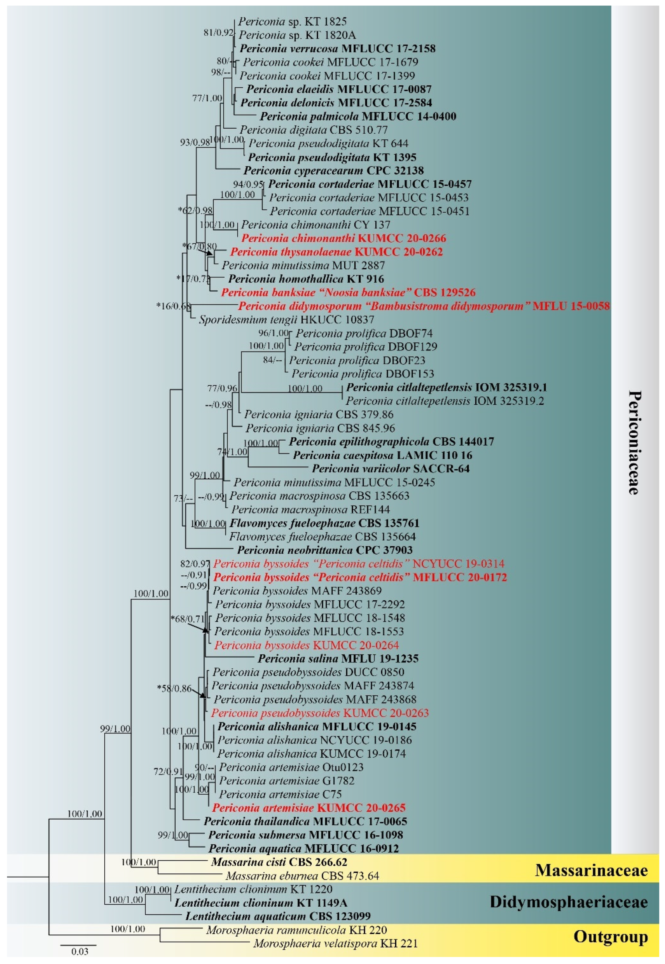
3.1. Phylogenetic Analyses
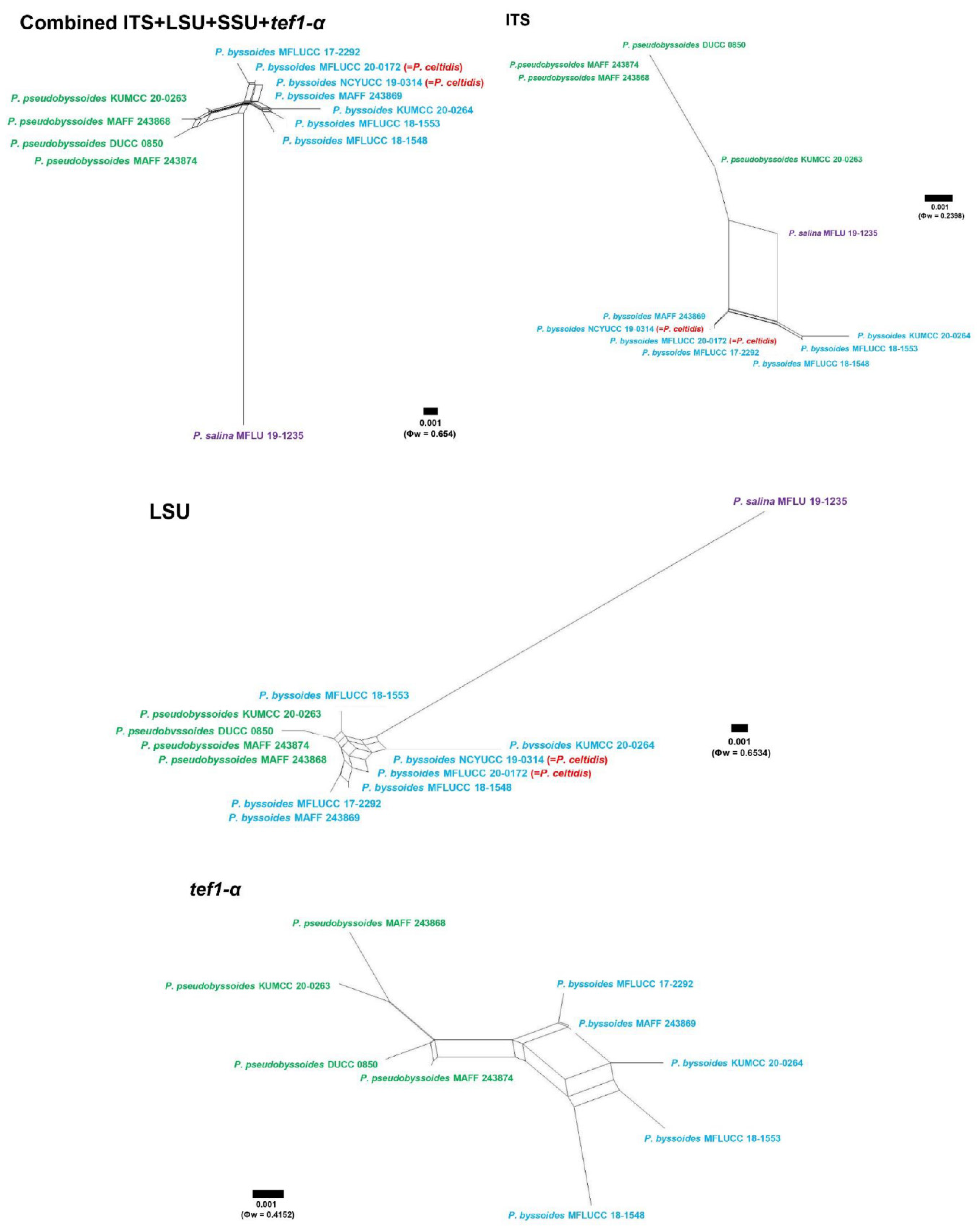
3.2. Genealogical Concordance Phylogenetic Species Recognition (GCPSR)
3.3. Taxonomy
4. Discussion
Author Contributions
Funding
Institutional Review Board Statement
Informed Consent Statement
Data Availability Statement
Acknowledgments
Conflicts of Interest
References
- Nannizzi, A. Repertorio Sistematico dei Miceti dell’ Uomo e degli Animali; SA Poligrafica Meinik Siena: Siena, Italy, 1934; Volume 4, pp. 1–557. [Google Scholar]
- Hongsanan, S.; Hyde, K.D.; Phookamsak, R.; Wanasinghe, D.N.; McKenzie, E.H.C.; Sarma, V.V.; Boonmee, S.; Jeewon, R.; Wijayawardene, N.; Devadatha, B.; et al. Refined families of Dothideomycetes: Dothideomycetidae and Pleosporomycetidae. Mycosphere 2020, 11, 1553–2107. [Google Scholar] [CrossRef]
- Farr, D.F.; Rossman, A.Y. Available online: https://nt.ars-grin.gov/fungaldatabases/ (accessed on 20 January 2022).
- Knapp, D.G.; Kovács, G.M.; Zajta, E.; Groenewald, J.Z.; Crous, P.W. Dark septate endophytic pleosporalean genera from semiarid areas. Persoonia 2015, 35, 87–100. [Google Scholar] [CrossRef] [PubMed] [Green Version]
- Sarkar, T.; Chakraborty, P.; Karmakar, A.; Saha, A.; Saha, D. First report of Periconia macrospinosa causing leaf necrosis of pointed gourd in India. J. Plant Pathol. 2019, 101, 1281. [Google Scholar] [CrossRef] [Green Version]
- Samarakoon, B.C.; Phookamsak, R.; Karunarathna, S.C.; Jeewon, R.; Chomnunti, P.; Xu, J.C.; Li, Y.J. New host and geographic records of five pleosporalean hyphomycetes associated with Musa spp. (Banana). Stud. Fungi 2021, 6, 92–115. [Google Scholar] [CrossRef]
- Kim, S.; Shin, D.S.; Lee, T.; Oh, K.B. Percinicins, two new fusicoccane diterpenes produced by an endophytic fungus Periconia sp. with antibacterial activity. J. Nat. Prod. 2004, 67, 448–450. [Google Scholar] [CrossRef] [PubMed]
- Harnpicharnchai, P.; Champreda, V.; Sornlake, W.; Eurwilaichitr, L. A thermotolerant β-glucosidase isolated from an endophytic fungi, Periconia sp., with a possible use for biomass conversion to sugars. Protein Expr. Purif. 2009, 67, 61–69. [Google Scholar] [CrossRef]
- Zhang, D.; Tao, X.; Chen, R.; Liu, J.; Li, L.; Fang, X.; Yu, L.Y.; Dai, J. Pericoannosin A, a polyketide synthase–nonribosomal peptide synthetase hybrid metabolite with new carbon skeleton from the endophytic fungus Periconia sp. Org. Lett. 2015, 17, 4304–4307. [Google Scholar] [CrossRef]
- Zhang, D.; Tao, X.; Liu, J.; Chen, R.; Zhang, M.; Li, L.; Fang, X.M.; Yu, L.; Dai, J. Periconiasin G, a new cytochalasan with unprecedented 7/6/5 tricyclic ring system from the endophytic fungus Periconia sp. Tetrahedron Lett. 2016, 57, 796–799. [Google Scholar] [CrossRef]
- Azhari, A.; Supratman, U. The Chemistry and Pharmacology of Fungal Genus Periconia: A Review. Sci. Pharm. 2021, 89, 34. [Google Scholar] [CrossRef]
- Tanaka, K.; Hirayama, K.; Yonezawa, H.; Sato, G.; Toriyabe, A.; Kudo, H.; Hashimoto, A.; Matsumura, M.; Harada, Y.; Kurihara, Y.; et al. Revision of the Massarineae (Pleosporales, Dothideomycetes). Stud. Mycol. 2015, 82, 75–136. [Google Scholar] [CrossRef] [Green Version]
- Saccardo, P.A. Sylloge Hyphomycetum. Sylloge Fungorum 1886, 4, 1–807. [Google Scholar]
- Crous, P.W.; Shivas, R.G.; Quaedvlieg, W.; van der Bank, M.; Zhang, Y.; Summerell, B.A.; Guarro, J.; Wingfield, M.; Wood, A.; Alfenas, A.; et al. Fungal planet description sheets: 214–280. Persoonia 2014, 32, 184–306. [Google Scholar] [CrossRef] [PubMed]
- Adamčík, S.; Cai, L.; Chakraborty, D.; Chen, X.H.; Cotter, H.V.T.; Dai, D.Q.; Dai, Y.C.; Das, K.; Deng, C.; Ghobad-Nejhad, M.; et al. Fungal biodiversity profiles 1–10. Cryptogam. Mycol. 2015, 36, 121–166. [Google Scholar] [CrossRef]
- Crous, P.W.; Groenewald, J.Z.; Shivas, R.G.; Edwards, J.; Seifert, K.A.; Alfenas, A.C.; Alfenas, R.F.; Burgess, T.; Carnegie, A.; Hardy, G.; et al. Fungal Planet description sheets: 69–91. Persoonia 2011, 26, 108–156. [Google Scholar] [CrossRef] [PubMed]
- Wijayawardene, N.N.; Hyde, K.D.; Al-Ani, L.K.T.; Tedersoo, L.; Haelewaters, D.; Kunhiraman, C.; Rajeshkumar, K.C.; Zhao, R.L.; Aptroot, A.; Leontyev, D.V.; et al. Outline of Fungi and fungi-like taxa. Mycosphere 2020, 11, 1060–1456. [Google Scholar] [CrossRef]
- Liu, N.G.; Hongsanan, S.; Yang, J.; Bhat, D.J.; Liu, J.K.; Jumpathong, J.J.; Liu, Z. Periconia thailandica (Periconiaceae), a new species from Thailand. Phytotaxa 2017, 323, 253–263. [Google Scholar] [CrossRef]
- Thambugala, K.M.; Wanasinghe, D.N.; Phillips, A.J.L.; Camporesi, E.; Bulgakov, T.S.; Phukhamsakda, C.; Ariyawansa, H.A.; Goonasekara, I.D.; Phookamsak, R.; Dissanayake, A.; et al. Mycosphere notes 1–50: Grass (Poaceae) inhabiting Dothideomycetes. Mycosphere 2017, 8, 697–796. [Google Scholar] [CrossRef]
- Hyde, K.D.; Chaiwan, N.; Norphanphoun, C.; Boonmee, S.; Camporesi, E.; Chethana, K.W.T.; Dayarathne, M.C.; de Silva, N.; Dissanayake, A.J.; Ekanayaka, A.H.; et al. Mycosphere notes 169–224. Mycosphere 2018, 9, 271–430. [Google Scholar] [CrossRef]
- Jayasiri, S.C.; Hyde, K.D.; Jones, E.B.G.; McKenzie, E.H.C.; Jeewon, R.; Phillips, A.J.L.; Bhat, D.J.; Wanasinghe, D.N.; Liu, J.K.; Lu, Y.Z.; et al. Diversity, morphology and molecular phylogeny of Dothideomycetes on decaying wild seed pods and fruits. Mycosphere 2019, 10, 1–186. [Google Scholar] [CrossRef]
- Phookamsak, R.; Hyde, K.D.; Jeewon, R.; Bhat, D.J.; Jones, E.B.G.; Maharachchikumbura, S.S.N.; Raspé, O.; Karunarathna, S.C.; Wanasinghe, D.; Hongsanan, S.; et al. Fungal diversity notes 929–1035: Taxonomic and phylogenetic contributions on genera and species of fungi. Fungal Divers. 2019, 95, 1–273. [Google Scholar] [CrossRef] [Green Version]
- Tode, H.J. Fungi Mecklenburgenses Selecti. Fasc. II, Generum Novorum Appendicem; Kessinger Publishing, LLC.: Lüneburg, Germany, 1791; Volume 2, pp. 1–67. [Google Scholar]
- Markovskaja, S.; Kacergius, A. Morphological and molecular characterisation of Periconia pseudobyssoides sp. nov. and closely related P. byssoides. Mycol. Prog. 2014, 13, 291–302. [Google Scholar] [CrossRef]
- Saccardo, P.A. Fungi Gallici lecti a cl. viris P. Brunaud, Abb. Letendre, A. Malbranche, J. Therry, vel editi in Mycotheca Gallica C. Roumeguèri. Series II. Michelia 1880, 2, 39–135. [Google Scholar]
- Hughes, S.J. Revisiones Hyphomycetum aliquot cum appendice de nominibus rejiciendis. Can. J. Bot. 1958, 36, 727–836. [Google Scholar] [CrossRef]
- Benjamin, C.R.; Hesseltine, C.W. Studies on the genus Phycomyces. Mycologia 1959, 51, 751–771. [Google Scholar] [CrossRef]
- Malloch, D.W. Scopinella sphaerophila. Fungi Canadenses 1976, 83, 1–2. [Google Scholar]
- Hawksworth, D.L. Three new genera of lichenicolous fungi. Bot. J. Linn. Soc. 1977, 75, 195–209. [Google Scholar] [CrossRef]
- Illman, W.I.; White, G.P. The synnematous hyphomycete Morrisographium pilosum (Earle) Moreletsynonymous with a ‘Coelomycete’, ‘Cornularia’ persicae’ (Schw.) Sacc. Mycotaxon 1984, 19, 145–150. [Google Scholar]
- Aptroot, A. A world revision of Massarina (Ascomycota). Nova Hedwig. 1998, 66, 89–162. [Google Scholar] [CrossRef]
- Okada, G.; Jacobs, K.; Kirisits, T.; Louis-Seize, G.W.; Seifert, K.A.; Sugita, T.; Takematsu, A.; Wingfield, M.J. Epitypification of Graphium penicillioides Corda, with comments on the phylogeny and taxonomy of graphium-like synnematous fungi. Stud. Mycol. 2000, 45, 169–188. [Google Scholar]
- Partridge, E.C.; Morgan-Jones, G. Notes on hyphomycetes LXXXVIII: New genera in which to classify Alysidium resinae and Pycnostysanus azaleae, with a consideration of Sorocybe. Mycotaxon 2002, 83, 335–352. [Google Scholar]
- Schubert, K.; Braun, U.; Groenewald, J.Z.; Crous, P.W. Cladosporium leaf-blotch and stem rot of Paeonia spp. caused by Dichocladosporium chlorocephalum gen. nov. Stud. Mycol. 2007, 58, 95–104. [Google Scholar] [CrossRef]
- Chlebicki, A. Cephalotrichum stemonitis as a biofilm inhabitant in the gold mine in Poland. Acta Mycol. 2008, 43, 67–70. [Google Scholar] [CrossRef]
- Kiyuna, T.; An, K.D.; Kigawa, R.; Sano, C.; Miura, S. Molecular assessment of fungi in “black spots” that deface murals in the Takamatsuzuka and Kitora Tumuli in Japan: Acremonium sect. Gliomastix including Acremonium tumulicola sp. nov. and Acremonium felinum comb. nov. Mycoscience 2011, 52, 1–17. [Google Scholar] [CrossRef]
- Videira, S.I.R.; Groenewald, J.Z.; Nakashima, C.; Braun, U.; Barreto, R.W.; de Wit, P.J.G.M.; Crous, P.W. Mycosphaerellaceae—Chaos or clarity? Stud. Mycol. 2017, 87, 257–421. [Google Scholar] [CrossRef]
- Yin, M.L.; Wingfield, M.J.; Zhou, X.D.; de Beer, Z.W. Phylogenetic re-evaluation of the Grosmannia penicillata complex (Ascomycota, Ophiostomatales), with the description of five new species from China and USA. Fungal Biol. 2020, 124, 110–124. [Google Scholar] [CrossRef]
- Species Fungorum. Available online: http://www.speciesfungorum.org/Names/Names/Names.asp (accessed on 20 January 2022).
- Tennakoon, D.S.; Kuo, C.H.; Maharachchikumbura, S.S.N.; Thambugala, K.M.; Gentekaki, E.; Phillips, A.J.L.; Bhat, D.J.; Wanasinghe, D.N.; de Silva, N.I.; Promputtha, I.; et al. Taxonomic and phylogenetic contributions to Celtis formosana, Ficus ampelas, F. septica, Macaranga tanarius and Morus australis leaf litter inhabiting microfungi. Fungal Divers. 2021, 108, 1–215. [Google Scholar] [CrossRef]
- Carmarán, C.C.; Novas, M.V. A review of Spegazzini taxa of Periconia and Sporocybe after over 115 years. Fungal Divers. 2003, 14, 67–76. [Google Scholar]
- Index Fungorum. Bambusistroma Didymosporum D.Q. Dai & K.D. Hyde. 2015. Available online: http://www.indexfungorum.org/Names/NamesRecord.asp?RecordID=551028 (accessed on 12 January 2022).
- Hyde, K.D.; Dong, Y.; Phookamsak, R.; Jeewon, R.; Bhat, D.J.; Jones, E.B.G.; Liu, N.G.; Abeywickrama, P.D.; Mapook, A.; Wei, D.; et al. Fungal diversity notes 1151–1276: Taxonomic and phylogenetic contributions on genera and species of fungal taxa. Fungal Divers. 2020, 100, 5–277. [Google Scholar] [CrossRef] [Green Version]
- Dai, D.Q.; Phookamsak, R.; Wijayawardene, N.N.; Li, W.J.; Bhat, D.J.; Xu, J.C.; Taylor, J.E.; Hyde, K.D.; Chukeatirote, E. Bambusicolous fungi. Fungal Divers. 2017, 82, 1–105. [Google Scholar] [CrossRef]
- Vilgalys, R.; Hester, M. Rapid genetic identification and mapping of enzymatically amplified ribosomal DNA from several Cryptococcus species. J. Bacteriol. 1990, 172, 4238–4246. [Google Scholar] [CrossRef] [Green Version]
- White, T.J.; Bruns, T.; Lee, S.J.W.T.; Taylor, J.W. Amplification and direct sequencing of fungal ribosomal RNA genes for phylogenetics. In PCR Protocols: A Guide to Methods and Applications; Academic Press: Cambridge, MA, USA, 1990; Volume 18, pp. 315–322. [Google Scholar]
- Rehner, S. Primers for Elongation Factor 1-Alpha (EF1-Alpha). 2001. Available online: http://ocid.NACSE.ORG/research/ (accessed on 23 September 2021).
- Tibpromma, S.; Hyde, K.D.; Jeewon, R.; Maharachchikumbura, S.S.N.; Liu, J.K.; Bhat, D.J.; Jones, E.B.G.; McKenzie, E.H.C.; Camporesi, E.; Bulgakov, T.; et al. Fungal diversity notes 491–602: Taxonomic and phylogenetic contributions to fungal taxa. Fungal Divers. 2017, 83, 1–261. [Google Scholar] [CrossRef]
- Hall, T.A. BioEdit: A user-friendly biological sequence alignment editor and analysis program for Windows 95/98/NT. Nucleic Acids Symp. Ser. 1999, 41, 95–98. [Google Scholar] [CrossRef]
- Crous, P.W.; Wingfield, M.J.; Lombard, L.; Roets, F.; Swart, W.J.; Alvarado, P.; Carnegie, A.J.; Moreno, G.; Luangsa-Ard, J.; Thangavel, R.; et al. Fungal Planet description sheets: 951–1041. Persoonia 2019, 43, 223–425. [Google Scholar] [CrossRef]
- Calvillo-Medina, R.P.; Cobos-Villagran, A.; Raymundo, T. Periconia citlaltepetlensis sp. nov. (Periconiaceae, Pleosporales): A psychrotolerant fungus from high elevation volcanic glacier (Mexico). Phytotaxa 2020, 459, 235–247. [Google Scholar] [CrossRef]
- Phukhamsakda, C.; McKenzie, E.H.C.; Phillips, A.J.L.; Jones, E.B.G.; Bhat, D.J.; Stadler, M.; Bhunjun, C.S.; Wanasinghe, D.N.; Thongbai, B.; Camporesi, E.; et al. Microfungi associated with Clematis (Ranunculaceae) with an integrated approach to delimiting species boundaries. Fungal Divers. 2020, 102, 1–203. [Google Scholar] [CrossRef]
- Katoh, K.; Rozewicki, J.; Yamada, K.D. MAFFT online service: Multiple sequence alignment, interactive sequence choice and visualization. Brief. Bioinform. 2019, 20, 1160–1166. [Google Scholar] [CrossRef] [Green Version]
- Stamatakis, A.; Hoover, P.; Rougemont, J. A rapid bootstrap algorithm for the RAxML web servers. Syst. Biol. 2008, 57, 758–771. [Google Scholar] [CrossRef]
- Stamatakis, A. RAxML Version 8: A tool for Phylogenetic Analysis and Post-Analysis of Large Phylogenies. Bioinformatics 2014, 30, 1312–1313. [Google Scholar] [CrossRef]
- Miller, M.A.; Pfeiffer, W.; Schwartz, T. Creating the CIPRES Science Gateway for inference of large phylogenetic trees. In Proceedings of the 2010 Gateway Computing Environments Workshop (GCE), New Orleans, LA, USA, 14 November 2010; pp. 1–8. [Google Scholar]
- Nylander, J.A.A. MrModeltest v2. Program Distributed by the Author; Evolutionary Biology Centre, Uppsala University: Uppsala, Sweden, 2004. [Google Scholar]
- Rannala, B.; Yang, Z. Probability distribution of molecular evolutionary trees: A new method of phylogenetic inference. J. Mol. Evol. 1996, 43, 304–311. [Google Scholar] [CrossRef]
- Zhaxybayeva, O.; Gogarten, J.P. Bootstrap, Bayesian probability and maximum likelihood mapping: Exploring new tools for comparative genome analyses. Genomics 2002, 3, 1–15. [Google Scholar] [CrossRef] [PubMed] [Green Version]
- Rambaut, A.; Drummond, A.J.; Xie, D.; Baele, G.; Suchard, M.A. Posterior summarisation in Bayesian phylogenetics using Tracer 1.7. Syst. Biol. 2018, 67, 901–904. [Google Scholar] [CrossRef] [PubMed] [Green Version]
- Bruen, T.C.; Philippe, H.; Bryant, D. A simple and robust statistical test for detecting the presence of recombination. Genetics 2006, 172, 2665–2681. [Google Scholar] [CrossRef] [PubMed] [Green Version]
- Huson, D.H. SplitsTree: Analyzing and visualizing evolutionary data. Bioinformatics 1998, 14, 68–73. [Google Scholar] [CrossRef]
- Huson, D.H.; Bryant, D. Application of phylogenetic networks in evolutionary studies. Mol. Biol. Evol. 2006, 23, 254–267. [Google Scholar] [CrossRef]
- Quaedvlieg, W.; Binder, M.; Groenewald, J.Z.; Summerell, B.A.; Carnegie, A.J.; Burgess, T.I.; Crous, P.W. Introducing the Consolidated Species Concept to resolve species in the Teratosphaeriaceae. Persoonia 2014, 33, 1–40. [Google Scholar] [CrossRef] [Green Version]
- Jeewon, R.; Hyde, K.D. Establishing species boundaries and new taxa among fungi: Recommendations to resolve taxonomic ambiguities. Mycosphere 2016, 7, 1669–1677. [Google Scholar] [CrossRef]
- Vu, D.; Groenewald, M.; de Vries, M.; Gehrmann, T.; Stielow, B.; Eberhardt, U.; Al-Hatmi, A.; Groenewald, J.Z.; Cardinali, G.; Houbraken, J.; et al. Large-scale generation and analysis of filamentous fungal DNA barcodes boosts coverage for kingdom fungi and reveals thresholds for fungal species and higher taxon delimitation. Stud. Mycol. 2019, 92, 135–154. [Google Scholar] [CrossRef]
- Rodrigues, A.; Mueller, U.G.; Ishak, H.D.; Bacci, M.; Pagnocca, F.C. Ecology of microfungal communities in gardens of fungus-growing ants (Hymenoptera: Formicidae): A year-long survey of three species of attine ants in Central Texas. FEMS Microbiol. Ecol. 2011, 78, 244–255. [Google Scholar] [CrossRef] [Green Version]
- Wu, W.P.; Zhuang, W.Y. Sporidesmium, Endophragmiella and related genera from China. Fungal Divers. Res. Ser. 2005, 15, 1–351. [Google Scholar]
- Shenoy, B.D.; Jeewon, R.; Wu, W.P.; Bhat, D.J.; Hyde, K.D. Ribosomal and RPB2 DNA sequence analyses suggest that Sporidesmium and morphologically similar genera are polyphyletic. Mycol. Res. 2006, 110, 916–928. [Google Scholar] [CrossRef] [PubMed]
- Bovio, E.; Garzoli, L.; Poli, A.; Prigione, V.; Firsova, D.; McCormack, G.P.; Varese, G.C. The culturable mycobiota associated with three Atlantic sponges, including two new species: Thelebolus balaustiformis and T. spongiae. Fuse 2018, 1, 141–167. [Google Scholar] [CrossRef] [PubMed] [Green Version]
- Corda, A.C.J. Icones fungorum hucusque cognitorum. J. Nat. Hist. 1837, 1, 1–32. [Google Scholar]
- Dayarathne, M.C.; Jones, E.B.G.; Maharachchikumbura, S.S.N.; Devadatha, B.; Sarma, V.V.; Khongphinitbunjong, K.; Chomnunti, P.; Hyde, K.D. Morpho-molecular characterization of microfungi associated with marine based habitats. Mycosphere 2020, 11, 1–188. [Google Scholar] [CrossRef]


| Species Name | Strain/Voucher No. | GenBank Accession Numbers | |||
|---|---|---|---|---|---|
| ITS | LSU | SSU | tef1-α | ||
| Flavomyces fulophazae | CBS 135664 | KP184000 | KP184039 | KP184081 | / |
| Flavomyces fulophazae | CBS 135761 | NR_137960 | NG_058131 | NG_061191 | / |
| Lentithecium aquaticum | CBS 123099 | NR_160229 | NG_064211 | NG_016507 | GU349068 |
| Lentithecium clioninum | KT 1149A | LC014566 | AB807540 | AB797250 | AB808515 |
| Lentithecium clioninum | KT 1220 | LC014567 | AB807541 | AB797251 | AB808516 |
| Massarina cisti | CBS 266.62 | / | AB807539 | AB797249 | AB808514 |
| Massarina eburnea | CBS 473.64 | / | GU301840 | GU296170 | GU349040 |
| Morosphaeria ramunculicola | KH 220 | / | AB807554 | AB797264 | AB808530 |
| Morosphaeria velatispora | KH 221 | LC014572 | AB807556 | AB797266 | AB808532 |
| Periconia alishanica | KUMCC 19-0174 | MW063167 | MW063231 | / | MW183792 |
| Periconia alishanica | MFLUCC 19-0145 | MW063165 | MW063229 | / | MW183790 |
| Periconia alishanica | NCYUCC 19-0186 | MW063166 | MW063230 | / | MW183791 |
| Periconia aquatica | MFLUCC 16-0912 | KY794701 | KY794705 | / | KY814760 |
| Periconia artemisiae | KUMCC 20-0265 | MW448657 | MW448571 | MW448658 | MW460898 |
| Periconia banksiae (as Noosia banksiae) | CBS 129526 | / | NG_064279 | / | / |
| Periconia byssoides | KUMCC 20-0264 | MW444854 | MW444855 | MW444856 | MW460895 |
| Periconia byssoides | MAFF 243869 | LC014582 | AB807569 | AB797279 | AB808545 |
| Periconia byssoides | MFLUCC 17-2292 | MK347751 | MK347968 | MK347858 | MK360069 |
| Periconia byssoides | MFLUCC 18-1548 | MK347794 | MK348013 | MK347902 | MK360070 |
| Periconia byssoides | MFLUCC 18-1553 | MK347806 | MK348025 | MK347914 | MK360068 |
| Periconia byssoides (as Periconia celtidis) | MFLUCC 20-0172 | MW063162 | MW063226 | / | / |
| Periconia byssoides (as Periconia celtidis) | NCYUCC 19-0314 | MW063163 | MW063227 | / | / |
| Periconia caespitosa | LAMIC 110 16 | MH051906 | MH051907 | / | / |
| Periconia chimonanthi | KUMCC 20-0266 | MW448660 | MW448572 | MW448656 | MW460897 |
| Periconia citlaltepetlensis | IOM 325319.1 | MH890645 | MT625978 | / | / |
| Periconia citlaltepetlensis | IOM 325319.2 | MT649221 | MT649216 | / | / |
| Periconia cookei | MFLUCC 17-1399 | MG333490 | MG333493 | / | MG438279 |
| Periconia cookei | MFLUCC 17-1679 | / | MG333492 | / | MG438278 |
| Periconia cortaderiae | MFLUCC 15-0451 | KX965734 | KX954403 | KX986346 | KY429208 |
| Periconia cortaderiae | MFLUCC 15-0453 | KX965733 | KX954402 | / | KY320574 |
| Periconia cortaderiae | MFLUCC 15-0457 | KX965732 | KX954401 | KX986345 | KY310703 |
| Periconia cyperacearum | CPC 32138 | NR_160357 | NG_064549 | / | / |
| Periconia delonicis | MFLUCC 17-2584 | / | NG_068611 | NG_065770 | MK360071 |
| Periconia didymosporum (as Bambusistroma didymosporum) | MFLU 15-0058 | KP761734 | KP761731 | KP761738 | KP761728 |
| Periconia digitata | CBS 510.77 | LC014584 | AB807561 | AB797271 | AB808537 |
| Periconia elaeidis | MFLUCC 17-0087 | MG742713 | MH108552 | MH108551 | / |
| Periconia epilithographicola | CBS 144017 | NR_157477 | / | / | / |
| Periconia homothallica | KT 916 | AB809645 | AB807565 | AB797275 | / |
| Periconia igniaria | CBS 379.86 | LC014585 | AB807566 | AB797276 | AB808542 |
| Periconia igniaria | CBS 845.96 | LC014586 | AB807567 | AB797277 | AB808543 |
| Periconia macrospinosa | CBS 135663 | KP183999 | KP184038 | KP184080 | / |
| Periconia macrospinosa | REF144 | JN859364 | JN859484 | / | / |
| Periconia minutissima | MFLUCC 15-0245 | KY794703 | KY794707 | / | / |
| Periconia minutissima | MUT 2887 | MG813227 | / | / | / |
| Periconia neobrittanica | CPC 37903 | NR_166344 | NG_068342 | / | / |
| Periconia palmicola | MFLUCC 14-0400 | / | NG_068917 | MN648319 | MN821070 |
| Periconia prolifica | DBOF23 | JQ724384 | / | / | / |
| Periconia prolifica | DBOF74 | JQ724435 | / | / | / |
| Periconia prolifica | DBOF129 | JQ724490 | / | / | / |
| Periconia prolifica | DBOF153 | JQ724513 | / | / | / |
| Periconia pseudobyssoides | DUCC 0850 | MG333491 | MG333494 | / | MG438280 |
| Periconia pseudobyssoides | KUMCC 20-0263 | MW444851 | MW444852 | MW444853 | MW460894 |
| Periconia pseudobyssoides | MAFF 243868 | LC014587 | AB807568 | AB797278 | AB808544 |
| Periconia pseudobyssoides | MAFF 243874 | LC014588 | AB807560 | AB797270 | AB808536 |
| Periconia pseudodigitata | KT 644 | LC014589 | AB807562 | AB797272 | AB808538 |
| Periconia pseudodigitata | KT 1395 | NR_153490 | NG_059396 | NG_064850 | AB808540 |
| Periconia salina | MFLU 19-1235 | MN047086 | MN017846 | MN017912 | / |
| Periconia sp. | C75 | MK304380 | / | / | / |
| Periconia sp. | CY 137 | HQ607981 | / | / | / |
| Periconia sp. | G1782 | MK247789 | / | / | / |
| Pericona sp. | KT 1825 | / | AB807573 | AB797283 | AB808549 |
| Pericona sp. | KT 1820A | / | AB807572 | AB797282 | AB808548 |
| Pericona sp. | Out0123 | MT908499 | / | / | / |
| Periconia submersa | MFLUCC 16-1098 | KY794702 | KY794706 | / | KY814761 |
| Periconia thailandica | MFLUCC 17-0065 | KY753887 | KY753888 | KY753889 | / |
| Periconia thysanolaenae | KUMCC 20-0262 | MW442967 | MW444850 | MW448659 | MW460896 |
| Periconia variicolor | SACCR-64 | DQ336713 | / | / | / |
| Periconia verrucosa | MFLUCC 17-2158 | MT310617 | MT214572 | MT226686 | MT394631 |
| Sporidesmium tengii | HKUCC 10837 | / | DQ408559 | / | / |
| Species Name | Morphological Characteristics In Vitro | |
|---|---|---|
| Conidiogenous Cells | Conidia | |
| Periconia artemisiae (KUN-HKAS 107384) | 2.5–4 × 1–2 μm, mono- to polyblastic, with 1–2 conidiogenous loci, lateral, or integrated, lateral and terminal, brown to dark brown, inconspicuous, giving rise to solitary conidia, or in short chains. | 5–8 × 4–5 μm globose, brown to dark brown, aseptate, smooth to slightly verruculose. |
| Periconia banksiae (≡ Noosia banksiae, CBS H-20587) | Solitary, lateral, or integrated, inconspicuous, lateral and terminal, with small, pimple-like pores of up to 0.5 µm diam. | Dimorphic: primary conidia (4–)7–10(–13) × (3.5–)4(–5) µm, aseptate, globose to fusoid-ellipsoidal, subhyaline to brown, smooth to verruculose with age, solitary or in short, branched chains. Secondary conidia 5–15 × 4–5 µm, phragmosporous, brown, verruculose, arising from disarticulating hyphal cells, initially in short chains, forming directly on conidiogenous cells when mature. |
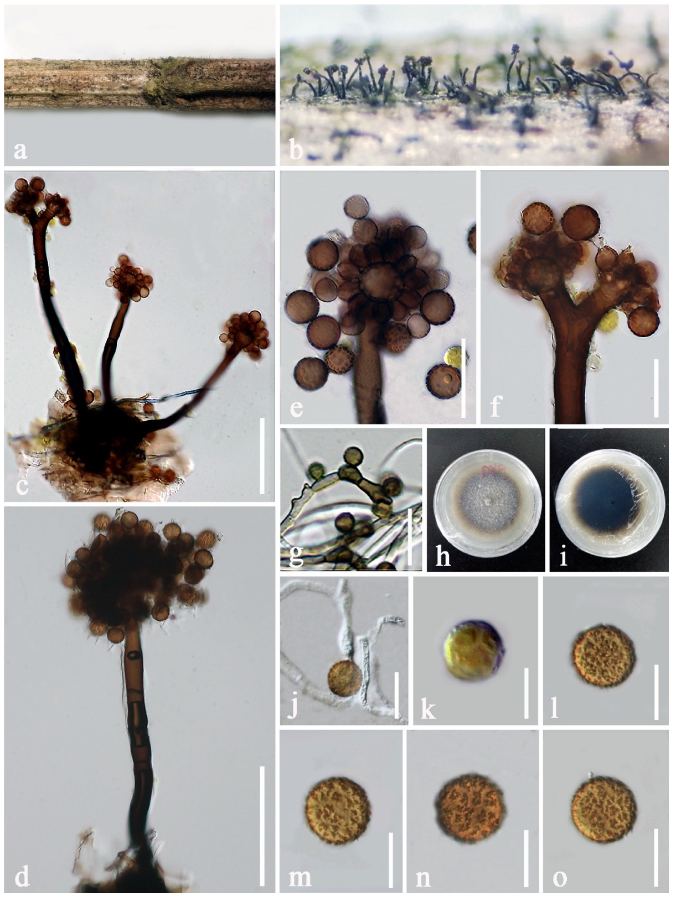
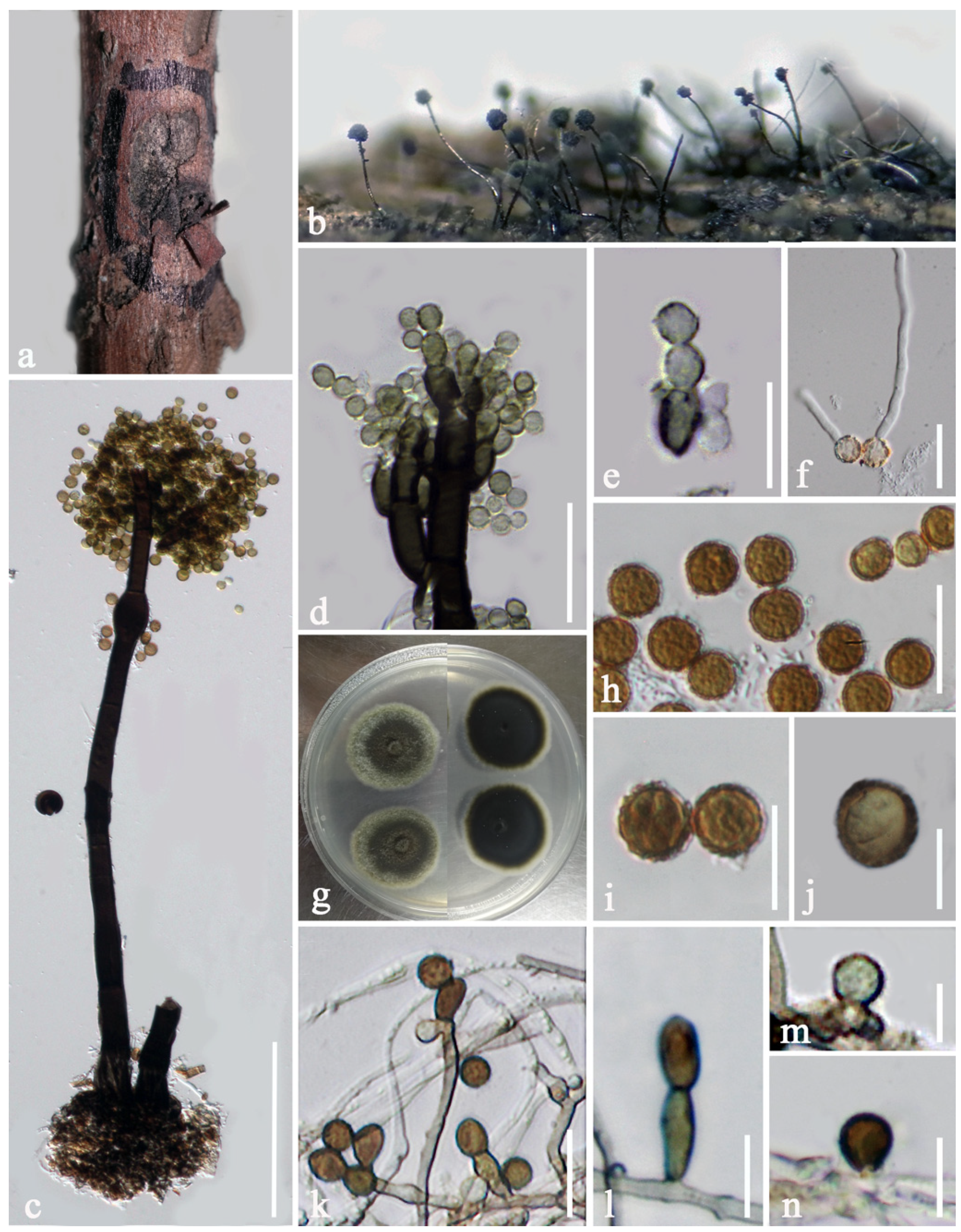
| P. chimonanthi (KUN-HKAS 107380) | 7–10 × 4.5–6 μm, polyblastic, solitary, erect, lateral and terminal, cylindrical to irregular, luteous to brown, discrete or integrated, determinate, or inconspicuous, percurrent proliferations, with 1–3 conidiogenous loci. | 6–8 × 6–8 μm, globose to oblong, or ellipsoidal, subhyaline to brown or dark brown, smooth to verruculose, solitary or in short chains. |
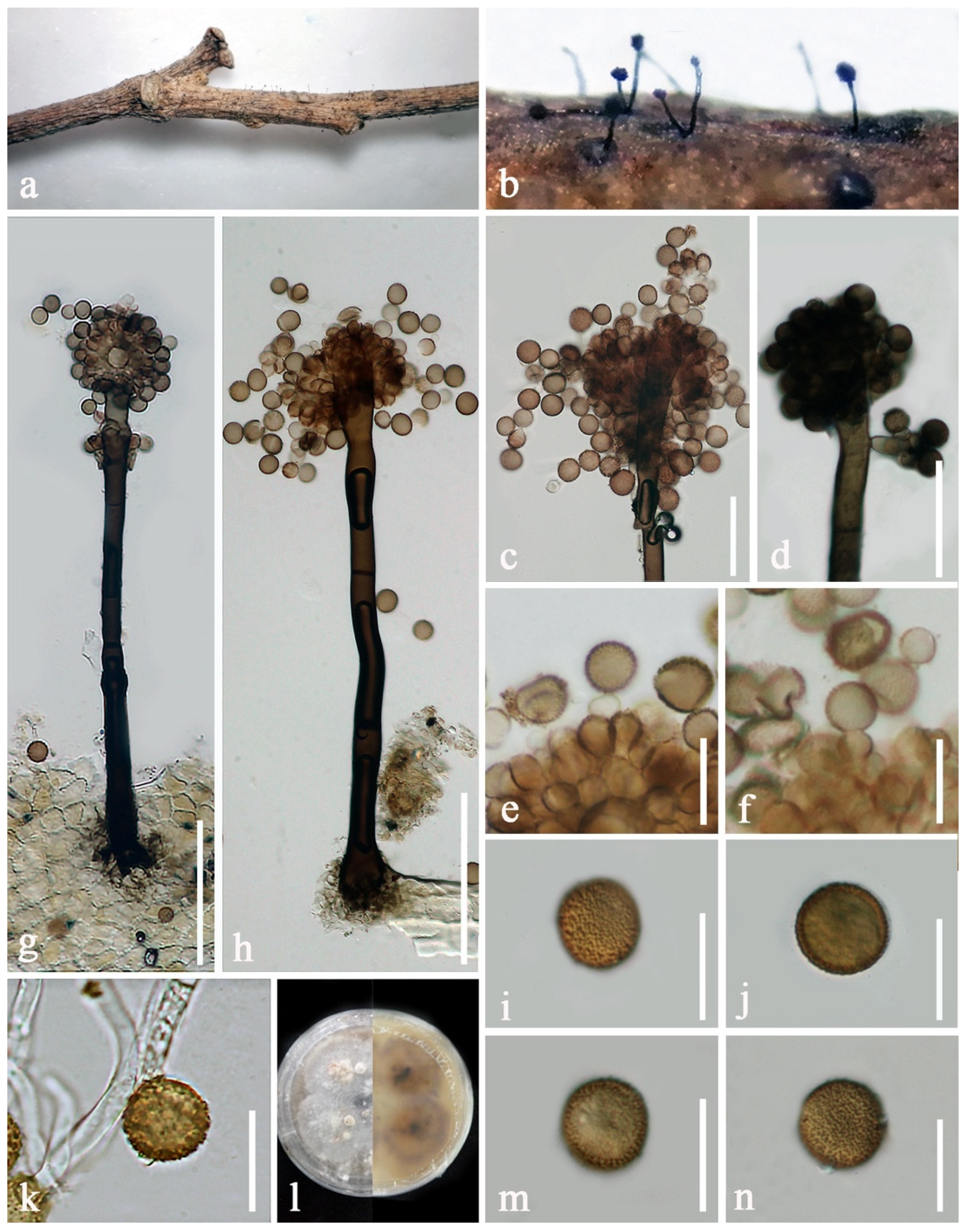
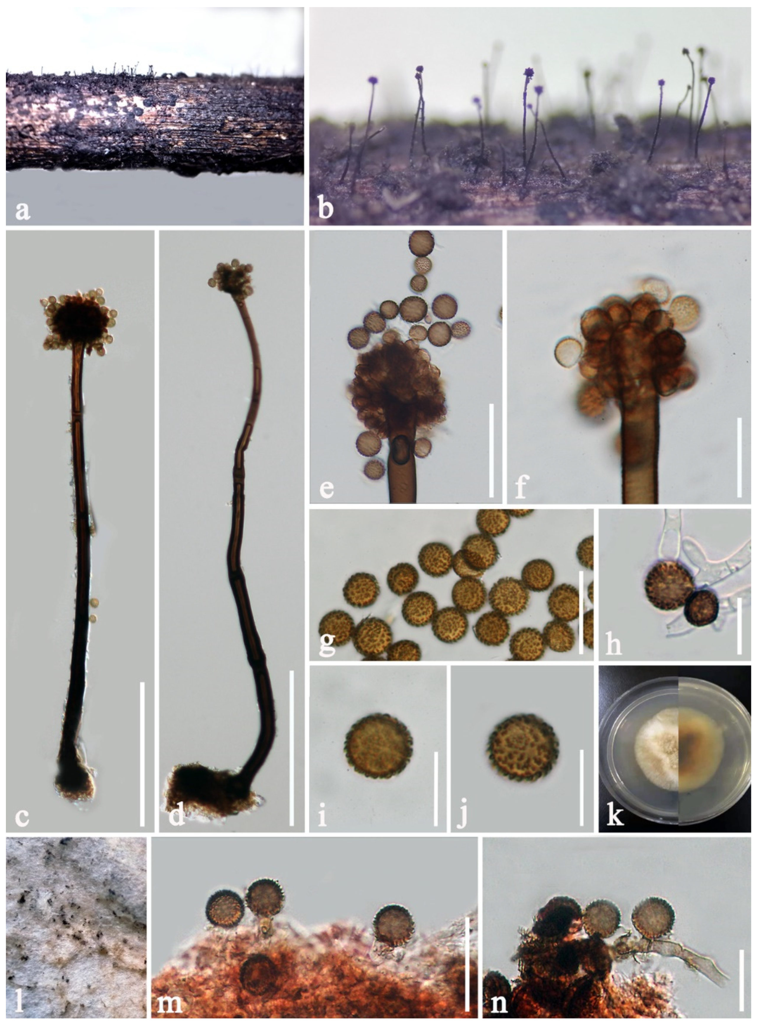
| P. pseudobyssoides (KUN-HKAS 107382) | 4–6 × 2.5–4 μm, monoblastic, solitary, terminal, inconspicuous, with small, pimple-like pores, arising from brown and verruculose hyphae. | 13–16 × 12–15 μm, solitary, globose, reddish-brown, aseptate, verruculose. |
| Species Name | Strain Number | Nucleotide Base Differences | |||||||||||||||||||||
|---|---|---|---|---|---|---|---|---|---|---|---|---|---|---|---|---|---|---|---|---|---|---|---|
| LSU (Base Position) | ITS (Base Position) | tef1-α (Base Position) | |||||||||||||||||||||
| 361 | 431 | 432 | 468 | 485 | 633 | 42 | 48 | 459 | 495 | 69 | 105 | 237 | 309 | 328 | 336 | 426 | 435 | 567 | 660 | 723 | 915 | ||
| Periconia byssoides | KUMCC 20-0264 | T | G | A | A | T | C | - | T | T | T | T | T | T | T | G | T | G | C | T | T | G | T |
| Periconia byssoides | MAFF 243869 | T | T | T | G | T | C | - | C | C | C | T | C | T | T | G | C | G | T | T | T | G | T |
| Periconia byssoides | MFLUCC 17-2292 | T | T | T | A | T | T | - | C | C | C | T | C | T | T | G | C | G | T | T | G | G | T |
| Periconia byssoides | MFLUCC 18-1548 | T | G | A | A | T | C | - | T | T | C | T | T | C | C | A | T | G | T | C | T | A | C |
| Periconia byssoides | MFLUCC 18-1553 | C | G | A | A | T | C | A | T | T | C | C | T | T | T | G | T | A | T | C | T | G | T |
| Periconia celtidis | MFLUCC 20-0172 | T | T | T | G | C | C | - | C | C | C | Sequence unavailable | |||||||||||
| Periconia celtidis | NCYUCC 19-0314 | T | T | T | G | C | C | - | C | C | C | Sequence unavailable | |||||||||||
| Taxa | Morphological Characteristics | References | ||
|---|---|---|---|---|
| Periconia byssoides Strain No. | Conidiophores | Conidiogenous Cells | Conidia | |
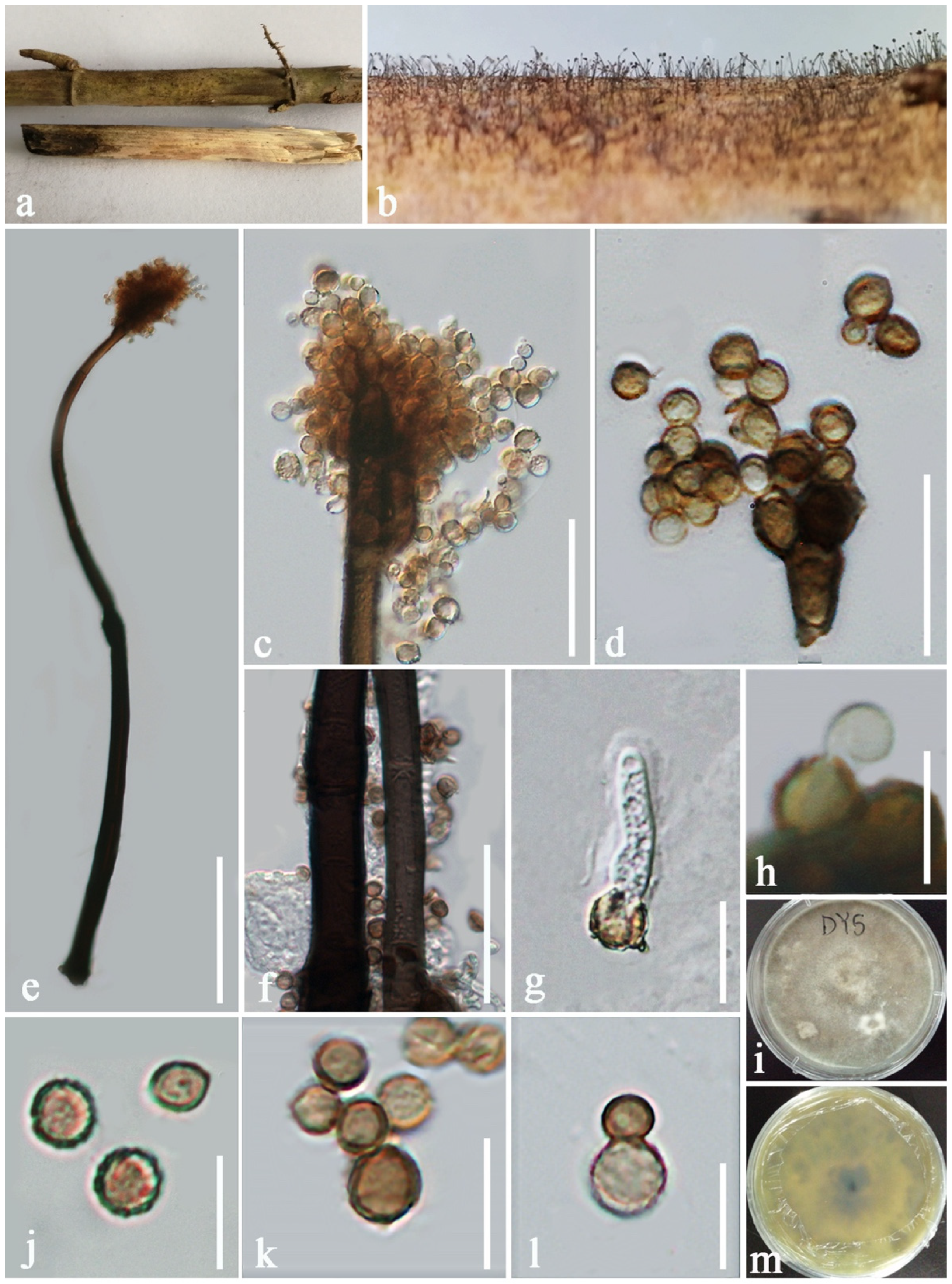
| KUMCC 20-0264 | 355–635 × 12.5–17 μm, macronematous, mononematous, solitary, rarely 1−2 together on stroma, 1−3-septate, dark brown to black at the base, paler brown towards the apex, unbranched, rarely branched at the apex, knot-like near the base, smooth or slightly rough, thick-walled. | 6–9 × 5–8 μm, polyblastic, discrete, terminal, subglobose to ellipsoidal, light brown, located at nodose apices of conidiophores. | 13–15 × 12.5–14.5 μm, solitary or catenate, globose to subglobose, orangish brown to brown, aseptate, echinulate or verruculose | This study |
| BILAS 50335/ culture S2-11P (Sporulated on MEA) | Variable in length, simple, micro- or semi-macronematous, unbranched and branched, initially subhyaline to brownish, becoming dark brown at maturity, subhyaline or hyaline at the apex, septate, verruculose, formed singly or in small groups pushing through the weft of mycelium. | Mono- and polyblastic, discrete, determinate, terminal or lateral, subglobose, smooth to verruculose, pale brown, formed on an apical cell and in the collar region around the septa. | (11.5–)12.5–15(−17) μm diam., globose, pale brown to brown, verruculose or verrucose, in acropetal chains (3–4 in number). | [24] |
| MFLUCC 17-2292 | 350–420 × 4.5–5.5 μm, macronematous, mononematous, single or rarely 2–3 together on stroma, brown to dark brown, erect, or bent, septate, smooth, thick-walled. | Monoblastic, discrete on stipe. | 9–12 × 8–12 μm, catenate, globose, brown to dark brown, aseptate, verruculose. | [21] |
| MFLUCC 19-0134 | 300–370 × 4–5 μm, macronematous, mononematous, unbranched, erect, single, light brown to dark brown, septate, smooth to minutely verruculose, thick-walled. | Monoblastic, proliferating, hyaline, terminal, blunt end, ovoid to globose, thick-walled. | 11–13 × 10–12 μm, solitary, subglobose to globose, light brown to dark brown, aseptate, finely verruculose. | [40] |
| MFLUCC 20-0172 (as P. celtidis) | 300–380 × 3.5–4.8 μm, macronematous, mononematous, unbranched, erect, single, greyish brown to dark brown, septate, smooth to minutely verruculose, thick-walled. | Monoblastic, proliferating, hyaline, terminal, blunt end, ovoid to globose, thick-walled. | 8–10 × 9–10 μm diam., solitary, subglobose to globose, light brown to dark brown, aseptate, verruculose. | [40] |
| Species Name | Strain Number | Nucleotide Base Differences | |||||||||||||
|---|---|---|---|---|---|---|---|---|---|---|---|---|---|---|---|
| ITS (Base Position) | LSU (Base Position) | tef1-α (Base Position) | |||||||||||||
| 76 | 419 | 522 | 371 | 331 | 405 | 471 | 564 | 570 | 579 | 648 | 720 | 725 | 764 | ||
| Periconia pseudobyssoidesT | BILAS 50334 | - | T | G | Sequence unavailable | Sequence unavailable | |||||||||
| Periconia pseudobyssoides | DUCC 0850 | - | T | G | C | G | T | C | T | T | C | C | T | G | C |
| Periconia pseudobyssoides | KUMCC 20-0263 | A | C | A | T | A | C | C | C | T | T | T | T | A | C |
| Periconia pseudobyssoides | MAFF 243868 | - | T | G | C | A | T | T | C | T | C | T | C | A | G |
| Periconia pseudobyssoides | MAFF 243874 | - | T | G | C | G | T | C | C | C | C | C | T | A | C |
Publisher’s Note: MDPI stays neutral with regard to jurisdictional claims in published maps and institutional affiliations. |
© 2022 by the authors. Licensee MDPI, Basel, Switzerland. This article is an open access article distributed under the terms and conditions of the Creative Commons Attribution (CC BY) license (https://creativecommons.org/licenses/by/4.0/).
Share and Cite
Yang, E.-F.; Phookamsak, R.; Jiang, H.-B.; Tibpromma, S.; Bhat, D.J.; Karunarathna, S.C.; Dai, D.-Q.; Xu, J.-C.; Promputtha, I. Taxonomic Reappraisal of Periconiaceae with the Description of Three New Periconia Species from China. J. Fungi 2022, 8, 243. https://doi.org/10.3390/jof8030243
Yang E-F, Phookamsak R, Jiang H-B, Tibpromma S, Bhat DJ, Karunarathna SC, Dai D-Q, Xu J-C, Promputtha I. Taxonomic Reappraisal of Periconiaceae with the Description of Three New Periconia Species from China. Journal of Fungi. 2022; 8(3):243. https://doi.org/10.3390/jof8030243
Chicago/Turabian StyleYang, Er-Fu, Rungtiwa Phookamsak, Hong-Bo Jiang, Saowaluck Tibpromma, Darbhe J. Bhat, Samantha C. Karunarathna, Dong-Qin Dai, Jian-Chu Xu, and Itthayakorn Promputtha. 2022. "Taxonomic Reappraisal of Periconiaceae with the Description of Three New Periconia Species from China" Journal of Fungi 8, no. 3: 243. https://doi.org/10.3390/jof8030243