Review

Insight into Biological Control Potential of Hirsutella citriformis against Asian Citrus Psyllid as a Vector of Citrus Huanglongbing Disease in America

by
Orquídea Pérez-González
,
Ricardo Gomez-Flores
and
Patricia Tamez-Guerra
*
Departamento de Microbiología e Inmunología-Laboratorio de Inmunología y Virología, Facultad de Ciencias Biológicas, Universidad Autónoma de Nuevo León, UANL, San Nicolás de los Garza, Nuevo León 66455, Mexico
*
Author to whom correspondence should be addressed.
J. Fungi 2022, 8(6), 573; https://doi.org/10.3390/jof8060573
Submission received: 29 April 2022 / Revised: 17 May 2022 / Accepted: 18 May 2022 / Published: 27 May 2022
(This article belongs to the Topic Fungal Diversity)

Abstract

:
Studies on Hirsutella citriformis Speare are scarce. Among these, some reports have focused on phenotypic identification, based on its morphological structure and morphometric characteristics. This fungus is known to control economically important citrus crop pests. In recent years, H. citriformis has received increased attention as a control agent for the Asian citrus psyllid Diaphorina citri Kuwayama (Hemiptera: Liviidae), which causes the Huanglongbing (HLB) disease. Unfortunately, formal H. citriformis strains characterization is marginal, which mainly involves the role of biologically active exudates (metabolites) produced during their growth. Information regarding their mode of action and biocontrol potential is limited. However, epizootics reports of this fungus, under suitable environmental conditions for its development (25 °C to 28 °C and ~80% relative humidity), have demonstrated its parasitization efficacy. Therefore, it becomes challenging to determine whether H. citriformis strains may be developed as commercial products. In this review, we showed relevant information on isolation and bioassay strategies of H. citriformis to evaluate potential biocontrol strains under laboratory and field conditions in America.

1. Introduction

In recent years, it has become critical to find environmentally friendly and viable alternatives to reduce the negative impact of chemicals against insect pests on crops worldwide. Entomopathogenic fungi were one of the first microorganisms used for biological control of pests and about 100 genera and 700 species of fungi with entomopathogenic potential have been registered to date. Species used in mycoinsecticide formulations include Beauveria bassiana (Balsamo) Vuillemin, B. brongniartii (Sacc.) Petch, Metarhizium anisopliae (Metchnikoff) Sorokin, Cordyceps confragosa (formaly Lecanicillium lecanii, Hirsutella thompsonii Fisher, and Cordyceps fumosorosea Wize (formaly Isaria fumosorosea) [1,2]. In this regard, from more than 171 of fungi-formulated products, 33.9% includes B. bassiana or M. anisopliae, and 5.8% and 4.1% involve C. fumosorosea Wize (formaly Isaria fumosorosea) and B. brongniartii, respectively [3,4]. Within this extensive range of fungi genera, some are generalists such as Beauveria and Metarhizium, which infect a wide range of hosts and are the most commercially applied, whereas others are selective or specialists that only affect certain insects and are less commercially used. This is the case of Moelleriella libera Webber (formal Aschersonia aleyrodis), which infects only the tobacco whitefly Bemisia tabaci (Hemiptera: Aleyrodidae), M. rileyi (Farlow) Samson (formerly Nomuraea rileyi), which infects only Lepidoptera larvae, and H. thompsonii, which mostly infects mites [5]. Among the specialist fungi, H. citriformis is a poorly studied fungus that has recently attracted attention due to its potential to cause epizootics by the Asian citrus psyllid Diaphorina citri, which is the vector of the Huanglongbing (HLB) disease) [6,7,8]. In this review, we conducted a literature review of the most relevant and up-to-date information on H. citriformis, including identification by traditional and modern methods, diverse biocontrol strategy reports, and laboratory and field studies in America.

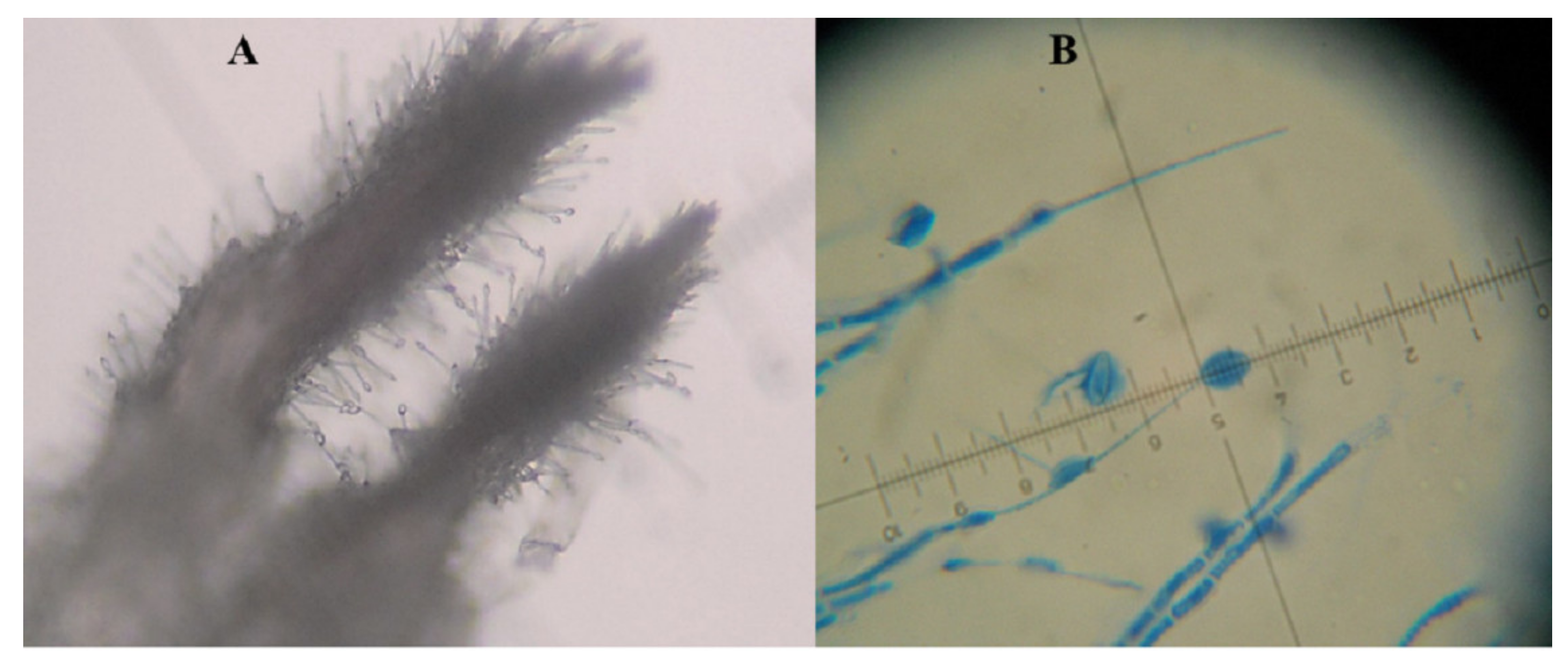
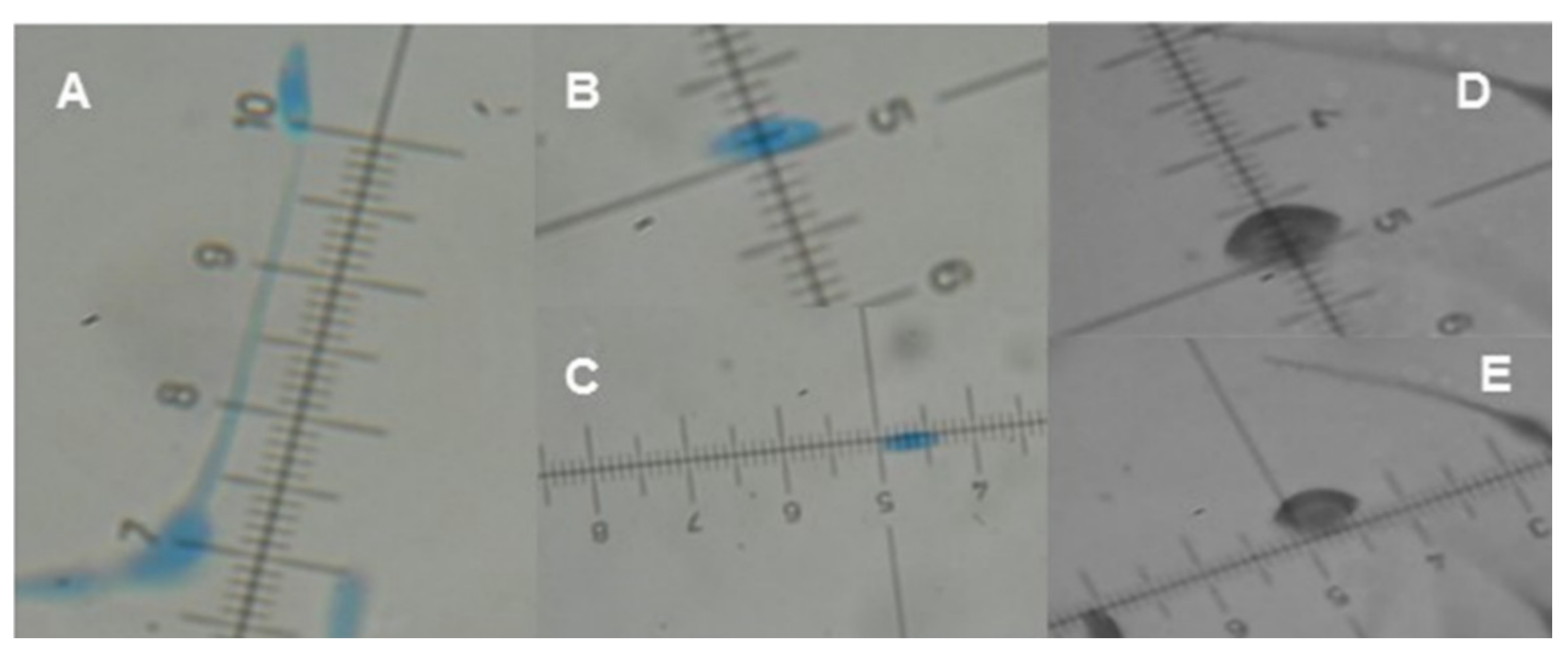
2. Hirsutella citriformis Identification

H. citriformis was first described in 1920 by Speare, followed by Petch in 1923 [9], and Main in 1951 [10]. It commonly infects Hemiptera insect species, and its morphology has been previously reported [6,7,10,11,12,13,14]. This fungus is classified within the Ascomycota Division, Pezizomycotina subphylum, Sordariomycetes class (Sung et al. 2007), Hypocreomycetidae subclass, Hypocreales Order, Ophiocordycipitaceae family, Hirsutella genus, and citriformis Species. Members of this group are the anamorphs of Cordyceps, Ophiocordyceps, or Torrubiella [15].
The typical H. citriformis mycelium is septate, fine, and hyaline and may present synnemata and phialides (conidiogenous cells). It has a spherical base with elongated necks that bear a single conidium at the tip, which has an elongated, allantoic, cimbiform, or fusiform shape (orange segment shape), covered with an ovoid mucilaginous material (Figure 1A). Some H. citriformis species are slow-growing fungi and produce limited conidia [6,7,12,14].
The most comprehensive description of H. citriformis, which was considered a reference of morphometric characterization until 2014, was reported by Mains in 1951 [10]. To date, this is the only Hirsutella species reported parasitizing D. citri [6,7,8,12] but phialide and conidium diameters of such species [10] are different from those of D. citri-infecting Hirsutella strains in Mexico (Figure 1B).
In this regard, H. citriformis reproductive structure measurements directly obtained from the host [7,16,17] or reproductive structures developed in culture medium [6,12,18] are different from previous reports on these species [10] (Figure 2). In host-derived species, isolates are found in different hemipteran species and geographic regions, which may result in diverse fungal structures development. It is expected that structures dimensions developed in culture media would be similar but we observed that even isolates from the same country showed differences in their size, even if cultivated in the same culture medium. This observation was among isolates from insects parasitized by fungus collected in different months in states with different temperature and humidity conditions.
Hirsutella citriformis synnema dimensions on their hosts are variable, which may relate to the host insect’s size [10] (Table 1).

3. Hirsutella citriformis Origin and Distribution

Previous reports have shown that H. citriformis origin was in the Asian continent [9,11], with distribution to other tropical or subtropical climate geographical regions. It is possible that human activity through commercial transportation of agricultural products worldwide (mainly maritime) had played a significant role in H. citriformis parasitized-insects spreading to other regions, including Cuba [19], Argentina [13]), and the United States of America [7,8] (Table 2).

4. Insects Associated with H. citriformis

Hirsutella citriformis was first reported by Speare in 1920 [11], followed by Peath in 1923 [9]. It was further detected in the 1940s in different insect species [10]. In the 21st century, most reports of H. citriformis isolates were exclusively from D. citri insects (Table 3). It is a typically synnematous species that infects a wide range of hemipterans, including insect vectors of plant diseases. It has been detected in hemipterans in Asian countries [6,9,11], South America [13,19], the Caribbean [17,21], and the United States of America [7,8]. H. citriformis parasitizes several insect populations, causing epizootics under suitable environmental conditions for its development [6,7,8,21].
In Mexico, H. citriformis has been isolated from D. citri Kuwayama (Hemiptera: Liviidae) carcasses, whose first isolate was reported in Veracruz state in 2008, after which it has been detected in different locations, mainly in Mexican citrus areas [12,18,25,27,28] (Table 4).
H. citriformis-parasitized insects in citrus ecosystems have allowed the frequent isolation of various strains of this fungus. For this reason, in recent years, H. citriformis has received significant interest as a potential biocontrol agent against the Asian citrus psyllid, present in most citrus growing regions worldwide.

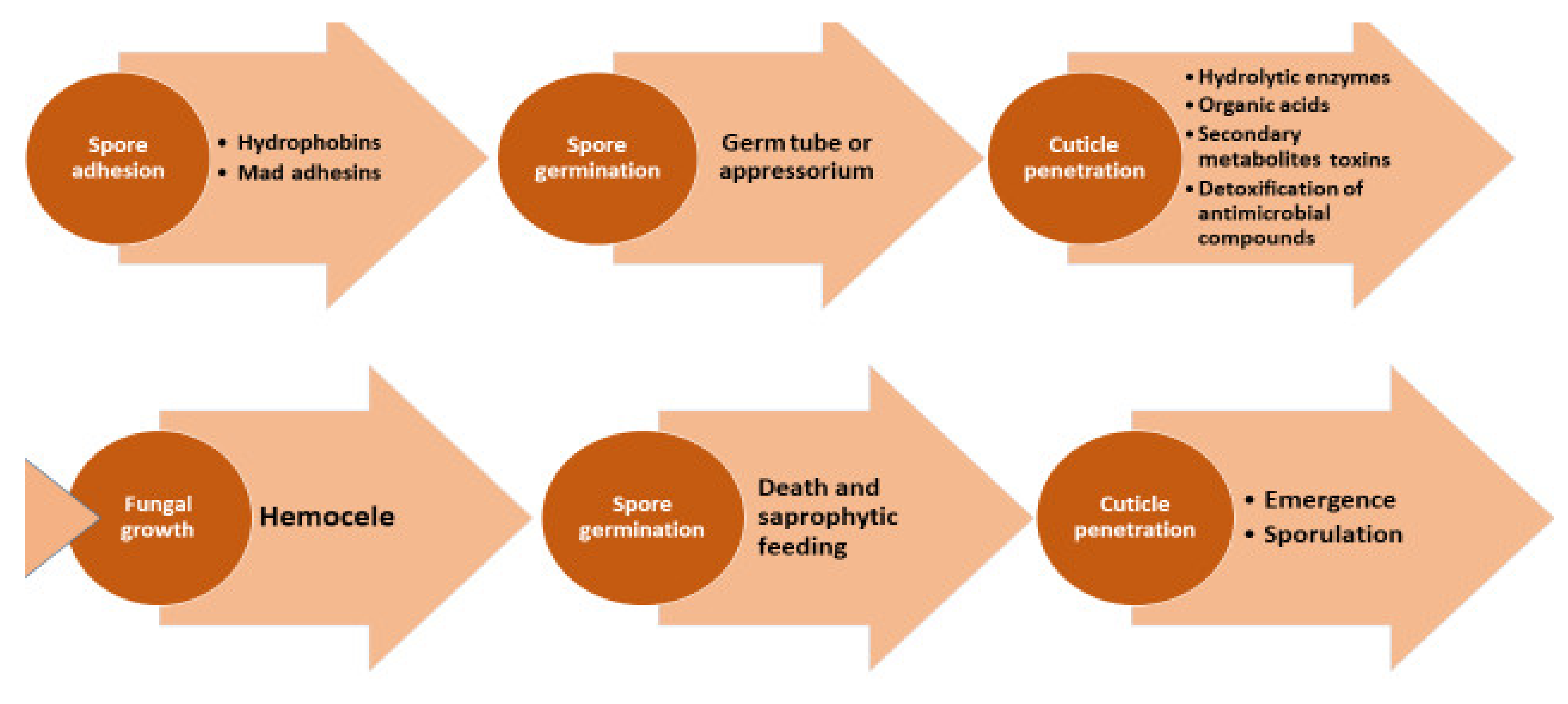
5. H. citriformis Mode of Action

The precise mode of action of H. citriformis is unknown but might be similar than that reported by Boucias et al. on Hirsutella genera [29]. The infection process begins after susceptible insects contact conidia, which adhere to their cuticle external surface and penetrate the hemocele, where they convert the tissues into biomass and develop mycelium inside (Figure 3).
After the insect dies, mycelium protrudes, showing H. citriformis characteristic structures. Its conidium binding to the insect is facilitated by a mucilaginous envelope. Culturing on artificial agar medium has demonstrated mycelial compatibility among strains, anastomosis occurrence, and one nucleus/conidium [30]. Furthermore, fungus vegetative stages have been studied in D. citri hemocele, where numerous hyphal bodies have been observed, showing a morphology close to the vegetative cells produced by other entomopathogenic Hypocreales fungi [29]. The H. citriformis mucilage that surrounds the conidium has the same protective potential than that reported for H. satumaensis at temperatures above or below the optimum, or after exposure to ultraviolet radiation by extended periods, cold stress, or high osmotic pressure [31]. In other fungi, the mucilage role is not clear but its contribution to host adhesion has been reported in nematodes [32].

6. Adverse Effects of Environmental Factors on H. citriformis

Although it is important to understand how environment decreases H. citriformis stability and viability in field, few studies on this matter have been reported. H. citriformis behavior and persistence under natural conditions are unknown, for which it is important to develop strategies to prevent mortality or improve its dissemination under field conditions to achieve a successful biocontrol product. In general, temperature, relative humidity (RH), and solar radiation are the most important abiotic environmental factors affecting entomopathogenic fungi germination, vegetative growth, and conidia viability [33].

6.1. Temperature

Temperature reduces H. citriformis fungus viability and affects conidium germination, development, and survival. Its optimal development temperature is 25 ± 2 °C, which is delayed and inhibited at lower and higher temperatures, respectively [34]. Pérez-González et al. [14] also reported strain-related differences based on the geographical region of isolation. They observed high-temperature resistance among strains isolated from a region that maintains elevated temperature and humidity throughout the year [14]. Furthermore, Orduño-Cruz et al. [34] showed that conidium germination was affected by the incubation temperature. In this regard, conidia have a higher germination percentage at 25 °C in a shorter incubation time, whereas at higher or lower temperatures, longer incubation time was required. They also reported that germination time varied depending on the isolate [34].

6.2. Relative Humidity

RH is one of the most important environmental factors altering the efficacy and survival of an entomopathogen. Hirsutella citriformis distribution studies under field conditions have shown that fungal infection of insects was higher in the months of high humidity and optimal temperature. High mortality of psyllid adults was reported in Réunion Island by H. citriformis, when the RH was higher than 88% [23]. Furthermore, maximum infection rates of 52.2% and 82.9% in D. citri by H. citriformis was observed in two Indonesian orange orchards, during September, February, and July, when the RH was higher than 80% [6]. H. citriformis naturally infesting D. citri populations was also observed at a RH higher than 80%, which was crucial for their growth and sporulation [21]. By 2016 in Colima, Mexico, an overall incidence of 58.2% of H. citriformis in citrus trees infested by D. citri was detected by PCR [28]. Similarly, D. citri mortality rate by H. citriformis was 23%, as reported in a two-year field test in Florida [8]. Authors observed up to 75% psyllid carcasses mummified and covered by synnemata, during the fall and winter months but absent during spring, probably due to a low RH [8]. Dead psyllids remained on citrus leaves during 68 days on average [8], thus indicating high fungus dissemination during template and high humidity seasons. In addition, authors did not recommend applying copper hydroxide, petroleum oil, or elemental sulfur at high rates to avoid killing this entomopathogenic fungus. In a follow-up study, authors indicated that D. citri adults killed by H. citriformis was as low as three to four percent on young leaves but six times higher than on mature leaves, suggesting the use of copper sulfate pentahydrate, aluminum tris, and alpha-keto/humic acids along with H. citriformis [35].
Higher infectivity of the fungus in the months in which the temperature ranged from 23 °C to 26 °C and the RH was higher than 80% was reported [26]. Moreover, H. citriformis presence on D. citri adults in Tamaulipas, Mexico was detected at temperatures between 25 °C and 28 °C and ~80% RH [24].

6.3. Solar Radiation

Several reports showed that entomopathogenic fungus conidia are inactivated in hours or days by direct sunlight [36]. However, the effect of solar radiation on H. citriformis conidia viability is unknown. Nevertheless, this fungus naturally adheres to mummified carcasses on the underside of citrus leaves, where they remain for more than two months and maintain infectivity to D. citri adults [8]. Another characteristic of this fungus is its location in the mummified carcasses, keeping in a ready-to-infect position. Healthy adults, looking to mate, may be attracted to the mummified carcass, either by visual or chemical signals [8].

7. Metabolites Production

Most microorganisms, including fungi, produce a wide variety of compounds or toxins (secondary metabolites) that have diverse activities. Unlike entomopathogenic fungi such as B. bassiana and Metarhizium, scarce information regarding metabolites produced by Hirsutella is available. It has been reported that different Hirsutella species, after growing in submerged fermentation, produce a variety of metabolites, some of which have activity against insects and mites (Table 5).
The best-characterized toxin is the Hirsutellin A (HTA), which is an isolated and sequenced protein produced by H. thompsonii [37,38]. It also produces oosporin, which reduces eggs production in mite females [40]. Hirsutellines production has been also reported by H. nivea [45], which possess antibacterial activity against Mycobacterium tuberculosis. Furthermore, exopolysaccharides from Hirsutella sp. have antibacterial activity against Gram-positive bacteria such as Bacillus subtilis and Micrococcus tetragenus [46]. Several benzene and phthalic acid compounds with potential insecticidal activity were detected in H. citriformis culture supernatants [49] (Table 5).
It has been also reported the presence of droplet exudates after growing H. citriformis strains on commercial agar media [50]. To date, these metabolites have not been studied to establish their toxicity against other organisms, such as insect pests. In fact, polysaccharides produced by other entomopathogenic fungi may stimulate fungus infection [51], whereas proteins promote fungus adherence, infection, and virulence [52]. We have observed that Mexican H. citriformis strains produce and exudate droplets, which showed at least three colors (light yellow, reddish brown, and crystalline), when grown on commercial potato dextrose agar media, supplemented with yeast extract at 1% w/v (Figure 4).
The initial metabolites analysis indicated that light yellow and reddish-brown color exudates are of proteinaceous nature, using the Bradford method [53], whereas the use of the DuBois method [53] showed that the crystalline exudates were carbohydrates [14]. H citriformis exudate metabolites are of mucilaginous-like consistency, showing the presence of ~60 kDa, 20 kDa, 14 kDa, and 2 kDa to 5 kDa proteins, as measured by polyacrylamide gel electrophoresis (Figure 5).
Based on these results, the molecular mass of the ~14 KDa protein might correspond to hirsutellin. We have collected and purified mucilaginous metabolites from H. citriformis liquid cultures, tested them under laboratory and field conditions against D. citri adults, and demonstrated their toxicity to D. citri adults (unpublished data).

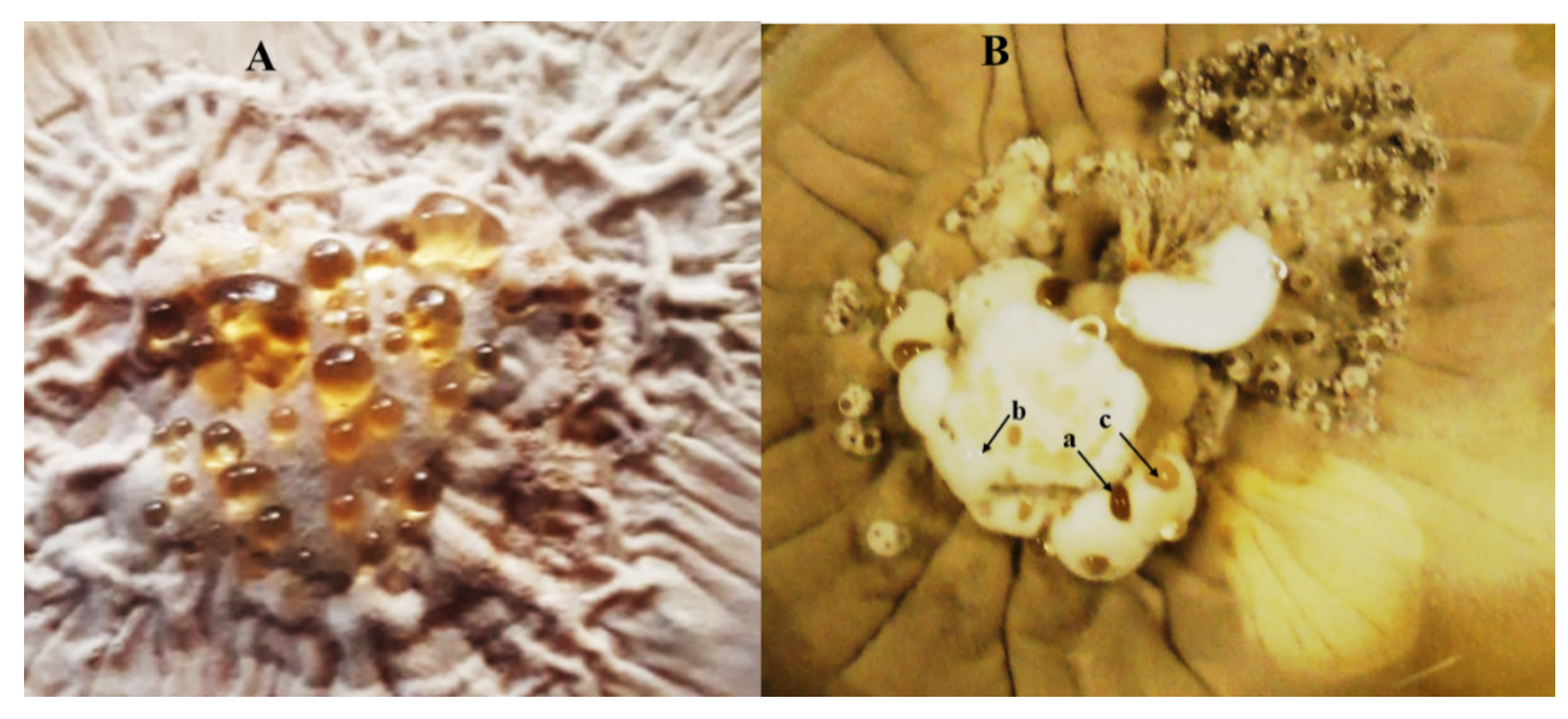
8. Hirsutella citriformis Epizootics on Diaphorina citri

H. citriformis causes epizootics on D. citri in various citrus regions, where high humidity is a determining factor for this fungus to infect and disseminate. In this regard, psyllids control by H. citriformis at RH higher than 80% has been reported [54]. In Cuba, D. citri nymphs and adults infected with H. citriformis in Mexican lemon trees (Citrus aurantifolia Swingle) and in Murraya paniculata (Lin) Jack plants (limonaria) were observed [17], whereas in Mexico and other countries, detection of H. citriformis infecting D. citri, where the RH was close to or higher than 80%, was also reported. In the municipality of Tuxpan, Veracruz, Mexico, an infection rate of 21.1% of D. citri by Hirsutella sp., where the RH was from 50% to 60%, was detected [25]. In addition, H. citriformis infecting D. citri in southern Tamaulipas, Mexico (municipalities of Ciudad Victoria, Mante, Gómez Farías, and Llera) was observed, where up to 43% of insects on orange trees and 95% on M. paniculata were mycosed [12]. Epizootics of H. citriformis infecting D. citri in Rio Bravo, Tamaulipas, Mexico, were reported in September and November, where the RH was close to 80% and temperature averaged 26 °C [24]. Moreover, a high prevalence of D. citri mycosed insects by H citriformis in the citrus regions of Troncones, Misantla, and Iztacuaco in Tlapacoyan, Veracruz, Mexico, where the RH was higher than 80% and temperature averaged 25 °C, was observed [26].
These reports confirm that temperatures ranging from 23 °C to 28 °C and RH close or higher than 70% improves H. citriformis infection and dissemination on D. citri.

9. Assessments of H. citriformis Biocontrol against D. citri and Bactericera cockerelli

To determine the pathogenicity of H. citriformis, laboratory bioassays have shown the potential of H. citriformis conidia to infect and kill D. citri and B. cockerelli (Šulc) (Hemiptera: Triozidae) adults, which became infected after fungus contact [7,12,37,55]. It is recognized that H. citriformis requires longer periods to cause insect death, compared with other entomopathogenic fungi, but limited studies have evaluated the role of this fungus to infect and kill D. citri nymphs. Experiments where nymphs were exposed to mycosed adults by contact resulted in H. citriformis infection and mortality at the significant rates of 75% and 10%, respectively [7,56]. In addition, conidia application by spraying was evaluated under laboratory conditions [57], showing that adults’ mortality was lower than that following direct application of conidia. After using this spraying technique but adding an adherent to the suspension, mortality rates were closer to those achieved by this fungus after direct application.
H. citriformis virulence was evaluated against D. citri adults, spraying selected strains in a semi-field bioassay, evidencing a mortality close to 50% and their dispersion to other citrus areas [58]. A similar study was developed in Campeche (18°50′11″ N 90°24′12″ O), Chiapas (16°24′36″ N 92°24′31″ O), Colima (19°14′37″ N 103°43′51″ O), and Quintana Roo (19°36′00″ N 87°55′00″ O) Mexico, showing that H. citriformis was a promising alternative to use against Hemiptera insects in integrated pest management programs [59].
In a northeast area in Montemorelos, Nuevo Leon, Mexico (25°11′14″ N 99°49′36″ O), we performed two bioassays against D. citri adults in September 2020 and September 2021, applying formulated conidia with two different gummous metabolites as adherents (Acacia sp. gum and H. citriformis produced gum). We demonstrated that Hirstuella gum control (without conidia) treatment resulted in close to 50% mortality, whereas the mortality by formulation with conidia and Hirsutella gum treatment was close to 80%. These results showed that H. citriformis-produced gum is toxic to D. citri (unpublished data).

10. Effects on Predators and Other Non-Target Arthropods

Unlike other entomopathogenic fungi such as B. bassiana and M. anisopliae that have a wide range of hosts, including 100 arthropod species and representing a potential treat to beneficial insects, H. citriformis has potential to infect Hemiptera insects. Nevertheless, it has only been isolated from this type of insect, representing a low-risk biocontrol entomopathogen to beneficial insects. To demonstrate H. citriformis safety against two of the most important predators in biological control, conidia were applied on Hippodamia convergens Guerin-Meneville (Coleoptera: Coccinellidae) adults and Chrysoperla rufilabris Burmeister (Neuroptera: Chrysopidae) nymphs, showing that treated insects were uninfected by H. citriformis, thus indicating that this fungus was not pathogenic against such predators [55].

11. Conclusions

Hirsutella citriformis is a fungus first described in 1908. Since then, few studies have been developed to better understand its morphological, growth, and genetic characteristics. From 2000, it has been reported as a biocontrol agent against the insect vector of the HLB disease. D. citri is currently considered the most important pest to citrus areas worldwide and numerous studies have been reported. Relevant H. citriformis characteristics include (a) slow growth and poor conidia production; (b) presence of synnemata; (c) its phialides have a spherical base, with elongated necks that have a single conidium at the tip, which has an elongated, allantoic, cimbiform, or fusiform shape, and it is covered by a mucilaginous layer; (d) it has been found parasitizing only Hemiptera class insects worldwide and appears to be safe for predatory beneficial insects; (e) it requires warm temperatures and high RH (higher than 70%) for its development; (f) it produces a variety of secondary metabolite, whose biological and chemical nature has not been yet elucidated; (g) its secondary metabolites mode of action against D. citri adults is unknown; (h) it causes epizootics on infected insects, under environmental conditions suitable for its development; and (i) it has shown pathogenicity against B. cockerelli and D. citri, under laboratory and field conditions. Based on pathogenicity results, we may expect that H. citriformis possesses potential as bioinsecticide under an inoculative-augmentative biological control scheme. However, factors that may limit the formation of epizootics should be studied.
Prevailing environmental conditions in the treated area are decisive for the establishment of a further epizootic. Therefore, areas recording long periods (at least one month) of high humidity (higher than 70%) and temperatures between 23 °C and 28 °C should be selected for H. citriformis application. In Mexico and other countries, citrus growing areas with these environmental conditions that allow H. citriformis development may be the target for its application as biological agent to reduce D. citri vector populations.

Author Contributions

Conceptualization, O.P.-G. and P.T.-G.; methodology, O.P.-G., P.T.-G. and R.G.-F.; software, P.T.-G.; validation, O.P.-G. and P.T.-G.; formal analysis, O.P.-G. and P.T.-G.; investigation, O.P.-G. and P.T.-G.; resources, O.P.-G., P.T.-G. and R.G.-F.; data curation, O.P.-G. and P.T.-G.; writing—original draft preparation, O.P.-G. and P.T.-G.; writing—review and editing, O.P.-G., P.T.-G. and R.G.-F.; visualization, P.T.-G.; supervision, O.P.-G. and P.T.-G.; project administration, O.P.-G. and P.T.-G.; funding acquisition, O.P.-G. and P.T.-G. All authors have read and agreed to the published version of the manuscript.

Funding

Authors thank CONACYT-MEXICO for grant A1-S-33091 to O.P.-G. Authors also thank Sistema Nacional de Investigadores (SNI-CONACYT, Mexico) for financial support through SNI-73211 to O.P.-G., SNI-9942 to R.G.-F. and SNI-16614 to P.T.-G.

Institutional Review Board Statement

This review does not require ethical approval.

Informed Consent Statement

Not applicable.

Data Availability Statement

Not applicable.

Conflicts of Interest

The authors declare no conflict of interest.

References

  1. Ekesi, S.; Maniania, N.K. Use of Entomopathogenic Fungi in Biological Pest Management; Ekesi, S., Maniania, N.K., Eds.; Research Signpost Editorial: Trivandrum, India, 2007; pp. 57–90. Available online: https://www.researchgate.net/publication/297758806 (accessed on 15 April 2022).
  2. Al-Ani, L.K.T. Entomopathogenic fungi in IP landscape. In Intellectual Property Issues in Microbiology; Singh, H., Keswani, C., Singh, S., Eds.; Springer: Singapore, 2019. [Google Scholar] [CrossRef]
  3. Moorhouse, E.R.; Gillespie, A.T.; Sellers, E.K.; Charnley, A.K. Influence of fungicides and insecticides on the entomogenous fungus Metarhizium anisopliae a pathogen of the vine weevil, Otiorhynchus sulcatus. Biocontrol Sci. Technol. 1992, 2, 49–58. [Google Scholar] [CrossRef]
  4. De Faria, M.R.; Wraight, S.P. Mycoinsecticides and Mycoacaricides: A comprehensive list with worldwide coverage and international classification of formulation types. Biol. Control 2007, 43, 237–256. [Google Scholar] [CrossRef]
  5. Maina, U.M.; Galadima, I.B.; Gamba, F.M.; Zakaria, D. A review on the use of entomopathogenic fungus in the management of insect pest of field crops. J. Entomol. Zool. Stud. 2018, 6, 27–32. [Google Scholar]
  6. Subandiyah, S.; Nikon, N.; Sato, H.; Wagiman, F.; Tsuyumu, S.; Fakatsu, T. Isolation and characterization of two entomopathogenic fungi attacking Diaphorina citri (Homoptera, Psilloidea) in Indonesia. Mycoscience 2000, 41, 509–513. [Google Scholar] [CrossRef]
  7. Meyer, J.M.; Hoy, M.A.; Boucias, D.G. Morphological and molecular characterization of a Hirsutella species infecting the Asian citrus psyllid, Diaphorina citri Kuwayama (Hemiptera: Psyllidae), in Florida. J. Invertebr. Pathol. 2007, 95, 101–109. [Google Scholar] [CrossRef]
  8. Hall, D.G.; Hentz, M.G.; Meyer, J.M.; Kriss, A.B.; Gottwald, T.R.; Boucias, D.G. Observations on the entomopathogenic fungus Hirsutella citriformis attacking adult Diaphorina citri (Hemiptera: Psyllidae) in a managed citrus grove. BioControl 2012, 57, 663–675. [Google Scholar] [CrossRef]
  9. Petch, T. The genus Hirsutella Petch. Trans. Br. Mycol. Soc. 1923, 9, 93–94. [Google Scholar] [CrossRef]
  10. Mains, E.B. Entomogenous species of Hirsutella, Tilachlidium and Synnematium. Mycologia 1951, 43, 691–718. [Google Scholar] [CrossRef]
  11. Speare, A.T. On certain entomogenous fungi. Slycologia 1920, 12, 62–76. [Google Scholar] [CrossRef]
  12. Casique-Valdes, R.; Reyes-Martinez, A.Y.; Sanchez-Peña, S.R.; Bidochka, M.J.; Lopez-Arroyo, J.I. Pathogenicity of Hirsutella citriformis (Ascomycota: Cordycipitaceae) to Diaphorina citri (Hemiptera: Psyllidae) and Bactericera cockerelli (Hemiptera: Triozidae). Fla. Entomol. 2011, 94, 703–705. [Google Scholar] [CrossRef]
  13. Toledo, A.V.; Simurro, M.E.; Balatti, P.A. Morphological and molecular characterization of a fungus, Hirsutella sp., isolated from planthoppers and psocids in Argentina. J. Insect Sci. 2013, 13, 18. [Google Scholar] [CrossRef] [PubMed] [Green Version]
  14. Perez-Gonzalez, O.; Rodriguez-Guerra, R.; Lopez-Arroyo, J.I.; Sandoval-Coronado, C.F.; Maldonado-Blanco, M.G. Radial growth, sporulation, and virulence of Mexican isolates of Hirsutella citriformis against Diaphorina citri. Southwest Entomol. 2015, 40, 111–120. [Google Scholar] [CrossRef]
  15. Tanada, Y.; Kaya, H.K. Fungal Infections. In Insect Pathology; Tanada, Y., Kaya, H.K., Eds.; Academic Press Inc.: Cambridge, MA, USA, 1993; pp. 318–387. [Google Scholar]
  16. Hywel-Jones, N.L. Hirsutella species associated with hoppers (Homoptera) in Thailand. Mycol. Res. 1997, 101, 1202–1206. [Google Scholar] [CrossRef] [Green Version]
  17. Álvarez, J.F.; Montes de Oca, F.N.; Grillo-Ravelo, H. Hongos entomopatógenos de Diaphorina citri Kirk. (Homoptera: Psyllidae) en Jovellanos, Matanzas. Centro Agrícola 2003, 30, 87. (In Spanish) [Google Scholar]
  18. Perez-Gonzalez, O.; Rodriguez-Villarreal, R.A.; Lopez-Arroyo, J.I.; Maldonado-Blanco, M.G.; Rodríguez-Guerra, R. Mexican strains of Hirsutella isolated from Diaphorina citri (Hemiptera: Liviidae): Morphologic and molecular characterization. Fla. Entomol. 2015, 98, 290–297. [Google Scholar] [CrossRef]
  19. Kondo, T.; González, G.; Tauber, C.; Guzmán-Sarmiento, Y.C.; Vinasco-Mondragon, A.F.; Forero, D. A checklist of natural enemies of Diaphorina citri Kuwayama (Hemiptera: Liviidae) in the department of Valle del Cauca, Colombia, and the world. Insecta Mundi 2015, 0457, 1–14. Available online: http://centerforsystematicentomology.org/ (accessed on 20 May 2022).
  20. Reddy, N.; Mahesh, G.; Priya, M.; Singh, R.S.; Manjunatha, L. Hirsutella. In Beneficial Microbes in Agro-Ecology; Academic Press: Cambridge, MA, USA; pp. 817–831. [CrossRef]
  21. Étienne, J.; Quilici, S.; Marival, D.; Franck, A. Biological control of Diaphorina citri (Hemiptera: Psyllidae) in Guadeloupe by imported Tamarixia radiata (Hymenoptera: Eulophidae). Fruits 2001, 56, 307–315. [Google Scholar] [CrossRef] [Green Version]
  22. Sajap, A.S. Prevalence of an entomopathogenic fungus, Hirsutella citriformis on Leucaena Psyllid, Heterapsylla cubana, in Malaysia. Pertanika J. Trop. Agric. Sci. 1993, 16, 95–99. [Google Scholar]
  23. Aubert, B. Trioza erytreae Del Guercio and Diaphorina citri Kuwayama (Homoptera: Psylloidea), the two vectors of citrus greening disease: Biological aspects and possible control strategies. Fruits 1987, 42, 149–162. [Google Scholar]
  24. Reyes-Rosas, M.A.; Loera-Gallardo, J.; López-Arroyo, J.I. Comparison of the chemical and natural control of Asian citrus psyllid Diaphorina citri. Rev. Mex. Cienc. Agric. 2013, 4, 95–501. Available online: http://www.redalyc.org/articulo.oa?id=263127562001 (accessed on 20 May 2022).
  25. Henríquez-Liria, D.J.; Navarro-Morales, S.; Bastardo, R.H.; Reyes-Valentín, M. Aspectos ecológicos de Diaphorina citri Kuwayama, 1908, en el cultivo de cítricos en Villa Altagracia y El Seibo, República Dominicana. Novit. Caribaea 2016, 10, 52–62. [Google Scholar] [CrossRef] [Green Version]
  26. Godoy-Ceja, C.A.; Cortez-Madrigal, H. Potential of Aclepias curassavica L. (Apocynaceae) in the biological control of pests. Rev. Mex. Cienc. Agric. 2018, 9, 303–315. [Google Scholar] [CrossRef] [Green Version]
  27. Ayala-Zermeño, M.A.; Gallou, A.; Berlanga-Padilla, A.M.; Serna-Domínguez, M.G.; Arredondo-Bernal, H.C.; Montesinos-Matías, R. Characterisation of entomopathogenic fungi used in the biological control programme of Diaphorina citri in Mexico. Biocontrol Sci. Technol. 2015, 25, 1192–1207. [Google Scholar] [CrossRef]
  28. Suaste-Dzul, A.; Gallou, A.; Félix-Portillo, M.; Moreno-Carrillo, G.; Sánchez-González, J.; Palomares-Pérez, M.; Arredondo-Bernal, H. Seasonal incidence of ‘Candidatus Liberibacter asiaticus’ (Rhizobiales: Rhizobiaceae) in Diaphorina citri (Hemiptera: Liviidae) in Colima, Mexico. Trop. Plant Pathol. 2017, 42, 410–415. [Google Scholar] [CrossRef]
  29. Boucias, D.G.; Scharf, D.W.; Breaux, S.E.; Purcell, D.H.; Mizell, R.E. Studies on the fungi associated with the glassy-winged sharpshooter Homalodisca coagulata with emphasis on a new species Hirsutella homalodiscae nom. prov. BioControl 2007, 52, 231–258. [Google Scholar] [CrossRef]
  30. Pérez-González, O.; Gomez-Flores, R.; Tamez-Guerra, P. Mycelial compatibility, anastomosis, and nuclear characterization of Mexican Hirsutella citriformis strains isolated from Diaphorina citri. PeerJ 2021, 9, e11080. [Google Scholar] [CrossRef]
  31. Qu, J.; Zou, X.; Yu, J.; Zhou, Y. The conidial mucilage, natural film coatings, is involved in environmental adaptability and pathogenicity of Hirsutella satumaensis Aoki. Sci. Rep. 2017, 7, 1301. [Google Scholar] [CrossRef]
  32. Jansson, H.B.; Friman, E. Infection-related surface proteins on conidia of the nematophagous fungus Drechmeria coniospora. Mycol. Res. 1999, 103, 249–256. [Google Scholar] [CrossRef]
  33. Vidal, C.; Fargues, J. Climatic constraints for fungal biopesticides. In Use of Entomopathogenic Fungi in Biological Pest Management; Ekesi, S., Maniania, N.K., Eds.; Research Signpost Editorial: Trivandrum, India, 2007; pp. 39–55. [Google Scholar]
  34. Orduño-Cruz, N.; Guzmán-Franco, A.W.; Rodríguez-Leyva, E.; Alatorre-Rosas, R.; González-Hernández, H.; Mora-Aguilera, G.; Rodríguez-Maciel, J.C. In vitro selection of a fungal pathogen for use against Diaphorina citri. Biol. Control 2015, 90, 6–15. [Google Scholar] [CrossRef]
  35. Hall, D.G.; Richardson, M.L.; Ammar, E.D.; Halbert, S.E. Asian citrus psyllid, Diaphorina citri, vector of citrus huanglongbing disease. Entomol. Exp. Appl. 2013, 146, 207–223. [Google Scholar] [CrossRef]
  36. Gardner, W.A.; Sutton, R.M.; Noblet, R. Persistence of Beauveria bassiana, Nomuraea rileyi, and Nosema necatrix on soybean foliage. Environ. Entomol. 1977, 6, 616–618. [Google Scholar] [CrossRef]
  37. Liu, W.Z.; Boucias, D.G.; McCoy, C.W. Extraction and characterization of the insecticidal toxin Hirsutellin A produced by Hirsutella thompsonii var. thompsonii. Exp. Mycol. 1995, 19, 254–262. [Google Scholar] [CrossRef] [PubMed]
  38. Boucias, D.G.; Farmerie, W.G.; Pendland, J.C. Cloning and sequencing of cDNA of the insecticidal toxin Hirsutellin A. J. Invertebr. Pathol. 1998, 72, 258–261. [Google Scholar] [CrossRef] [PubMed]
  39. Quesada-Sojo, K.A.; Rivera-Méndez, W. Hirsutella, agente biocontrolador de ácaros e insectos de importancia agronómica. Revista Tecnología en Marcha 2016, 29, 85–93. [Google Scholar] [CrossRef]
  40. Rosas-Acevedo, J.L.; Boucias, D.G.; Lezama, R.; Sims, K.; Pescador, A. Exudate from sporulating cultures of Hirsutella thompsonii Fisher inhibit oviposition by the two-spotted spider mite Tetranychus urticae Koch. Exp. Appl. Acarol. 2003, 29, 213–222. [Google Scholar] [CrossRef]
  41. Wang, B.; Wu, W.; Liu, X. Purification and characterization of a neutral serine protease with nematicidal activity from Hirsutella rhossiliensis. Mycopathologia 2007, 163, 169–176. [Google Scholar] [CrossRef]
  42. Mazet, I.; Vey, A. Hirsutellin A, a toxic protein produced in vitro by Hirsutella thompsonii. Microbiology 1995, 141, 1343–1348. [Google Scholar] [CrossRef] [Green Version]
  43. Herrero-Galan, E.; Lacadena, J.; Martinez-del-Pozo, A.; Boucias, D.G.; Olmo, N.; Oñaderra, M.; Gavilanes, J. The insecticidal protein Hirsutellin A from the mite fungal pathogen Hirsutella thompsonii is a ribotoxin. Proteins 2008, 72, 217–228. [Google Scholar] [CrossRef]
  44. Maimala, S.; Tartar, A.; Boucias, D.; Chandrapaty, A. Detection of the toxin Hirsutellin A from Hirsutella thompsonii. J. Invertebr. Pathol. 2002, 80, 112–126. [Google Scholar] [CrossRef]
  45. Madla, S.; Isaka, M.; Wongsa, P. Modification of culture conditions for production of the anti-tubercular hirsutellones by the insect pathogenic fungus Hirsutella nivea BCC2594. Lett. Appl. Microbiol. 2008, 47, 74–78. [Google Scholar] [CrossRef]
  46. Ramachandran, A.; Thangappan, B.S.; Ponmurugan Ponnusamy, P. Evaluation of secondary metabolites of Hirsutella citriformis against Udaspes folus infecting Curcuma longa L. J. Pharm. Res. 2013, 7, 7–14. [Google Scholar] [CrossRef]
  47. Meng, L.; Sun, S.; Shen, Z.; Wang, P.; Jiang, X. Antioxidant activity of polysaccharides produced by Hirsutella sp. and relation with their chemical characteristics. Carbohydr. Polym. 2015, 117, 452–457. [Google Scholar] [CrossRef] [PubMed]
  48. Leung, P.; Zhao, P.; Wu, J.Y. Chemical properties and antioxidant activity of exopolysaccharides from mycelial culture of Cordyceps sinensis fungus Cs-HK1. Food Chem. 2009, 114, 1251–1256. [Google Scholar] [CrossRef]
  49. Li, R.; Jiang, X.; Guan, H. Optimization of mycelium biomass and exopolysaccharides production by Hirsutella sp. In submerged fermentation and evaluation of exopolysaccharides antibacterial activity. Afr. J. Biotechnol. 2010, 9, 195–202. [Google Scholar] [CrossRef]
  50. Samson, R.A.; McCoy, C.W.; O’Donnell, K.L. Taxonomy of the acarine parasite Hirsutella thompsonii. Mycologia 1980, 72, 359–377. [Google Scholar] [CrossRef]
  51. Loughheed, T.C.; MacLeod, D.M. Extracellular metabolic products of a Hirsutella species. Nature 1958, 182, 114–115. [Google Scholar] [CrossRef]
  52. Blango, M.G.; Kniemeyer, O.; Brakhage, A.A. Conidial surface proteins at the interface of fungal infections. PLoS Pathog. 2019, 15, e1007939. [Google Scholar] [CrossRef] [Green Version]
  53. Bradford, M.M. A rapid and sensitive method for the quantitation of microgram quantities of protein utilizing the principle of protein-dye binding. Anal. Biochem. 1976, 72, 248–254. [Google Scholar] [CrossRef]
  54. Palma-Ramos, A.; Moreno-Aparicio, A.M.; Castrillón-Rivera, L.E.; Castañeda-Sánchez, J.I.; Muñoz-Duarte, A.R.; Mendoza-Pérez, F.; Padilla-Desgarennes, C. Detecting enzymatic activity of β-glucuronidase polymorphonuclear neutrophil in human grains Nocardia brasiliensis actinomycetoma, in vitro. Dermatol. Rev. Mex. 2016, 60, 488–498. [Google Scholar]
  55. Pérez-Gonzalez, O.; Rodriguez-Guerra, R.; Lopez-Arroyo, J.I.; Sandoval-Coronado, C.F.; Maldonado-Blanco, M.G. Effect of Mexican Hirsutella citriformis (Hypocreales: Ophiocordycipitaceae) strains on Diaphorina citri (Hemiptera: Liviidae) and the predators Chrysoperla rufilabris (Neuroptera: Chrysopidae) and Hippodamia convergens (Coleoptera: Coccinellidae). Fla. Entomol. 2016, 99, 509–515. [Google Scholar] [CrossRef] [Green Version]
  56. Ibarra-Cortés, K.H.; González-Hernández, H.; Guzmán-Franco, A.W. Susceptibility of nymphs and adults of Diaphorina citri to the entomophathogenic fungus Hirsutella citriformis. Biocontrol Sci. Technol. 2017, 27, 433–438. [Google Scholar] [CrossRef]
  57. Halbert, S.E.; Manjunath, K.L. Asian citrus psyllids (Sternorrhyncha: Psyllidae) and greening disease of citrus: A literature review and assessment of risk in Florida. Fla. Entomol. 2004, 87, 330–353. [Google Scholar] [CrossRef]
  58. Pérez-González, O.; Sandoval-Coronado, C.F.; Maldonado-Blanco, M.G. Evaluation of Mexican strains of Hirsutella citriformis against Diaphorina citri in a semifield bioassay. Southwest Entomol. 2016, 41, 361–372. [Google Scholar] [CrossRef]
  59. Pérez-González, O.; Arredondo-Bernal, H.C.; Montesinos-Matías, R.; Mellín-Rosas, M.A.; Maldonado-Blanco, M.G. Effect of native strains of Hirsutella citriformis1 on Diaphorina citri2 adults under field conditions. Southwest Entomol. 2020, 45, 435–446. [Google Scholar] [CrossRef]
Figure 1. Hirsutella citriformis mycelium structures. (A) H. citriformis synnemata developing on Diaphorina citri and (B) phialide and conidia. Measurements are in micrometers.
Jof 08 00573 g001
Figure 2. Mexican Hirsutella strains morphological characteristics. (A) Phialide length, (B) conidium width, (C) conidium length, (D) conidium width, including the mucilaginous envelope, and (E) conidium length, including the mucilaginous envelope. Measurements are in micrometers.
Jof 08 00573 g002
Figure 3. Hirsutella thompsonii mode of action.
Jof 08 00573 g003
Figure 4. Different exudates produced by Mexican Hirsutella citriformis strains grown on potato dextrose agar supplemented with 0.1% yeast extract (PDAY). (A) H. citriformis strain producing mostly light brown exudates and (B) H. citriformis strain producing (a) deep brown, (b) crystalline, and (c) light brown exudates.
Jof 08 00573 g004
Figure 5. Polyacrylamide gel electrophoresis of a Mexican Hirsutella citriformis strain sample. Lane 1 = mycelium; lane 2 = supernatant; lane 3 = mycelium; lane 4S = gummous material dissolved in solvent and sonicated (five pulses/40 W/30 sec). This material was slightly fragmented; lane 4C = the gummous material was cut, and a fragment was directly deposited as sample; lane 5 = solvent. Note that samples dissolved in a solvent were off the gel lane.
Jof 08 00573 g005
Table 1. Most representative reported Hirsutella citriformis structure measurements (µm).
ReferenceSource 1Phialide LengthConidiumMucus Diameter
TotalBaseSterigmaLengthDiameter
[11]H 5.5–8.51.5–1.8
[10]H36.0–54.06.0–14.030.0–40.0 5.0–8.02.0–2.5
[16]H18.5–52.0 3.5–5.01.0–1.5
[6]CM27.5–62.35.1–9.422.4–52.9 6.4–7.62.1–2.8
[17]H 16.8–23.6 6.8–9.11.5–2.3
[7]H 17.5 ± 1.9 5.9 ± 0.82.6 ± 0.3
[12]H 6.8–7.01.5–2.0
[13]H35.6–55.4 28.7–47.5 5.9–7.92.0–3.0
[13]H22.4–34.7 16.8–28.0 5.6–7.82.2–2.8
[14]CM26.0–42.04.0–8.520.0–38.0 5.4–6.31.6–2.0
1 H = host; CM = culture media.
Table 2. Hirsutella citriformis geographic distribution.
CountryRegionReference
CubaJovellanos, Matanzas[17]
Carmelina in Cienfuegos; Morón and Ceballos in Ciego de Ávila; San Antonio de los Baños in La Habana Cabrera et al., 2004, cited by [20]
Jaguey El Grande, Matanzas[19]
ArgentinaLos Hornos and La Plata, Buenos Aires[13]
United StatesHawaii[11]
Polk, Marion, and Hendry, Florida[7]
Indian River, Florida[8]
Table 3. Hirsutella citriformis isolation reports from different insects belonging to the Hemiptera order.
Parasitized Insect (Family)LocationReference
Fulgoridea family LatreilleNew Zeeland[11]
Ricania discalis Germar (Ricaniidae)New Zeeland
Perkinsiella sacharicida Kirkaldy (Hemíptera: Cicadellidae)Hawaii
Siphanta acuta Walker (Hemiptera: Flatidae)Hawaii
Pentatomidae family LeachIndia[9]
Bothriocera venosa Fowler (Cixiidae)Puerto RicoGregory & Martorell 1940, cited by [10]
Leptopharsa constricta Champion (Tingidae)United States[10]
Corythuca ulmi Osborn & Drake (Tingidae)United States[10]
Heteropsylla cubana Crawford (Psyllidae)Malaysia[22]
Oliarus dimidiatus Berg (Cixiidae)Argentina[23]
[13]
Diaphorina citri Kuwayama (Liviidae)Rénion island, France[17]
Cuba[6]
[19]
Indonesia[21]
Isla Guadalupe[7]
Florida[8]
Veracruz, MX[24]
Tamaulipas, MX[12]
Tamaulipas, MX[25]
Veracruz & Puebla, MX[26]
Veracrux, MX[27]
Campeche, MX[14]
Colima, MX
Chiapas, MX
Quintana Roo, MX
San Luis Potosí, MX
Tabasco, MX
Veracruz, MX
Yucatán, MX
Hidalgo, MXPérez-González et al., (unpublished data)
Oaxaca, MX
Table 4. States and municipalities of Mexico reporting Hirsutella citriformis.
StateMunicipalityReference
CampecheEdzna[18]
ChiapasTapachula[18]
ColimaTecomán[18]
HidalgoSanta AnaPérez-González et al., (unpublished data)
OaxacaMelchor OcampoPérez-González et al., (unpublished data)
PueblaLa Legua and La Garita[26]
Quintana RooNuevo Israel[18]
San Luis PotosíXolol[18]
TabascoHuimanguillo[18]
TamaulipasRio Bravo[24]
Gómez Farías and Llera[12]
VeracruzTlapacoyan[18]
Troncones, Ixtacuaco and El Lindero[26]
Tuxpan[25]
YucatánMococha[18]
Table 5. Toxins produced by Hirsutella species.
Hirsutella SpeciesToxinType of ToxinToxin Size/YieldActivityReference
H. thompsonii strain JAB-04Hirsutellin ADot-blot anti HtA16.3 kDa/0.12 mg/mLInsecticidal vs. Galleria mellonella[37,38]
thompsoniiHirsutellin A
Hirsutellin B
Ribotoxin130 amino-acidsInsecticidal vs. Galleria mellonella
Insecticidal vs. Drosophila melanogaster
[39]
H. thompsonii strain HtM120I Hirsutellin ADot-blot anti HtA16 kDa
(200–400 ng/mL)
Insecticidal vs. Tetranychus urticae[40]
H. thompsoniiCulture exudatesPhomalactones?Undetermined Reduces eggs production in mites[40]
H. rhossilensisHnspSAAPF-pNA protease activity & others30 kDaNematicide[41]
H. thompsoniiHirsutellin AW Blot NH2 terminal16 kDaInsecticidal vs. Aedes aegypti, Galleria mellonella[42]
H. thompsoniiHirsutellin ARibotoxin130 aaUnidentified[43]
H. thompsoniiHirsutellin ADot-blot anti HtA481–936 ng/mLInsecticidal vs. Galleria mellonella[44]
H. nivea strain BCC2594Hirsutellones A, B, C and DAlkaloids29.9, 1696, 22.6 and 15.7 mg/LAntibacterial against Mycobacterium tuberculosis[45]
Unidentified sp.Exo-polysaccharide (EPS)Mannose (man), galactose (gal), glucose (glc)23 kDaGram (+) bacteria,
Bacillus subtilis, Micrococcus tetragenus
[46]
H. beakdumountainEPSEPS 1 y 2: gal, glc, man
EPS3: man, glc
EPS1 = 43 kDa
EPS2 = 19.5 kDa
EPS3 = 4.7 kDa
Potential to eliminate hydroxyl radicals[47]
Intracellular polysaccharideIPS 1 y 2: gal, glc, man
IPS 3: man, glc
IPS1 = 23.1 kDa
IPS2 = 21.5 kDa
IPS3 = 10.4 kDa
Potential to eliminate hydroxyl radicals[47]
H. sinensisEPSPolysaccharides (65–70%) + protein (25%)5 kDa–200 kDaAntioxidant activity[48]
H. satumaensis AokiMucilage from aerial conidiaProtein content, oligo-saccharides + man0.12 mg/mLInsecticidal vs. Galleria mellonella Plutella xylostella[30]
H. citriformisCulture supernatantBenzene and phthalic acidUnidentifiedInsecticidal activity[49]
Publisher’s Note: MDPI stays neutral with regard to jurisdictional claims in published maps and institutional affiliations.

