Article

Lignicolous Freshwater Fungi from Plateau Lakes in China (I): Morphological and Phylogenetic Analyses Reveal Eight Species of Lentitheciaceae, Including New Genus, New Species and New Records

1
College of Agriculture and Biological Science, Dali University, Dali 671003, China
2
Center of Excellence in Fungal Research, Mae Fah Luang University, Chiang Rai 57100, Thailand
3
School of Science, Mae Fah Luang University, Chiang Rai 57100, Thailand
4
Center for Yunnan Plateau Biological Resources Protection and Utilization, College of Biological Resource and Food Engineering, Qujing Normal University, Qujing 655011, China
5
School of Food and Pharmaceutical Engineering, Guizhou Institute of Technology, Guiyang 550003, China
6
Co-Innovation Center for Cangshan Mountain and Erhai Lake Integrated Protection and Green Development of Yunnan Province, Dali University, Dali 671003, China
*
Author to whom correspondence should be addressed.
J. Fungi 2023, 9(10), 962; https://doi.org/10.3390/jof9100962
Submission received: 5 August 2023 / Revised: 3 September 2023 / Accepted: 21 September 2023 / Published: 25 September 2023
(This article belongs to the Special Issue Freshwater Fungal Diversity)

Abstract

:
During the investigation of lignicolous freshwater fungi in plateau lakes in Yunnan Province, China, eight Lentitheciaceae species were collected from five lakes viz. Luguhu, Qiluhu, Xingyunhu, Cibihu, and Xihu lake. Based on morphological characters and phylogenetic analysis of combined ITS, LSU, SSU, and tef 1-α sequence data, a new genus Paralentithecium, two new species (Paralentithecium suae, and Setoseptoria suae), three new records (Halobyssothecium phragmitis, H. unicellulare, and Lentithecium yunnanensis) and three known species viz. Halobyssothecium aquifusiforme, Lentithecium pseudoclioninum, and Setoseptoria bambusae are reported.

1. Introduction

Lignicolous freshwater fungi are those fungi that grow on submerged woody debris in freshwater habitats, including lentic (e.g., lakes, ponds, swamps, and pools), lotic (e.g., rivers, streams, creeks, brooks), and other habitats (e.g., cooling tower, tree holes) [1,2,3]. They play an important role in the material and energy cycle of freshwater ecosystems [4,5,6,7,8]. Lignicolous freshwater fungi are a highly diverse group, with the majority belonging to Dothideomycetes and Sordariomycetes (Ascomycota), and a few species in Eurotiomycetes and Orbiliomycetes [3,9,10,11,12]. Lignicolous freshwater fungi have been investigated worldwide, but mainly in lotic habitats of tropical, subtropical, and temperate regions [10,13,14,15,16], with a few from lentic habitats [17]. Those fungi in lentic habitats are poorly studied. This study collects submerged decaying wood from plateau lakes in Yunnan, China, to investigate the species diversity of lignicolous freshwater fungi in the lakes.
Yunnan Province is in the southwest of China, it is a low-latitude, high-altitude inland province, and is one of the biodiversity hotspots in the Yunnan–Guizhou Plateau [18]. Yunnan has three climatic zones, tropical (southwest, south, and southeast borders), subtropical (west, middle, and east), and temperate regions (high-elevation area in the northwest) [19]. The special geographical location and climatic conditions endow Yunnan with abundant natural resources. There are plateau cold-resistant biomes in the west and tropical biomes in the south and southwest. Plateau lakes are an important part of terrestrial lakes and an important part of regional water cycling. They are distributed at higher altitudes, have a large number, and have a wide basin area. They are sensitive to climate change and have made outstanding contributions to coping with global climate change and shaping regional biodiversity patterns [20,21]. There have been several biological studies conducted on plateau lakes in Yunnan, such as waterbirds [22,23], invasive fish [24], water plants [25,26,27], and microorganisms [2,17,28,29,30,31,32]. Yunnan has abundant lignicolous freshwater fungi resources, and from 1986 to 2021, a total of 281 lignicolous freshwater fungi taxa have been reported. These species were mainly reported in lotic habitats (rivers, streams), with only 53 (19%) species from plateau lakes [12].
Species of Lentitheciaceae from freshwater habitats are mainly in Halobyssothecium, Lentithecium, Setoseptoria, and Tingoldiago [14,15,33,34,35,36,37]. The family was introduced by Zhang et al. [35] to accommodate those lentitheciaceous taxa that have narrow peridia, fusiform to broadly cylindrical pseudoparaphyses, hyaline ascospores with 1–3-transverse septa and containing refractive globules, surrounded by a mucilaginous sheath or extended appendage-like sheaths and asexual morphs are stagonospora-like or dendrophoma-like [14,38,39,40]. Currently, more than 100 species are reported in Lentitheciaceae. The last treatment of Lentitheciaceae was provided by Wijayawardene et al. [41] with acceptance of 18 genera: Crassoascoma [42], Darksidea [43], Groenewaldia [44], Halobyssothecium [45], Katumotoa [46], Keissleriella [47], Lentithecium [35], Murilentithecium [40], Neolentithecia [48], Neoophiosphaerella [34], Phragmocamarosporium [49], Pleurophoma [50,51], Poaceascoma [52], Pseudokeissleriella [53], Pseudomurilentithecium [54], Setoseptoria [37], Tingoldiago [55], and Towyspora [56].
We are investigating the diversity of lignicolous freshwater fungi from plateau lakes in Yunnan Province, and 13 collections of lentitheciaceous-like taxa were obtained. Based on morphological and multigene phylogenetic analysis, a new genus Paralentithecium is introduced to accommodate P. aquaticum, and a new taxon P. suae, Setoseptoria suae sp. nov. and new records Halobyssothecium and Lentithecium are described and illustrated. The sexual morph of Halobyssothecium phragmitis is also introduced.

2. Materials and Methods

2.1. Samples Collection

The fresh samples were submerged in lake water with a diameter of less than 2 cm and a length of more than 20 cm, including tree trunks, branches, twigs, and rotten branches of grasses. The specimens in this study were collected from Dali City (Cibihu, Xihu, and Erhai Lakes), Lijiang City (Luguhu Lake), and Yuxi City (Xingyunhu and Qiluhu Lakes) in Yunnan. The collected samples were placed in plastic ziplock bags and were taken back to the laboratory for processing.

2.2. Sample Processing and Cultivation

The samples were brought to the laboratory in ziplock bags to avoid moisture loss and then trimmed to 15 cm in length with pruning scissors. Each sample with a label number that is attached to the end of the sample with a thumbtack (Figure 1a). In addition, plastic boxes with the size of 24 cm × 16 cm × 6 cm were prepared. First, rinse the inside of the plastic box with sterile water, then wipe the entire plastic box with 75% alcohol. After drying, put two layers of sterilized tissues on the bottom of the box, lay three sterilized straws on the tissues to prevent the sample from directly touching the sterilized tissues, and add an appropriate amount of sterile water (the water soaks sterile tissues, but accumulates at the bottom), and then arrange the processed samples horizontally on the straw, ten samples in each plastic box, and label the boxes in obvious places (Figure 1b,c). The samples were placed on a culture rack and incubated at room temperature for one week (Figure 1d).

2.3. Morphological Studies and Isolation

Macromorphological characters of samples were observed using Optec SZ 760 compound stereomicroscope (Chongqing Optec Instrument Co., Ltd., Chongqing, China). The temporarily prepared microscope slide was placed under a Nikon ECLIPSE Ni-U compound stereomicroscope (Nikon, Tokyo, Japan) for observation and microscopic morphological photography. The morphology of colonies on native substrates was photographed with a Nikon SMZ1000 stereo-zoom microscope. Indian ink was used to reveal the presence of a gelatinous sheath around the ascospores or conidia. The measurements of photomicrographs were obtained using Tarosoft (R) Image Frame Work version 0.9.7. Images were edited with Adobe Photoshop CS5 Extended version 12.0.0.0 software (Adobe Systems, San Jose, CA, USA).
Single spore isolations were performed as follows: the tip of a sterile toothpick dipped in sterile water was used to capture the conidia of the target colony directly from the specimen; the conidia were then streaked on the surface of water agar (WA, Composition: Agar 20 g/L, Chloramphenicol 0.1 g/L) or potato dextrose agar (PDA, CM123, Composition: Potato infusion 5.0 g/L, Dextrose 20 g/L, Agar 20 g/L, Chloramphenicol 0.1 g/L, from Beijing Bridge Technology Co., Ltd., Beijing, China) and incubated at room temperature overnight. The single germinated conidia were transferred to fresh PDA medium and incubated at room temperature. A few of the remaining germinated spores in the media plate were separated along with agar using a needle and transferred onto water-mounted glass slides for photographs to capture the germination position of the germ tubes.
After finalizing the observation and isolation, the specimens were dried under natural light, wrapped in absorbent paper, and placed in a ziplock bag with mothballs. Specimens were deposited in the herbarium of Kunming Institute of Botany, Academia Sinica (KUN-HKAS). The living cultures were deposited in the China General Microbiological Culture Collection Center (CGMCC) and Kunming Institute of Botany Culture Collection (KUNCC). MycoBank numbers are registered in the MycoBank database (https://www.mycobank.org/Registration%20home (accessed on 4 August 2023)). Entries will be added to the Greater Mekong Subregion database [57].

2.4. DNA Extraction, PCR Amplification and Sequencing

DNA extraction, PCR amplification, sequencing, and phylogenetic analysis were done following the methods of Dissanayake et al. [58]. Mycelia for DNA extraction from each isolate was grown on PDA for 3–4 weeks at room temperature. Total genomic DNA was extracted from 100 to 300 mg axenic mycelium via scraping from the edges of the growing culture using a sterile scalpel and transferred to a 1.5 mL microcentrifuge tube using sterilized inoculum needles. The mycelium was ground to a fine powder with liquid nitrogen or quartz sand to break the cells for DNA extraction. When the cultures could not be maintained with some of the collected samples, fruiting structures (20–50 mg) were removed from the natural substrate using a sterile scalpel placed on sterile paper and then transferred to a 1.5 mL microcentrifuge tube. DNA was extracted with the TreliefTM Plant Genomic DNA Kit (TSP101) following the manufacturer’s guidelines.
Four gene regions, ITS, LSU, SSU, and tef 1-α were amplified using ITS5/ITS4 [59], LR0R/LR5 [60], NS1/NS4 [59], and EF1-983F/EF1-2218R [61] primer pairs, respectively. The PCR mixture contained 12.5 µL of 2× Power Taq PCR MasterMix (a premix and ready-to-use solution, including 0.1 Units/µL Taq DNA Polymerase, 500 µm dNTP Mixture each (dATP, dCTP, dGTP, dTTP), 20 mm Tris–HCl pH 8.3, 100 Mm KCl, 3 mM MgCl2, stabilizer, and enhancer), 1 µL of each primer including forwarding primer and reverse primer (10 µm), 1 µL template DNA extract and 9.5 µL deionized water. The PCR thermal cycling conditions of ITS and SSU were as follows: 94 °C for s min, followed by 35 cycles of denaturation at 94 °C for 30 s, annealing at 56 °C for 50 s, elongation at 72 °C for 1 min, and a final extension at 72 °C for 10 min; LSU and tef 1-α were as follows: 94 °C for 3 min, followed by 35 cycles of denaturation at 94 °C for 30 s, annealing at 55 °C for 50 s, elongation at 72 °C for 1 min, and a final extension at 72 °C for 10 min. PCR products were then purified using minicolumns, purification resin, and buffer according to the manufacturer’s protocols (Amersham product code: 27-9602-01). The sequences were carried out at Beijing Tsingke Biological Engineering Technology and Services Co., Ltd. (Beijing, China).

2.5. Phylogenetic Analyses

ITS, LSU, SSU, and tef 1-α sequence data used for phylogenetic analysis are selected based on the preliminary identification results and the related publications [14,15]. The sequences were aligned using MAFFT online service: multiple alignment program MAFFT v.7 (http://mafft.cbrc.jp/alignment/server/index.html (accessed on 30 August 2023)) [62], and sequence trimming was performed with trimAl v1.2 for Windows, and all parameters were set by default (http://trimal.cgenomics.org for specific operation steps (accessed on 30 August 2023)) [63]. The sequence dataset was combined using SquenceMatrix v.1.7.8 [64]. FASTA alignment formats were changed to PHYLIP and NEXUS formats by the website: ALignment Transformation EnviRonment (ALTER) (http://sing.ei.uvigo.es/ALTER/ (accessed on 30 August 2023)) [65]. The alignments and phylogenetic trees were deposited in TreeBASE (http://www.treebase.org/ (accessed on 31 August 2023), accession number: 30729-30733).
The single-gene phylogenetic tree was obtained based on maximum likelihood (ML) only, and the multigene phylogenetic tree was obtained based on maximum likelihood (ML) and Bayesian criterion (BI). ML tree and BI tree were run on the CIPRES Science Gateway portal [66,67,68,69]. MrModeltest v. 2.3 [70] was run under the AIC (Akaike Information Criterion) implemented in PAUP v. 4.0b10. to evaluate the best-fit model in both ML and BI analyses. ML analyses for the datasets were performed with RAxML-HPC2 on XSEDE v. 8.2.10 [66] using the determined best-fit substitution model with 1000 bootstrap iterations. The BI analysis was computed with MrBayes v. 3.2.6 [69]. Six simultaneous Markov chains were run with a suitable number of generations, and trees were sampled every 100th generation, ending the run automatically when the standard deviation of split frequencies dropped below 0.01. Alignment gaps were treated as missing characters in the analysis of the combined data set, where they will occur in relatively conserved regions. Trees were inferred using the heuristic search option with 1000 random sequence additions, with maxtrees set at 1000. Phylogenetic trees were visualized using FigTree v1.4.0 (http://tree.bio.ed.ac.uk/software/figtree/ (accessed on 31 August 2023)), editing and typesetting using Adobe Illustrator (AI) (Adobe Systems Inc., San Jose, CA, USA). The new sequences were submitted in GenBank, and the strain information used in this paper is provided in Table 1.

3. Results

3.1. Phylogenetic Analysis
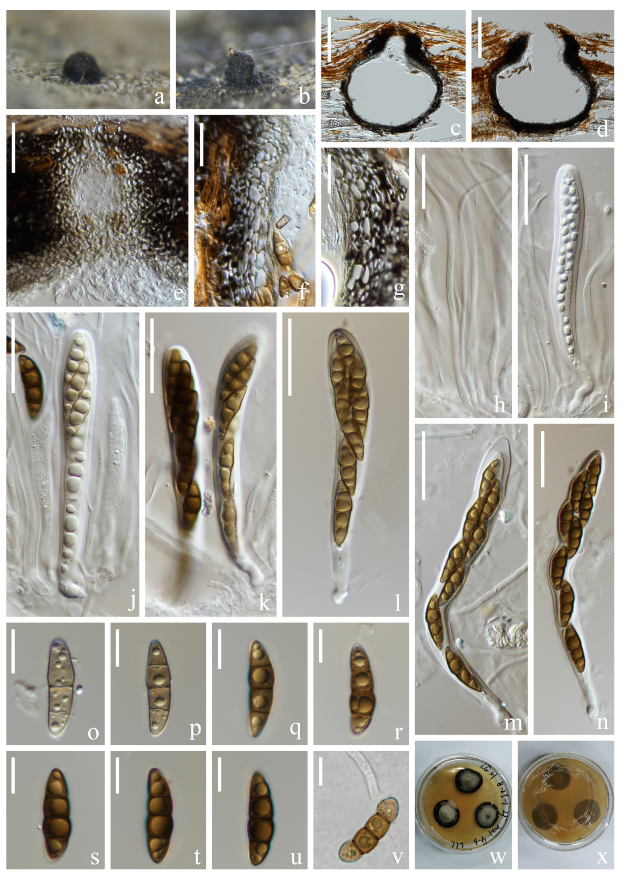
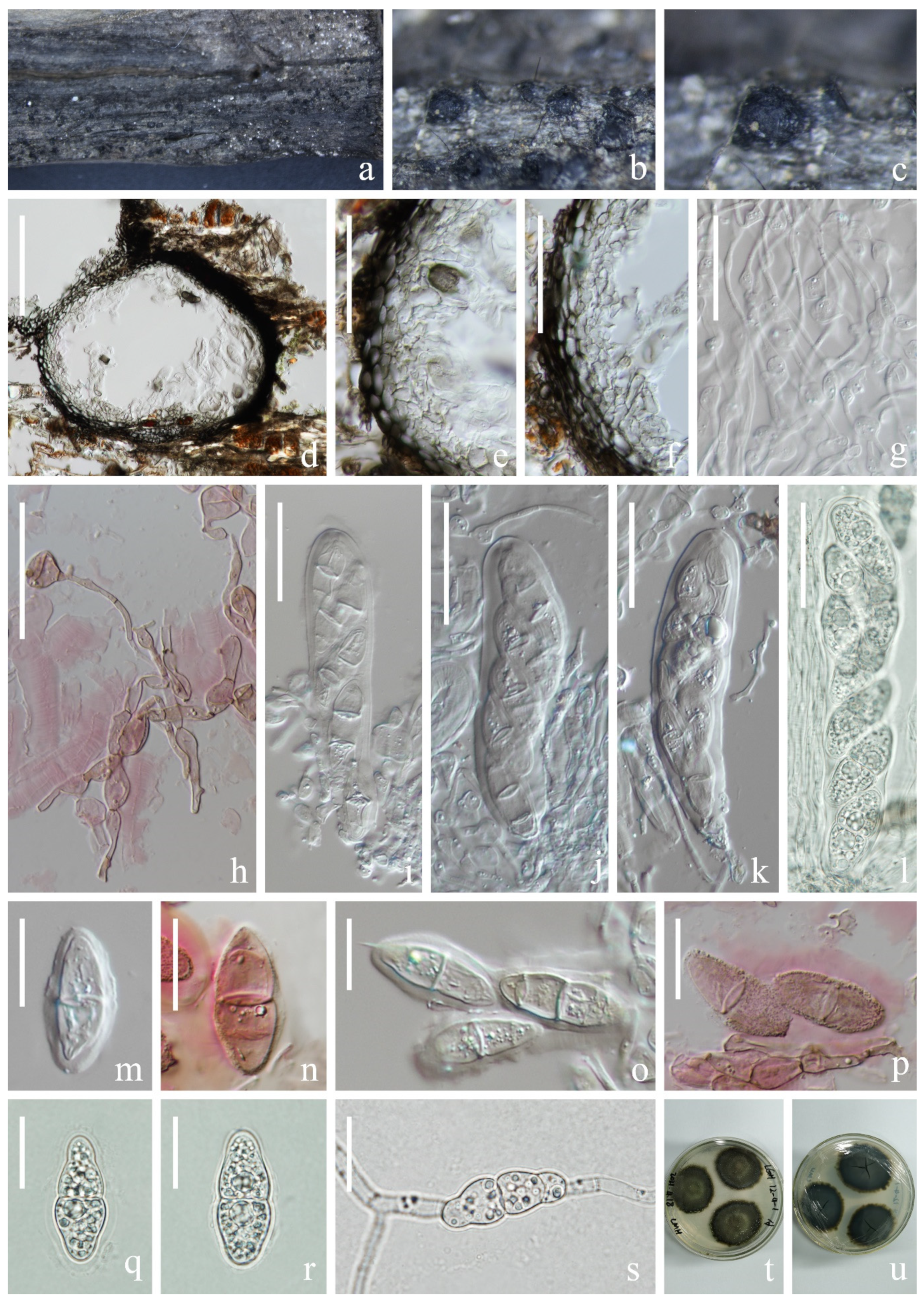
The combined ITS, LSU, SSU, and tef 1-α dataset comprises 147 taxa, including nine genera of Lentitheciaceae, with Pleomonodictys capensis (CBS 968.97) and P. descalsii (CBS 142298) as outgroup taxa (Figure 2). The dataset comprised 3777 characters (LSU: 1285 bp; SSU: 1021 bp; ITS: 539 bp; tef 1-α: 932 bp, including gaps). Maximum likelihood (ML) analysis and Bayesian analysis produced similar topologies that were consistent across the major clades. The likelihood of the final tree is evaluated and optimized under GAMMA. The best RAxML tree with a final likelihood value of −31,318.755060 is presented (Figure 2). The matrix had 1636 distinct alignment patterns, with 27.52% undetermined characters or gaps. Estimated base frequencies were as follows: A = 0.239720, C = 0.248808, G = 0.272414, T = 0.239058; substitution rates AC = 1.212047, AG = 2.534776, AT = 1.388124, CG = 1.249521, CT = 7.002685, GT = 1.000000, α = 0.226056, Tree-Length: 3.286461. Bayesian analyses generated 4412 trees (average standard deviation of split frequencies: 0.009960) from which 3310 were sampled after 25% of the trees were discarded as burn-in. The alignment contained a total of 1441 unique site patterns. Bootstrap support values with an ML greater than 75%, and Bayesian posterior probabilities (PP) greater than 0.97 are given above the nodes.
The multigene phylogenetic analyses showed that the 13 fresh collections clustered within Lentitheciaceae. Five known species, Halobyssothecium aquifusiforme (KUNCC 22-12665), H. phragmitis (KUN-HKAS 127181), H. unicellulare (KUNCC 22-12413), Lentithecium pseudoclioninum (KUNCC 22-12414 and KUNCC 22-12415), L. yunnanensis (KUNCC 22-124201, KUNCC 22-12421 and KUNCC 22-12422) and Setoseptoria bambusae (KUNCC 22-12416, KUNCC 22-12417 and KUNCC 22-12418) clustered with their ex-type strains, respectively. Paralentithecium suae (KUNCC 22-12412) clustered sister to P. aquaticum (CBS 123099) in an independent clade within Lentitheciaceae. Setoseptoria suae (KUNCC 22-12419) was placed sister to S. phragmitis (CBS 114804 and CBS 114966). Single-gene phylogenies are shown as Supplemental Materials (Figures S1–S4) because they resulted in being less informative and resolutive than those based on the four-loci concatenated tree.

3.2. Taxonomy

Halobyssothecium aquifusiforme J. Yang, Jian K. Liu & K.D. Hyde, Fungal Diversity 119: 39 (2023). Figure 3.
Index Fungorum number: IF559450; Facesoffungi number: FoF12783.
Saprobic on submerged decaying wood in a freshwater lake. Asexual morph: Undetermined. Sexual morph: Ascomata 354–382 µm high, 328–366 µm wide, immersed, clustered, sometimes solitary, scattered, subglobose or ellipsoidal, dark brown to black, carbonaceous, uniloculate, ostiolate. Ostiolar neck central, 86–114 µm long, 138–168 µm wide, papillate, rounded, short, dark brown, composed of several layers of pseudoparenchymatous cells. Peridium 22–35 µm thick, composed of several layers of pseudoparenchymatous cells, an outer layer composed of black cells, arranged in a textura angularis, inner layer composed of hyaline, flattened cells, arranged in a textura angularis. Pseudoparaphyses about 2 µm wide, branched, septate, hyaline, filamentous, anastomosing above the asci. Asci 97–129 × 13–16 µm ( x ¯ = 113 × 14 µm, n = 20) µm, 8-spored, clavate to subcylindrical, bitunicate, fissitunicate, apex rounded, short pedicellate, with an ocular chamber. Ascospores (20–)24–27 × 7–8 µm ( x ¯ = 25 × 8 µm, n = 40), overlapping, uniseriate to biseriate, central cells are brown to dark brown, 1-septate when young, 3-septate when mature, constricted at the septa, slightly curved, fusiform, guttulate, conical and narrowly rounded at the ends, one cell on the central septum side is swollen, lacking gelatinous sheaths or appendages.
Culture characteristics: Ascospore germinating on PDA within 12 h. Colonies on PDA reaching 3 cm diameter in 6 weeks at room temperature. Mycelium superficial, initially white, later becoming brown to black, with pale brown dense aerial mycelium on the surface, mastoid, marginal mycelium smooth, sparse, brown to black; from below, light brown at the center, dark brown at the margin.
Material examined: China, Yunnan Province, Dali City, Eryuan County, Cibihu Lake, 26°09′59″ N, 99°55′27″ E (2050 m), on unknown submerged decaying wood, 21 July 2021, S.P. Huang and L.L. Li, L788 (KUN-HKAS 124599), living cultures (KUNCC 22-12665).
Known host and distribution: China, Guizhou Province, Anshun City, Gaodang village, 26.071° N, 105.698° E, Suoluo River, on decaying wood submerged in a freshwater stream HKAS 112638, (holotype), HKAS 112641 (paratype) [15].
Notes: The phylogenetic analysis showed that our new strain, KUNCC 22-12665 clustered sister to Halobyssothecium aquifusiforme (GZCC 20-0481 and MFLUCC 19-0305) with 99% ML/1.00 PP supports (Figure 2). Our species is similar to H. aquifusiforme in having immersed, subglobose ascomata, and fusiform, guttulate, septate ascospores which are constricted at the septum [15]. We, therefore, identified our new collection as H. aquifusiforme and provided detailed descriptions and illustrations for it. Halobyssothecium aquifusiforme is an aquatic species that was collected on submerged decaying wood in a freshwater stream in Guizhou, China. Our two new collections were collected from a plateau lake in Yunnan.
Halobyssothecium phragmitis M.S. Calabon, E.B.G. Jones, S. Tibell & K.D. Hyde, Mycological Progress 20: 711 (2021). Figure 4.
Index Fungorum number: IF558090; Facesoffungi number: FoF 09431.
Saprobic on submerged decaying wood in a freshwater lake. Sexual morph: Ascomata 529–566 µm high, 545–691 µm wide, immersed or semi-immersed, solitary to gregarious, scattered, subglobose or ellipsoidal, dark brown, subcarbonaceous or coriaceous, uniloculate, with indistinct ostiolate. Ostiolar neck 172–265 µm high, 184–320 µm wide, central, papillate, rounded, short, dark brown, composed of several layers of pseudoparenchymatous cells. Peridium 26–77 µm thick, composed of several layers of pseudoparenchymatous cells, an outer layer composed of brown cells, arranged in a textura angularis and textura globulosa, and an inner layer composed of hyaline, flattened cells, arranged in a textura angularis. Pseudoparaphyses 2–3 µm wide, septate, hyaline, filiform, branched, anastomosing above the asci. Asci (102–)111–130(–137) × 10–12 µm ( x ¯ = 121 × 11 µm, n = 30), 8-spored, clavate to subcylindrical, bitunicate, fissitunicate, short pedicellate with an ocular chamber. Ascospores 22–27 × 5–6 µm ( x ¯ = 25 × 6 µm, n = 40), overlapping, uniseriate to biseriate, fusiform with narrow ends, cells swollen nearly central septum and gradually narrow toward ends, slightly curved, pale brown to dark brown and lightening from central cells to the end cells, 1-septate when young, 5-septate when mature, and constricted at the septa, lacking gelatinous sheaths or appendages. Asexual morph: Coelomycetes [14].
Material examined: China, Yunnan Province, Dali City, Eryuan County, Xihu Lake, 26°00′33″ N, 100°03′35″ E (1970 m), on unknown submerged decaying wood, 8 May 2021, S.P. Huang and L.L Li, L783 (KUN-HKAS 127181).
Known host and distribution: SWEDEN, Gotland, Kappelshamnsviken, on dead Phragmites culm (Poaceae), MFLU 20–0550 (holotype); ibid., Sudersand, on dead Phragmites (Poaceae) stem, MFLU 20–0552 (paratype) [14].
Notes: Halobyssothecium phragmitis was introduced by Calabon et al. [14], and only the asexual morph is known. This species was collected on Phragmites (Poaceae) culm in Europe. Phylogenetic analysis combined with ITS, LSU, SSU, and tef 1-α sequence data showed that our new collection (KUN-HKAS 127181) clustered with two strains of H. phragmitis (MFLUCC 20–0223 and MFLUCC 20–0225). The comparison of ITS, LSU, SSU, and tef 1-α sequences between our new collection (KUN-HKAS 127181) and the ex-type of H. phragmitis (MFLUCC 20–0226) showed 8 bp, 1 bp, 3 bp, and 3 bp differences, respectively. Morphologically, our new collection is similar to other sexual members of Halobyssothecium in having immersed or semi-immersed, subglobose or ellipsoidal, dark brown, subcarbonaceous or coriaceous ascomata, clavate to subcylindrical, bitunicate asci and 3-septate, fusiform ascospores [14,15,45,71]. Based on phylogenetic analysis and morphological evidence, we identified our new collection as H. phragmitis, and described its asexual morph. This is the first report of this species in China [14].
Halobyssothecium unicellulare (Abdel-Aziz) M.S. Calabon, K.D. Hyde & E.B.G. Jones, Mycological Progress 20: 715 (2021). Figure 5.
Index Fungorum number: IF558094; Facesoffungi number: FoF 09437
Saprobic on submerged decaying wood in a freshwater lake. Sexual morph: Undetermined. Asexual morph: Coelomycetes. Conidiomata 135–178 µm high, 205–242 µm wide, immersed to semi-immersed, most immersed, clustered, sometimes solitary, scattered, subglobose or ellipsoidal, uniloculate, dark brown to black, carbonaceous, short ostiolate, papillate, rounded. Conidiomatal walls 14–31 µm thick, composed of several layers of hyaline to black–brown cells of textura angularis. Conidiophores are reduced to conidiogenous cells. Conidiogenous cells 5–12 × 3–5 µm ( x ¯ = 8 × 4 µm, n = 30), hyaline, thin-walled, holoblastic, smooth, subglobose to pear-shaped, swollen at the base, sometimes one conidiogenous cell producing two conidia. Conidia 9–11 × 4–5 µm ( x ¯ = 10 × 5 µm, n = 60), subglobose, ovate, clavate, ellipsoid, allantoid or irregular, hyaline, aseptate, several small to one big guttulate, smooth-walled.
Culture characteristics: Conidia germinating on PDA within 12 h and germ tubes produced from one end of the conidia. Colonies on PDA, circular, reaching 5 cm in one month at room temperature, flat surface, pale brown to brown in PDA medium. Mycelium superficial, white to brown, hairy, effuse with wavy edge, dense, circular, raised, undulate to filiform with age; reverse light brown in the middle, with a dark brown deposit on the outside.
Material examined: China, Yunnan Province, Dali City, Eryuan County, Xihu Lake, 26°00′33″ N, 100°03′35″ E (1970 m), on unknown submerged decaying wood, 8 May 2021, S.P. Huang and L.L Li, L412 (KUN-HKAS 124589), living cultures, KUNCC 22-12413.
Known host and distribution: EGYPT, Sohag City, on decayed wood submerged in the River Nile, CBS H-22674 (holotype) [72].
Notes: The multigene phylogenetic analysis showed that our new collection (KUNCC 22-12413) clustered with the ex-type strain of Halobyssothecium unicellulare (MD 6004) with 91% ML/1.00 PP support (Figure 2). Morphologically, our new collection fits well with the original description of H. unicellulare [72]. The nucleotide comparison of LSU and SSU sequence data between our new collection (KUNCC 22-12413) and H. unicellulare (MD 6004) revealed 2 bp (including one gap) and 1 bp (including one gap) differences, respectively. We therefore identified it as H. unicellulare and it was reported from China for the first time.
Lentithecium pseudoclioninum Kaz. Tanaka & K. Hiray, Studies in Mycology 82: 99 (2015). Figure 6.
Index Fungorum number: IF811309; Facesoffungi number: FoF12785.
Saprobic on submerged decaying wood in a freshwater lake. Asexual morph: Undetermined. Sexual morph: Ascomata 201–310 µm high, 227–274 µm wide, black, semi-immersed, gregarious, erumpent, globose or subglobose, uniloculate, ostiolate. Ostiolar neck central, papillate, 92–110 µm long, 100–107 µm wide. Peridium 20–32 µm, thick-walled, brown to dark brown cells, composing several layers of pseudoparenchymatous cells of textura angularis, outer layers heavily pigmented, inner layers hyaline to pale brown, flattened. Pseudoparaphyses 2–3 µm wide, filamentous, branched septate. Asci 98–118 × 14–16 µm ( x ¯ = 108 × 15 µm, n = 30), 8-spored, bitunicate, fissitunicate, cylindric-clavate, slightly curved, pedicellate, apex rounded with a minute ocular chamber. Ascospores 28–32 × 8–10 µm ( x ¯ = 30 × 9 µm, n = 30), overlapping uni- to biseriate, narrowly fusiform, with a nearly median primary septum, constricted at the septum, hyaline, guttulate, usually with 2–4 larger guttules, asymmetrical, broadly fusiform, narrowly rounded at the ends, with a mucilaginous sheath.
Culture characteristics: Ascospore germinating on PDA within 12 h and germ tubes produced from the ends of the spore. Colonies on PDA, circular, reaching 5 cm in one month at room temperature, smooth surface, papillae, brown to dark brown. Mycelium superficial, hairy, smooth, circular, reverse grayish; reverse pale to brown, crack at the middle, flocculent at the edge.
Material examined: China, Yunnan Province, Dali City, Eryuan County, Xihu Lake, 26°00′33″ N, 100°03′35″ E (1970 m), on unknown submerged decaying wood, 8 May 2021, S.P. Huang and L.L Li, L413 (KUN-HKAS 124590), living cultures (KUNCC 22-12414); ibid., Erhai Lake, 26°00′32″ N, 100°03′35″ E (1970 m), on unknown submerged decaying wood, 01 April 2021, Z.Q. Zhang, L445 (KUN-HKAS 124593), living cultures (KUNCC 22-12415)
Known host and distribution: JAPAN, Aomori, Hirosaki, Aoki, Mohei pond, on submerged twigs of woody plant, KT 1113 (holotype) and KT 1111 (paratype); China, Guizhou Province, Weining City, Caohai National Nature Reserve, near 26.817° N, 104.217° E, on submerged decaying aquatic plants in Caohai lake, GZAAS 20-0378 [15].
Notes: Our two new collections are morphologically consistent with the holotype of Lentithecium pseudoclioninum [34]. In addition, phylogenetic analysis revealed that these two collections clustered with L. pseudoclioninum (Figure 2). Based on morphological and phylogenetic evidence, we identified our new collection as L. pseudoclioninum. Lentithecium pseudoclioninum has been collected on submerged twigs of woody plants in China and Japan [15,34]. Our two specimens were collected from a freshwater plateau lake in Yunnan, China.
Lentithecium yunnanensis W.H. Lu, Karun. & Tibpromma, Phytotaxa 554: 108 (2022). Figure 7.
Index Fungorum number: IF559622; Facesoffungi number: FoF 10778.
Saprobic on submerged decaying wood in a freshwater lake. Asexual morph: Undetermined. Sexual morph: Ascomata 246–285 µm high, 179–229 µm wide, immersed to semi-immersed, clustered, sometimes solitary, scattered, subglobose or ellipsoidal, dark brown to black, carbonaceous, uni- to bi-loculate, with indistinct ostiolate. Ostiolar neck central, papillate, 127–156 µm long, 96–110 µm wide. Peridium 11–21 µm thick, composed of several layers of pseudoparenchymatous cells, outer layer composed of back brown to brown cells, arranged in textura angularis, inner layer composed of hyaline cells, arranged in textura angularis. Pseudoparaphyses about 2 µm wide, hyaline, filamentous, branched, septate, globose to subglobose swollen at the apex, sometimes swollen at the septum, anastomosing at the apex, embedded in a hyaline gelatinous matrix. Asci 98–117 × 14–15 µm ( x ¯ = 108 × 15 µm, n = 15), 8-spored, clavate to subcylindrical, bitunicate, apex rounded, short pedicellate with an ocular chamber. Ascospores 27–30 × 5–6 µm ( x ¯ = 28 × 6 µm, n = 30), overlapping, uniseriate to biseriate, hyaline, 1-septate, smooth, constricted at the septa, slightly curved, guttulate, lacking gelatinous sheaths or appendages.
Culture characteristics: Ascospore germinating on PDA within 12 h and germ tubes produced from both ends of the spore. Colonies on PDA, circular, reaching 6 cm in 45 days at room temperature, smooth surface, papillae, brown in PDA medium. Mycelium superficial, brown to dark brown, hairy, smooth, circular; reverse brown to dark brown, crack at the middle, flocculent at the edge.
Material examined: China, Yunnan Province, Dali City, Eryuan County, Xihu Lake, 26°17′37″ N, 99°58′33″ E (2100 m), on unknown submerged decaying wood, 22 July 2021, L.L. Li, L680 (KUN-HKAS 124598), living cultures (KUNCC 22-12420 = KUNCC 22-12422); ibid., 26°17′24″ N, 99°57′56″ E (2100 m), on unknown submerged decaying wood, 22 July 2021, X.J. Yuan, L679 (KUN-HKAS 124597), living culture (KUNCC 22-12421).
Known host and distribution: China, Yunnan, Kunming, Songhua Dam Reservoir, on dead culms of Artemisia sp., HKAS 123192 (holotype) [73].
Notes: Lentithecium yunnanensis is a terrestrial species introduced by Lu et al. [73] that occurs on dead culms of Artemisia sp. near humid places. We collected two Lentithecium-like collections from decaying wood submerged in Xihu Lake, Dali, Yunnan Province. Phylogenetic analysis showed that our two new collections clustered with two strains of L. yunnanensis (KUNCC 22-10776 and KUNCC 22-10776). In addition, the morphology of our two collections is similar to the holotype of L. yunnanensis in having semi-immersed to immersed, subglobose to globose ascomata with short ostioles, and hyaline, clavate to fusiform, septate ascospores. Therefore, the two new collections were identified as L. yunnanensis, which was reported from the freshwater habitat for the first time.
Paralentithecium H.W. Shen, K.D. Hyde & Z.L. Luo gen. nov.
MycoBank number: 849738.
Etymology: referring to the comparable morphological characters to that of Lentithecium.
Saprobic on submerged decaying wood in a freshwater lake. Asexual morph: Undetermined. Sexual morph: Ascomata immersed to semi-immersed, clustered, sometimes solitary, scattered, subglobose or ellipsoidal, dark brown to black, carbonaceous, uni- to bi-loculate, with indistinct ostiolate. Peridium thick, composed of several layers of pseudoparenchymatous cells, an outer layer composed of back brown to brown cells, arranged in textura angularis, and an inner layer composed of hyaline cells, arranged in textura angularis. Pseudoparaphyses thick, hyaline, filamentous, branched, septate, globose to subglobose swollen at the apex, sometimes swollen at the septum, anastomosing at the apex, embedded in a hyaline gelatinous matrix. Asci 8-spored, clavate to subcylindrical, bitunicate, apex rounded, short pedicellate with an ocular chamber. Ascospores overlapping, uniseriate to biseriate, hyaline, 1-septate, smooth, constricted at the septa, slightly curved, with gelatinous sheaths.
Type species: Paralentithecium aquaticum (Yin. Zhang, J. Fourn. & K.D. Hyde) H.W. Shen & Z.L. Luo.
Paralentithecium aquaticum (Yin. Zhang, J. Fourn. & K.D. Hyde) H.W. Shen & Z.L. Luo, comb. nov.
MycoBank number: MB 512791.
Basionym: Lentithecium aquaticum Yin. Zhang, J. Fourn. & K.D. Hyde, Fungal Diversity 38: 234 (2009).
Known host and distribution: FRANCE, Ariège, Rimont, Peyrau, on submerged wood of Fraxinus excelsior; on submerged wood of Alnus glutinosa; Le Baup brook, along D 18, on submerged wood of Platanus sp. [35].
Notes: Lentithecium aquaticum was introduced by Zhang et al. [35] based on phylogenetic analysis and morphological characteristics. The placement of this species was not stable and has been changed by several studies [10,45,71,74]. Previous phylogenetic analyses indicated that Lentithecium aquaticum did not cluster with other Lentithecium species, and it formed an individual lineage basal to Darksidea, Halobyssothecium and Lentithecium [10,14,34,41,48,71]. Furthermore, phylogenetic studies of Dayarathne et al. [45] and Devadatha et al. [71] showed that L. aquaticum clustered within Setoseptoria. Several other studies excluded L. aquaticum from Lentithecium [10,74]. The latest phylogenetic analysis based on combined ITS, LSU, SSU, and tef 1-α genes showed that L. aquaticum formed a separate lineage outside of Lentithecium [48]. Our phylogenetic analysis shows that L. aquaticum clusters with our new collection KUNCC 22-12412 and forms a distinct lineage within Lentitheciaceae with 100 ML/1.00 PP support (Figure 2). Therefore, we propose a new genus, Paralentithecium to accommodate Paralentithecium aquaticum (Lentithecium aquaticum) and a new species P. suae.
Paralentithecium suae H.W. Shen, K.D. Hyde & Z.L. Luo sp. nov. Figure 8.
MycoBank number: 849739; Facesoffungi number: FoF 14876.
Etymology: “suae” (Lat.) in memory of the Chinese mycologist Prof. Hong-Yan Su (4 April 1967–3 May 2022).
Holotype: KUN-HKAS 124587.
Saprobic on submerged decaying wood in a freshwater lake. Asexual morph: Undetermined. Sexual morph: Ascomata 212–253 µm high, 175–204 µm wide, immersed to semi-immersed, clustered, sometimes solitary, scattered, subglobose or ellipsoidal, dark brown to black, carbonaceous, uni- to bi-loculate, with indistinct ostiolate. Peridium 17–32 µm thick, composed of several layers of pseudoparenchymatous cells, outer layer composed of bark brown to brown cells, arranged in textura angularis, inner layer composed of hyaline cells, arranged in textura angularis. Pseudoparaphyses 2–3 µm wide, hyaline, filamentous, branched, septate, globose to subglobose swollen at the apex, sometimes swollen at the septum (6–10 µm wide), anastomosing at the apex, embedded in a hyaline gelatinous matrix. Asci 104–134 × 24–28 µm ( x ¯ = 119 × 26 µm, n = 25), 8-spored, clavate to subcylindrical, bitunicate, apex rounded, short pedicellate with an ocular chamber. Ascospores 28–34 × 11–14 µm ( x ¯ = 31 × 13 µm, n = 40), overlapping, uniseriate to biseriate, hyaline, 1-septate, broadly fusiform, smooth, constricted at the septa, slightly curved, guttulate, with gelatinous sheaths.
Culture characteristics: Ascospore germinating on PDA within 12 h and germ tubes produced from both ends of the spore. Colonies on PDA, circular, reaching 4–5 cm in one month at room temperature, smooth surface, papillae, brown to dark brown, olive green in PDA medium. Mycelium superficial, brown to dark brown, hairy, smooth, circular; reverse dark brown, crack at the middle, flocculent at the edge, dark brown with greenish.
Material examined: China, Yunnan Province, Lijiang City, Ninglang County, Luguhu Lake, 27°44′15″ N, 100°45′16″ E (2700 m), on unknown submerged decaying wood, 5 March 2021, Z.Q. Zhang and L. Sha, L184 (KUN-HKAS 124587, holotype), ex-type living cultures (CGMCC 3.24265 = KUNCC 22–12412).
Notes: In our phylogenetic analysis, Paralentithecium suae clustered with P. aquaticum with 100% ML/1.00 PP support (Figure 2). Comparison of ITS, LSU, SSU, and tef 1-α sequences between Paralentithecium suae and P. aquaticum revealed 11 bp, 4 bp, 4 bp, and 22 bp differences, respectively. Paralentithecium suae resembles P. aquaticum in having hyaline, 1-septate, broadly fusiform ascospores with gelatinous sheaths [35]. However, P. suae is distinct from P. aquaticum in having globose to subglobose pseudoparaphyses that are swollen at the apex and sometimes swollen at the septum. In contrast, the pseudoparaphyses of P. aquaticum are not swollen. In addition, ascospores of P. aquaticum contain four refractive globules, while P. suae has ascospores with many small guttules [35]. Therefore, we introduce P. suae as a new species.
Setoseptoria bambusae J. Yang, Jian K. Liu & K.D. Hyde, Fungal Diversity 119: 44 (2022). Figure 9.
Index Fungorum number: IF559452; Facesoffungi number: FoF12786.
Saprobic on submerged decaying wood in a freshwater lake. Asexual morph: Undetermined. Sexual morph: Ascomata 245–375 µm high, 194–296 µm wide, black, superficial to semi-immersed, gregarious, fully or partly erumpent, globose, uniloculate, ostiolate. Peridium 26–39 µm wide, thick, multi-layered, outer layer most heavily pigmented, comprising blackish to dark brown amorphous layer, middle layer heavily pigmented, inner layer, pale brown to hyaline, cells towards the inside lighter, flattened, thick-walled. Pseudoparaphyses 2–3 µm wide, filamentous, branched septate. Asci 113–128 × 15–19 µm ( x ¯ = 120 × 17 µm, n = 30), 8-spored, bitunicate, fissitunicate, clavate to cylindric-clavate, pedicellate, apex rounded with a minute ocular chamber. Ascospores 32–40 × 6–8 µm ( x ¯ = 36 × 7 µm, n = 20), overlapping uni- to biseriate, narrowly fusiform, with a nearly median primary septum, deeply constricted at the septum, hyaline, guttulate, asymmetrical, conical, and narrowly rounded at the ends.
Culture characteristics: Ascospore germinating on PDA within 12 h and germ tubes produced from one end of the spore. Colonies on PDA, circular, reaching 6 cm in 45 days at room temperature, smooth surface, papillae, pale brown in PDA medium. Mycelium superficial, grayish-brown to brown, hairy, smooth, circular; reverse pale brown at the edges, dark brown in the middle, flocculent at the edge.
Material examined: China, Yunnan Province, Yuxi City, Jiangchuan District, Xingyunhu Lake, 24°23′05″ N, 102°48′22″ E (1720 m), on unknown submerged decaying wood, 10 July 2021, H.W. Shen, L511 (KUN-HKAS 124592), living culture (KUNCC 22–12417); ibid., 24°23′05″ N, 102°48′22″ E (1720 m), on the submerged stem of Phragmites sp. (Poaceae), 10 July 2021, S. Luan, L579 (KUN-HKAS 124596), living culture (KUNCC 22–12418); ibid., on submerged stem of Phragmites sp. (Poaceae), 10 July 2021, Y.K. Jiang, L474 (KUN-HKAS 124591), living culture (KUNCC 22–12416).
Known host and distribution: China, Guizhou Province, Anshun City, Gaodang Village, 26.071° N, 105.698° E, Suoluo River, on decaying bamboo culms submerged in a freshwater stream, HKAS 112629 (holotype) [15].
Notes: Setoseptoria bambusae was introduced by Yang et al. [15] to accommodate two collections, GZCC 17–0044 (ex-type strain) and IFRD500-013 (previously identified as S. arundinaceae, without description). In this study, our four new collections clustered with the ex-type strain of S. bambusae with 100% ML/1.00 PP statistical support (Figure 2). Furthermore, our collections fit the morphological characteristics of S. bambusae except for the size of asci and ascospores, our isolate has shorter asci (113–128 vs. 130–180 µm) and longer ascospores (32–40 vs. 28–37 µm). Therefore, we identified them as S. bambusae. Our four new collections were collected from lentic freshwater habitats. The holotype was collected from lotic habitats.
Setoseptoria suae H.W. Shen, K.D. Hyde & Z.L. Luo sp. nov. Figure 10.
MycoBank number: 849740; Facesoffungi number: FoF 14877.
Etymology: “suae” (Lat.) in memory of the Chinese mycologist Prof. Hong-Yan Su (4 April 1967–3 May 2022).
Holotype: KUN-HKAS 124595.
Saprobic on submerged decaying wood in a freshwater lake. Sexual morph: Undetermined. Asexual morph: Conidiomata 383–512 µm high, 173–196 µm wide, solitary, scattered, semi-immersed to immersed in the host, pycnidial, subglobose to ellipsoidal, unilocular, black, ostiolate, apapillate. Ostiole short, centrally located. Conidiomatal wall 33–55 µm wide, thickening at the upper zone, thick-walled, composed of several layers of textura angularis, an outer layer comprising brown to dark brown cells, pigmented; inner layer comprising hyaline cells. Conidiophores reduced to conidiogenous cells. Conidiogenous cells (4–)7–15(–26) × 4–6 µm ( x ¯ = 11 × 5 µm, n = 25), arising from the inner layers of conidiomata, hyaline, enteroblastic, phialidic, determinate, ampuliform, subcylindrical to lageniform. Conidia 33–43 × 4–6 µm ( x ¯ = 38 × 5 µm, n = 50), subcylindrical, with obtuse to subobtuse ends, straight or slightly curved, hyaline, (1–)3-septate, euseptate, mostly with one large central guttule per cell when young, with many small guttules in each cell at maturity, slightly constricted at the septum, smooth-walled.
Culture characteristics: Conidia germinated on PDA within 12 h and germ tubes produced from the ends of the spore. Colonies on PDA, circular, reaching 6 cm in one month at room temperature, brown to dark brown. Mycelium superficial, brown to dark brown, hairy, smooth, circular; dark brown from below.
Material examined: China, Yunnan Province, Yuxi City, Tonghai County, Qiluhuhu Lake, 24°08′37″ N, 102°46′24″ E (1800 m), on submerged stem of Phragmites sp. (Poaceae), 11 July 2021, H.W. Shen, L570 (KUN-HKAS 124595, holotype), ex-type living cultures (CGMCC 3.24266 = KUNCC 22–12419).
Notes: Phylogenetic analysis showed that Setoseptoria suae clustered with S. phragmitis with 100% ML/0.99 PP statistical support (Figure 2). The comparison of ITS and LSU sequences between S. suae and S. phragmitis shows that the similarities are 96.9% (538/555 bp) and 99.9% (826/827 bp), respectively. Setoseptoria suae resembles S. phragmitis in having immersed, globose conidiomata, hyaline, subcylindrical, smooth, guttulate, (1–)3-septate conidia [37]. However, Setoseptoria suae can be distinguished from S. phragmitis by its larger conidia (33–43 × 4–6 µm vs. (19–)25–35(–38) × (3.5–)4 µm). In addition, the conidia of S. phragmitis mostly have one large central guttule per cell, while Setoseptoria suae has conidia with many small guttules in each cell. We, therefore, introduce S. suae as a new species.

4. Discussion

Yunnan, located on the Yunnan–Guizhou Plateau, is one of the global biodiversity hotspots with rich biological resources [18,19,75]. In recent years, research on lignicolous freshwater fungi in Yunnan has developed rapidly, and a large number of new species have been reported from lotic freshwater habitats such as streams and rivers [10,13,76,77,78,79,80,81]. A few studies have reported lignicolous freshwater fungi from lentic habitats in Yunnan Province. For example, Cai et al. [17] and Luo et al. [2] investigated lignicolous freshwater fungi in Fuxianhu and Dianchi Lakes, respectively. However, freshwater fungi in lentic habitats have not been updated recently. In this study, we investigate the freshwater fungi in Cibihu, Luguhu, Qiluhu, Xihu, and Xingyunhu lakes in Yunnan Province, one new genus, two new species, and three new records are reported, the results indicate that high undiscovered diversity of lignicolous freshwater fungi in lentic habitats.
Zhang et al. [36] provided the first multigene phylogenetic analysis of Pleosporales and introduced the family Lentitheciaceae which accepted the genera Lentithecium, Katumotoa, and Keissleriella. Dong et al. [10] treated the family with ten genera and this was followed by Wijayawardene et al. [41]. Previous studies based on morphology and phylogenetic analyses showed that the classification of Lentithecium, Keissleriella, and Setoseptoria is confusing as the placement of several taxa was problematic and has been transferred to different genera. For example, Suetrong et al. [82] transferred Keissleriella rara to Lentithecium as L. rarum; however, later studies showed that L. rarum clustered with K. trichophoricola in Keissleriella [14]. Similarly, Zhang et al. [35] transferred Keissleriella linearis to Lentithecium as L. lineare, Singtripop et al. [83] re-examined the type specimen of K. linearis (L. lineare) and transferred it to Keissleriella based on LSU phylogenetic analysis, and this was confirmed by subsequent phylogenetic studies [14,34,72]. The placements of Lentithecium species have been revised in recent years based on multigene phylogenetic studies [14,34,84]. Calabon et al. [14] transferred several Lentithecium species with brown and versicolored ascospores without sheaths and hyaline conidia to Halobyssothecium, including L. cangshanense, L. carbonneanum, L. kunmingense, L. unicellulare, and L. voraginesporum. Currently, 13 species are accepted in Halobyssothecium. In the present study, we report the sexual morph of H. phragmitis and provide detailed morphological descriptions for its sexual morph.

Supplementary Materials

The following supporting information can be downloaded at: https://www.mdpi.com/article/10.3390/jof9100962/s1.

Author Contributions

Conceptualization, Z.-L.L., S.B. and K.D.H.; methodology, H.-W.S. and X.-J.S.; formal analysis, H.-W.S.; investigation, H.-W.S., D.-F.B., X.-J.S. and X.-G.T.; resources, Z.-L.L. and S.B.; data curation, H.-W.S.; writing—original draft preparation, H.-W.S. and X.-G.T.; writing—review and editing, D.-F.B., S.B., X.-J.S., K.D.H. and Z.-L.L.; funding acquisition, Z.-L.L. All authors have read and agreed to the published version of the manuscript.

Funding

This research was funded by the National Natural Science Foundation of China (Project ID: 32060005) and the Yunnan Fundamental Research Project (202201AW070001).

Institutional Review Board Statement

Not applicable.

Informed Consent Statement

Not applicable.

Data Availability Statement

All sequences generated in this study were submitted to GenBank database.

Acknowledgments

Hong-Wei Shen thanks Long-Li Li, Qiu-Xia Yang, Sha Luan, and Si-Ping Huang for their help with sample collection, DNA extraction, and PCR amplification. Thanks to Rong-Ju Xu for his help in specimens and culture preservation.

Conflicts of Interest

The authors declare no conflict of interest.

References

  1. Wong, M.K.M.; Goh, T.K.; Hodgkiss, I.J.; Hyde, K.D.; Ranghoo, V.M.; Tsui, C.K.M.; Ho, W.H.; Wong, W.S.W.; Yuen, T.K. Role of Fungi in Freshwater Ecosystems. Biodivers. Conserv. 1998, 7, 1187–1206. [Google Scholar] [CrossRef]
  2. Luo, J.; Yin, J.F.; Cai, L.; Zhang, K.Q.; Hyde, K.D. Freshwater fungi in Lake Dianchi, a heavily polluted lake in Yunnan, China. Fungal Divers. 2004, 16, 93–112. [Google Scholar]
  3. Hyde, K.D.; Fryar, S.; Tian, Q.; Bahkali, A.H.; Xu, J.C. Lignicolous freshwater fungi along a north-south latitudinal gradient in the Asian/Australian region; can we predict the impact of global warming on biodiversity and function? Fungal Ecol. 2016, 19, 190–200. [Google Scholar] [CrossRef]
  4. Zare-Maivan, H.; Shearer, C.A. Extracellular enzyme production and cell wall degradation by freshwater lignicolous fungi. Mycologia 1988, 80, 365–375. [Google Scholar] [CrossRef]
  5. Yuen, T.K.; Hyde, K.D.; Hodgkiss, I.J. Physiological growth parameters and enzyme production in tropical freshwater fungi. Mater. Org. (Berl) 1998, 32, 2–16. [Google Scholar]
  6. Abdel-Raheem, A.; Shearer, C.A. Extracellular enzyme production by freshwater ascomycetes. Fungal Divers. 2002, 11, 1–19. [Google Scholar]
  7. Bucher, V.V.C.; Hyde, K.D.; Pointing, S.B.; Reddy, C.A. Production of wood decay enzymes, mass loss and lignin solubilization in wood by marine ascomycetes and their anamorphs. Fungal Divers. 2004, 15, 14. [Google Scholar]
  8. Duarte, S.; Fernandes, I.; Nogueira, M.J.; Cássio, F.; Pascoal, C. Temperature alters interspecific relationships among aquatic fungi. Fungal Ecol. 2013, 6, 187–191. [Google Scholar] [CrossRef]
  9. Dong, W.; Hyde, K.D.; Doilom, M.; Yu, X.D.; Bhat, D.J.; Jeewon, R.; Boonmee, S.; Wang, G.N.; Nalumpang, S.; Zhang, H. Pseudobactrodesmium (Dactylosporaceae, Eurotiomycetes, Fungi) a novel lignicolous genus. Front. Microbiol. 2020, 11, 456. [Google Scholar] [CrossRef]
  10. Dong, W.; Wang, B.; Hyde, K.D.; McKenzie, E.H.C.; Raja, H.A.; Tanaka, K.; Abdel-Wahab, M.A.; Abdel-Aziz, F.A.; Doilom, M.; Phookamsak, R.; et al. Freshwater Dothideomycetes. Fungal Divers. 2020, 105, 319–575. [Google Scholar] [CrossRef]
  11. Calabon, M.S.; Hyde, K.D.; Jones, E.B.G.; Luo, Z.L.; Dong, W.; Hurdeal, V.G.; Gentekaki, E.; Rossi, W.; Leonardi, M.; Thiyagaraja, V.; et al. Freshwater fungal numbers. Fungal Divers. 2022, 114, 3–235. [Google Scholar] [CrossRef]
  12. Shen, H.W.; Bao, D.F.; Bhat, D.J.; Su, H.Y.; Luo, Z.L. lignicolous freshwater fungi in Yunnan Province, China: An overview. Mycology 2022, 13, 119–132. [Google Scholar] [CrossRef] [PubMed]
  13. Luo, Z.L.; Hyde, K.D.; Liu, J.K.; Bhat, D.J.; Bao, D.F.; Li, W.L.; Su, H.Y. Lignicolous freshwater fungi from china ii: Novel Distoseptispora (Distoseptisporaceae) species from northwestern Yunnan Province and a suggested unified method for studying lignicolous freshwater fungi. Mycosphere 2018, 9, 444–461. [Google Scholar] [CrossRef]
  14. Calabon, M.S.; Jones, E.B.G.; Hyde, K.D.; Boonmee, S.; Tibell, S.; Tibell, L.; Pang, K.L.; Phookamsak, R. Phylogenetic assessment and taxonomic revision of Halobyssothecium and Lentithecium (Lentitheciaceae, Pleosporales). Mycol. Prog. 2021, 20, 701–720. [Google Scholar] [CrossRef]
  15. Yang, J.; Liu, L.L.; Jones, E.B.G.; Hyde, K.D.; Liu, Z.Y.; Bao, D.F.; Liu, N.G.; Li, W.L.; Shen, H.W.; Yu, X.D.; et al. Freshwater fungi from karst landscapes in China and Thailand. Fungal Divers. 2023, 119, 1–212. [Google Scholar] [CrossRef]
  16. Tsui, C.K.M.; Hyde, K.D.; Hodgkiss, I.J. Biodiversity of fungi on submerged wood in hong kong streams. Aquat. Microb. Ecol. 2000, 21, 289–298. [Google Scholar] [CrossRef]
  17. Cai, L.; Tsui, C.K.M.; Zhang, K.; Hyde, K.D. Aquatic fungi from lake Fuxian, Yunnan, China. Fungal Divers. 2000, 14, 57–70. [Google Scholar]
  18. Xu, Y.; Shen, Z.H.; Ying, L.X.; Wang, Z.H.; Huang, J.H.; Zang, R.G.; Jiang, Y.X. Hotspot analyses indicate significant conservation gaps for evergreen broadleaved woody plants in China. Sci. Rep. 2017, 7, 1859. [Google Scholar] [CrossRef]
  19. Qian, L.S.; Chen, J.H.; Deng, T.; Sun, H. Plant diversity in yunnan: Current status and future directions. Plant Divers. 2020, 42, 281–291. [Google Scholar] [CrossRef]
  20. Liu, S.R.; He, X.Y.; Yang, W.S.; Ren, G.P.; Li, Y.P.; Zhou, J.; Cai, Q.H.; Xiao, W. Spatial distribution and significance of high mountain micro-waterbodies in northwestern Yunnan, China. J. Hydroecol. 2017, 38, 18–23. [Google Scholar] [CrossRef]
  21. Liu, S.R.; De Yang, D.; Li, X.F.; Tan, L.; Sun, J.; He, X.Y.; Yang, W.S.; Ren, G.P.; Fornacca, D.; Cai, Q.H.; et al. Diversity in benthic and environmental characteristics on alpine micro-waterbodies and stream ecosystems in northwest Yunnan. Biodivers. Sci. 2019, 27, 1298–1308. [Google Scholar]
  22. Wang, R.X.; Yang, X.J. Waterbird composition and changes with wetland park construction at lake Dianchi, Yunnan-Guizhou plateau. Mt. Res. Dev. 2021, 41, R29–R37. [Google Scholar] [CrossRef]
  23. Lu, B.; Pan, M.; Li, B.; Yang, B.; Song, R.B.; Li, Y. Morphological characteristics and habitat demand analysis of common water birds in Dianchi Lake. Environ. Sci. Surv. 2022, 41, 15–19. [Google Scholar]
  24. Xiao, Q.Z.; Chen, L.J.; Qiu, Y.P.; Chen, G.Z. The alien fish Largemouth bass, Micropterus salmoides, found in Dianchi basin, Yunnan, China. Chin. J. Zool. 2020, 55, 834–835. [Google Scholar]
  25. Wu, Y.; Li, L.; Zheng, L.; Dai, G.; Ma, H.; Shan, K.; Wu, H.; Zhou, Q.; Song, L. Patterns of succession between bloom-forming cyanobacteria Aphanizomenon flos-aquae and Microcystis and related environmental factors in large, shallow Dianchi Lake, China. Hydrobiologia 2016, 765, 1–13. [Google Scholar] [CrossRef]
  26. Yang, W.; Deng, D.G.; Meng, X.L.; Zhang, S. Temporal and spatial variations of phytoplankton community structure in lake Erhai, a chinese plateau lake, with reference to environmental factors. Russ. J. Ecol. 2019, 50, 352–360. [Google Scholar] [CrossRef]
  27. Wang, H.; Wen, Z.; Zhang, Z.H.; Zhang, X.L.; Fu, H.; Cao, Y.; Ni, L.Y.; Cao, T.; Li, K.Y. Environmental vs. spatial drivers of submerged macrophyte community assembly in different seasons and water depths in a mesotrophic bay of Erhai Lake, China. Ecol. Indic. 2020, 117, 106696. [Google Scholar] [CrossRef]
  28. Han, L.; Li, Z.Y.; Guo, X.F.; Tan, J.L.; He, S.Z.; Cui, X.L.; Li, S.L. Hannaella dianchiensis sp. nov., a basidiomycetous yeast species isolated from lake water. Int. J. Syst. Evol. Microbiol. 2017, 67, 2014–2018. [Google Scholar] [CrossRef]
  29. Liu, X.Y. Studies of Aquatic Fungal Diversity in Erhai Lake and Morphology, Molecular Phylogenetic Systematics of Minimelanlocus. Master’s Thesis, Dali University, Dali, China, 2016. [Google Scholar]
  30. Han, L. Diversity and Spatial Distribution of Fungi in Dianchi Lake of Yunnan plateau. Ph.D. Dissertation, Yunnan University, Kunming, China, 2018. [Google Scholar]
  31. Chen, Z.B.; Xu, S.G.; Yu, L.; Liu, J.N.; Su, Y.; Shao, X.D.; Ruan, Y.N.; Wang, D.K. Bacterial diversity of surface sediment at Lake Dian in summer and winter seasons. Int. J. Agric. Biol. 2020, 23, 559–565. [Google Scholar]
  32. Zhang, Y.; Zuo, J.E.; Wang, S.K.; Salimova, A.; Li, A.J.; Li, L.L. Spatial distribution of nitrogen metabolism functional genes of Eubacteria and Archaebacteria in Dianchi Lake. Huanjing Kexue/Environ. Sci. 2020, 41, 2908–2917. [Google Scholar]
  33. Tanaka, K.; Hatakeyama, S.; Harada, Y. Three new freshwater ascomycetes from rivers in Akkeshi, Hokkaido, northern Japan. Mycoscience 2005, 46, 287–293. [Google Scholar] [CrossRef]
  34. Tanaka, K.; Hirayama, K.; Yonezawa, H.; Sato, G.; Toriyabe, A.; Kudo, H.; Hashimoto, A.; Matsumura, M.; Harada, Y.; Kurihara, Y.; et al. Revision of the Massarineae (Pleosporales, Dothideomycetes). Stud. Mycol. 2015, 82, 75–136. [Google Scholar] [CrossRef] [PubMed]
  35. Zhang, Y.; Wang, H.K.; Fournier, J.; Crous, P.W.; Jeewon, R.; Pointing, S.B.; Hyde, K.D. Towards a phylogenetic clarification of Lophiostoma/Massarina and morphologically similar genera in the Pleosporales. Fungal Divers. 2009, 38, 225–251. [Google Scholar]
  36. Zhang, Y.; Schoch, C.L.; Fournier, J.; Crous, P.W.; de Gruyter, J.; Woudenberg, J.H.C.; Hirayama, K.; Tanaka, K.; Pointing, S.B.; Spatafora, J.W.; et al. Multi-locus phylogeny of Pleosporales: A taxonomic, ecological and evolutionary re-evaluation. Stud. Mycol. 2009, 64, 85–102. [Google Scholar] [CrossRef] [PubMed]
  37. Quaedvlieg, W.; Verkley, G.J.M.; Shin, H.D.; Barreto, R.W.; Alfenas, A.C.; Swart, W.J.; Groenewald, J.Z.; Crous, P.W. Sizing up Septoria. Stud. Mycol. 2013, 75, 307–390. [Google Scholar] [CrossRef] [PubMed]
  38. Zhang, Y.; Crous, P.W.; Schoch, C.L.; Hyde, K.D. Pleosporales. Fungal Divers. 2012, 53, 1–221. [Google Scholar] [CrossRef] [PubMed]
  39. Hyde, K.D.; Jones, E.B.G.; Liu, J.K.; Ariyawansa, H.; Boehm, E.; Boonmee, S.; Braun, U.; Chomnunti, P.; Crous, P.W.; Dai, D.Q.; et al. Families of Dothideomycetes. Fungal Divers. 2013, 63, 1–313. [Google Scholar] [CrossRef]
  40. Wanasinghe, D.N.; Jones, E.B.G.; Camporesi, E.; Boonmee, S.; Ariyawansa, H.A.; Wijayawardene, N.N.; Mortimer, P.E.; Xu, J.; Yang, J.B.; Hyde, K.D. An exciting novel member of Lentitheciaceae in Italy from Clematis vitalba. Cryptogam. Mycol. 2014, 35, 323–337. [Google Scholar] [CrossRef]
  41. Wijayawardene, N.; Hyde, K.; Dai, D.; Sánchez-García, M.; Goto, B.; Saxena, R.; Erdoğdu, M.; Selçuk, F.; Rajeshkumar, K.; Aptroot, A.; et al. Outline of fungi and fungus-like taxa—2021. Mycosphere 2022, 13, 53–453. [Google Scholar] [CrossRef]
  42. Liu, Z.P.; Zhang, S.N.; Cheewangkoon, R.; Zhao, Q.; Liu, J.K. Crassoascoma gen. nov. (Lentitheciaceae, Pleosporales): Unrevealing microfungi from the Qinghai-Tibet plateau in China. Diversity 2022, 14, 15. [Google Scholar] [CrossRef]
  43. Knapp, D.G.; Kovács, G.M.; Zajta, E.; Groenewald, J.Z.; Crous, P.W. Dark septate endophytic pleosporalean genera from semiarid areas. Persoonia 2015, 35, 87–100. [Google Scholar] [CrossRef] [PubMed]
  44. Rajeshkumar, K.C.; Varma, R.K.; Sruthi, O.P.; Gautam, A.K.; Crous, P.W. Groenewaldia (Lentitheciaceae), a new corticolous fungal genus from India. Mycol. Prog. 2023, 22, 43. [Google Scholar] [CrossRef]
  45. Dayarathne, M.C.; Wanasinghe, D.N.; Jones, E.B.G.; Chomnunti, P.; Hyde, K.D. A novel marine genus, Halobyssothecium (Lentitheciaceae) and epitypification of Halobyssothecium obiones comb. nov. Mycol. Prog. 2018, 17, 1161–1171. [Google Scholar] [CrossRef]
  46. Tanaka, K.; Harada, Y. Bambusicolous fungi in Japan (6): Katumotoa, a new genus of phaeosphaeriaceous ascomycetes. Mycoscience 2005, 46, 313–318. [Google Scholar] [CrossRef]
  47. von Höhnel, F.X.R. Fragmente Zur Mykologie XXIII. Mitteilung, Nr. 1154 bis 1188. Sitz. Kais. Akad. Wiss. Math.-Nat. Kl. Abt. I 1919, 128, 535–625. Available online: https://archive.org/details/sbaww_128_0535-0625/mode/2up (accessed on 16 February 2023).
  48. Hyde, K.D.; Suwannarach, N.; Jayawardena, R.S.; Manawasinghe, I.S.; Liao, C.; Doilom, M.; Cai, L.; Zhao, P.; Buyck, B.; Phukhamsakda, C.; et al. Mycosphere notes 325–344—Novel species and records of fungal taxa from around the world. Mycosphere 2021, 12, 1101–1156. [Google Scholar] [CrossRef]
  49. Wijayawardene, N.N.; Hyde, K.D.; Bhat, D.J.; Goonasekara, I.D.; Nadeeshan, D.; Camporesi, E.; Schumacher, R.K.; Wang, Y. additions to brown spored coelomycetous taxa in Massarinae, Pleosporales: Introducing Phragmocamarosporium gen. nov. and Suttonomyces gen. nov. Cryptogam. Mycol. 2015, 36, 213–224. [Google Scholar] [CrossRef]
  50. de Gruyter, J.; Aveskamp, M.M.; Woudenberg, J.H.C.; Verkley, G.J.M.; Groenewald, J.Z.; Crous, P.W. Molecular phylogeny of Phoma and allied anamorph genera: Towards a reclassification of the Phoma complex. Mycol. Res. 2009, 113, 508–519. [Google Scholar] [CrossRef]
  51. Crous, P.W.; Wingfield, M.J.; Guarro, J.; Sutton, D.A.; Acharya, K.; Barber, P.A.; Boekhout, T.; Dimitrov, R.A.; Dueñas, M.; Dutta, A.K.; et al. Fungal Planet description sheets: 320–370. Persoonia 2015, 34, 167–266. [Google Scholar] [CrossRef]
  52. Phookamsak, R.; Manamgoda, D.S.; Li, W.J.; Dai, D.Q.; Singtripop, C.; Hyde, K.D. Poaceascoma helicoides gen et sp. nov., a new genus with scolecospores in Lentitheciaceae. Cryptogam. Mycol. 2015, 36, 225–236. [Google Scholar] [CrossRef]
  53. Yang, Y.; Zhang, S.N.; Yu, X.D.; Liu, J.K. Pseudokeissleriella bambusicola gen. et sp. nov. (Lentitheciaceae, Pleosporales) from bamboos in Sichuan Province, China. Phytotaxa 2022, 560, 263–273. [Google Scholar] [CrossRef]
  54. Hyde, K.D.; Dong, Y.; Phookamsak, R.; Jeewon, R.; Bhat, D.J.; Jones, E.B.G.; Liu, N.G.; Abeywickrama, P.D.; Mapook, A.; Wei, D.; et al. Fungal Diversity Notes 1151–1276: Taxonomic and phylogenetic contributions on genera and species of fungal taxa. Fungal Divers. 2020, 100, 5–277. [Google Scholar] [CrossRef]
  55. Hirayama, K.; Tanaka, K.; Raja, H.A.; Miller, A.N.; Shearer, C.A. A molecular phylogenetic assessment of Massarina ingoldiana sensu lato. Mycologia 2010, 102, 729–746. [Google Scholar] [CrossRef]
  56. Li, G.J.; Hyde, K.D.; Zhao, R.L.; Hongsanan, S.; Abdel-Aziz, F.A.; Abdel-Wahab, M.A.; Alvarado, P.; Alves-Silva, G.; Ammirati, J.F.; Ariyawansa, H.A.; et al. Fungal diversity notes 253–366: Taxonomic and phylogenetic contributions to fungal taxa. Fungal Divers. 2016, 78, 1–237. [Google Scholar] [CrossRef]
  57. Chaiwan, N.; Gomdola, D.; Wang, S.; Monkai, J.; Tibpromma, S.; Doilom, M.; Wanasinghe, D.N.; Mortimer, P.E.; Lumyong, S.; Hyde, K.D. https://gmsmicrofungi.org: An online database providing updated information of microfungi in the Greater Mekong Subregion. Mycosphere 2021, 12, 1513–1526. [Google Scholar] [CrossRef]
  58. Dissanayake, L.S.; Samarakoon, M.C.; Mortimer, P.E.; Lu, Y.Z.; Li, Q.R.; Hyde, K.D.; Kang, J.C. Morpho-molecular characterization of two novel amphisphaeriaceous species from Yunnan, China. Phytotaxa 2020, 446, 144–158. [Google Scholar] [CrossRef]
  59. White, T.J.; Bruns, T.; Lee, S.; Taylor, J. Amplification and direct sequencing of fungal ribosomal rna genes for phylogenetics. PCR Protoc. 1990, 18, 315–322. [Google Scholar]
  60. Vilgalys, R.; Hester, M. Rapid genetic identification and mapping of enzymatically amplified ribosomal dna from several cryptococcus species. J. Bacteriol. 1990, 172, 4238–4246. [Google Scholar] [CrossRef]
  61. Rehner, S. Primers for Elongation Factor 1-α (EF1-α); Insect Biocontrol Laboratory USDA, ARS, PSI: Beltsville, MD, USA, 2001; p. 4.
  62. Katoh, K.; Rozewicki, J.; Yamada, K.D. MAFFT online service: Multiple sequence alignment, interactive sequence choice and visualization. Brief. Bioinform. 2018, 20, 1160–1166. [Google Scholar] [CrossRef]
  63. Capella-Gutiérrez, S.; Silla-Martínez, J.M.; Gabaldón, T. TrimAl: A tool for automated alignment trimming in large-scale phylogenetic analyses. Bioinformatics 2009, 25, 1972–1973. [Google Scholar] [CrossRef]
  64. Vaidya, G.; Lohman, D.J.; Meier, R. Cladistics multi-gene datasets with character set and codon information. Cladistics 2011, 27, 171–180. [Google Scholar] [CrossRef] [PubMed]
  65. Glez-Peña, D.; Gómez-Blanco, D.; Reboiro-Jato, M.; Fdez-Riverola, F.; Posada, D. ALTER: Program-oriented conversion of dna and protein alignments. Nucleic Acids Res. 2010, 38, 14–18. [Google Scholar] [CrossRef] [PubMed]
  66. Stamatakis, A.; Hoover, P.; Rougemont, J. A rapid bootstrap algorithm for the raxml web servers. Syst. Biol. 2008, 57, 758–771. [Google Scholar] [CrossRef]
  67. Stamatakis, A. RAxML-VI-HPC: Maximum likelihood-based phylogenetic analyses with thousands of taxa and mixed models. Bioinformatics 2006, 22, 2688–2690. [Google Scholar] [CrossRef] [PubMed]
  68. Miller, M.A.; Pfeiffer, W.; Schwartz, T. Creating the CIPRES Science Gateway for inference of large phylogenetic Trees. In Proceedings of the 2010 Gateway Computing Environments Workshop (GCE), New Orleans, LA, USA, 14 November 2010. [Google Scholar]
  69. Ronquist, F.; Teslenko, M.; Van Der Mark, P.; Ayres, D.L.; Darling, A.; Höhna, S.; Larget, B.; Liu, L.; Suchard, M.A.; Huelsenbeck, J.P. Mrbayes 3.2: Efficient bayesian phylogenetic inference and model choice across a large model space. Syst. Biol. 2012, 61, 539–542. [Google Scholar] [CrossRef] [PubMed]
  70. Nylander, J.A.A. Modeltest v2. Program Distributed by the Author; Evolutionary Biology Centre, Uppsala University: Uppsala, Sweden, 2004. [Google Scholar]
  71. Devadatha, B.; Calabon, M.S.; Abeywickrama, P.D.; Hyde, K.D.; Jones, E.B.G. Molecular data reveals a new holomorphic marine fungus, Halobyssothecium estuariae, and the asexual morph of Keissleriella phragmiticola. Mycology 2020, 11, 167–183. [Google Scholar] [CrossRef]
  72. Hyde, K.D.; Hongsanan, S.; Jeewon, R.; Bhat, D.J.; McKenzie, E.H.C.; Jones, E.B.G.; Phookamsak, R.; Ariyawansa, H.A.; Boonmee, S.; Zhao, Q.; et al. Fungal diversity notes 367–490: Taxonomic and phylogenetic contributions to fungal taxa. Fungal Divers. 2016, 80, 1–270. [Google Scholar] [CrossRef]
  73. Lu, W.H.; Dai, D.Q.; Lu, L.; Liu, X.F.; Wei, X.M.; Karunarathna, S.C.; Tibpromma, S. Additions to Microfungi in China: Lentithecium yunnanensis sp. nov. Phytotaxa 2022, 554, 103–121. [Google Scholar] [CrossRef]
  74. Crous, P.W.; Wingfield, M.J.; Burgess, T.I.; Hardy, G.E.S.J.; Gené, J.; Guarro, J.; Baseia, I.G.; García, D.; Gusmão, L.F.P.; Thangavel, R.; et al. Fungal Planet description sheets: 716–784. Persoonia 2018, 40, 240–393. [Google Scholar] [CrossRef]
  75. Wijayawardene, N.N.; Dissanayake, L.S.; Li, Q.R.; Dai, D.Q.; Xiao, Y.; Wen, T.C.; Karunarathna, S.C.; Wu, H.X.; Zhang, H.; Tibpromma, S. Yunnan–Guizhou Plateau: A mycological hotspot. Phytotaxa 2021, 523, 1–31. [Google Scholar] [CrossRef]
  76. Su, H.Y.; Hyde, K.D.; Maharachchikumbura, S.S.N.; Ariyawansa, H.A.; Luo, Z.L.; Promputtha, I.; Tian, Q.; Lin, C.G.; Shang, Q.J.; Zhao, Y.C.; et al. The families Distoseptisporaceae fam. nov., Kirschsteiniotheliaceae, Sporormiaceae and Torulaceae, with new species from freshwater in Yunnan Province, China. Fungal Divers. 2016, 80, 375–409. [Google Scholar] [CrossRef]
  77. Bao, D.F.; McKenzie, E.H.C.; Bhat, D.J.; Hyde, K.D.; Luo, Z.L.; Shen, H.W.; Su, H.Y. Acrogenospora (Acrogenosporaceae, Minutisphaerales) appears to be a very diverse genus. Front. Microbiol. 2020, 11, 1606. [Google Scholar] [CrossRef] [PubMed]
  78. Bao, D.F.; Luo, Z.L.; Liu, J.K.; Bhat, D.J.; Sarunya, N.; Li, W.L.; Su, H.Y.; Hyde, K.D. Lignicolous freshwater fungi in china iii: Three new species and a new record of Kirschsteiniothelia from northwestern Yunnan Province. Mycosphere 2018, 9, 755–768. [Google Scholar] [CrossRef]
  79. Luo, Z.L.; Hyde, K.D.; Bhat, D.J.; Jeewon, R.; Maharachchikumbura, S.S.N.; Bao, D.F.; Li, W.L.; Su, X.J.; Yang, X.Y.; Su, H.Y. Morphological and molecular taxonomy of novel species Pleurotheciaceae from freshwater habitats in Yunnan, China. Mycol. Prog. 2018, 17, 511–530. [Google Scholar] [CrossRef]
  80. Luo, Z.L.; Hyde, K.D.; Liu, J.K.; Maharachchikumbura, S.S.N.; Jeewon, R.; Bao, D.F.; Bhat, D.J.; Lin, C.G.; Li, W.L.; Yang, J.; et al. Freshwater Sordariomycetes. Fungal Divers. 2019, 99, 451–660. [Google Scholar] [CrossRef]
  81. Dong, W.; Hyde, H.D.; Jeewon, R.; Doilom, M.; Yu, X.D.; Wang, G.N.; Liu, N.G.; Hu, D.M.; Nalumpang, S.; Zhang, H. Towards a natural classification of annulatascaceae-like taxa ⅱ: Introducing five new genera and eighteen new species from freshwater. Mycosphere 2021, 12, 1–88. [Google Scholar] [CrossRef]
  82. Suetrong, S.; Schoch, C.L.; Spatafora, J.W.; Kohlmeyer, J.; Volkmann-Kohlmeyer, B.; Sakayaroj, J.; Phongpaichit, S.; Tanaka, K.; Hirayama, K.; Jones, E.B.G. Molecular systematics of the marine Dothideomycetes. Stud. Mycol. 2009, 64, 155–173. [Google Scholar] [CrossRef]
  83. Singtripop, C.; Camporesi, E.; Ariyawansa, H.A.; Wanasinghe, D.N. Keissleriella dactylidis, sp. nov., from Dactylis glomerata and its phylogenetic placement chonticha. Sci. Asia 2015, 41, 295–304. [Google Scholar] [CrossRef]
  84. Tibell, S.; Tibell, L.; Pang, K.L.; Calabon, M.; Jones, E.B.G. Marine fungi of the Baltic Sea. Mycology 2020, 11, 195–213. [Google Scholar] [CrossRef]
Figure 1. (a) Sample with a label (arrow indicates sample number); (b) Samples in the plastic box; (c) Plastic box with labels (arrows indicate labels documenting detailed sampling sites and sample order); (d) The samples were incubated on the culture rack.
Jof 09 00962 g001
Figure 2. Maximum likelihood (ML) tree is based on combined LSU, SSU, ITS, and tef 1-α sequence data. Bootstrap support values with an ML greater than 70% and Bayesian posterior probabilities (PP) greater than 0.97 are given above the nodes, shown as “ML/PP”. The tree is rooted to Pleomonodictys capensis (CBS 968.97) and P. descalsii (CBS 142298). New species are indicated in red bold, new strains are indicated in blue, and type strains are in black bold.
Jof 09 00962 g002aJof 09 00962 g002b
Figure 3. Halobyssothecium aquifusiforme (KUN-HKAS 1124599). (a,b) Appearance of ascomata on the host; (c,d) Sections of ascomata; (e) Ostiole; (f,g) Section of peridium; (h) Pseudoparaphyses; (in) Asci; (ou) Ascospores; (v) Germinated conidium; (w,x) Colony on MEA, obverse (w) and reverse (x); Scale bar: (c,d) = 150 µm; (e,in) = 30 µm; (fh) = 20 µm; (ov) = 10 µm.
Jof 09 00962 g003
Figure 4. Halobyssothecium phragmitis (KUN-HKAS 124600, new geographic record). (a,b) Appearance of ascomata on the host; (c,d) Sections of ascomata; (e,f) Section of peridium; (g) Pseudoparaphyses; (h,i) Asci; (jn) Ascospores. Scale bar: (c,d) = 200 µm; (eg) = 20 µm; (h,i) = 40 µm; (jn) = 10 µm.
Jof 09 00962 g004
Figure 5. Halobyssothecium unicellulare (KUN-HKAS 124589, new geographic record). (a,b) Appearance of conidiomata on the host. (c,d) Sections of conidiomata. (e,f) Conidiomatal wall. (g,h) Developing conidia attach to conidiogenous cells. (il) Conidia. (m) Germinated conidium. (n) Colony on PDA, obverse (upper) and reverse (lower). Scale bar: (c,d) = 40 µm, (e,f) = 20 µm, (gm) = 10 µm.
Jof 09 00962 g005
Figure 6. Lentithecium pseudoclioninum (KUN-HKAS 124590). (a) Appearance of ascomata on the host; (b,c) Sections of ascomata; (d) Ostiole; (e,f) Section of peridium; (g) Pseudoparaphyses; (h,i) Asci; (jo) Ascospores; (p) Germinated conidium; (q) Colony on PDA, obverse (upper) and reverse (lower). Scale bar: (b,c) = 100 µm; (d) = 50 µm; (eg) = 20 µm; (h,i) = 30 µm; (jp) = 10 µm.
Jof 09 00962 g006
Figure 7. Lentithecium yunnanensis (KUN-HKAS 124597, new habitat records). (ac) Appearance of ascomata on the host; (d,e) Sections of ascomata; (f) Section of peridium; (g) Pseudoparaphyses; (h) Ascomata wall with hypha; (ik) Asci; (lp) Ascospores; (q) Germinated conidium; (r) Colony on PDA, obverse (upper) and reverse (lower). Scale bar: (d,e) = 100 µm; (f,g) = 20 µm; (ik) = 30 µm; (h,lq) = 10 µm.
Jof 09 00962 g007
Figure 8. Paralentithecium suae (KUN-HKAS 124587, holotype). (ac) Appearance of ascomata on the host; (d) Sections of ascomata; (e,f) Section of peridium; (g,h) Pseudoparaphyses; (il) Asci; (mr) Ascospores; (s) Germinated conidium; (t,u) Colony on PDA, obverse (t) and reverse (u). Scale bar: (d) = 100 µm; (el) = 40 µm; (ms) = 20 µm.
Jof 09 00962 g008
Figure 9. Setoseptoria bambusae (KUN-HKAS 124592). (ac) Appearance of ascomata on the host; (d,e) Sections of ascomata; (f,g) Section of peridium; (h) Pseudoparaphyses; (ik) Asci; (lo) Ascospores; (p,q) Ascospore stained in Indian ink; (r) Germinated conidium; (s) Colony on PDA, obverse (left) and reverse (right). Scale bar: (d,e) = 150 µm; (fk) = 30 µm; (h,i) = 30 µm; (lr) = 20 µm.
Jof 09 00962 g009
Figure 10. Setoseptoria suae (KUN-HKAS 124595, holotype). (ac) Appearance of conidiomata on the host; (d) Sections of conidiomata; (e,f) Section of peridium; (g,h) Conidiomata and conidiogenous cells; (io) Conidia; (p) Germinated conidium; (q) Colony on PDA, obverse (left) and reverse (right). Scale bar: (d) = 100 µm; (e,f) = 30 µm; (g,h) = 10 µm; (h,i) = 30 µm; (ip) = 10 µm.
Jof 09 00962 g010
Table 1. Taxa used in the phylogenetic analyses and their corresponding GenBank accession numbers.
SpeciesStrain/Voucher NumberGenBank Accession Number
LSUSSUITStef 1-α
Bambusicola bambusaeMFLUCC 11–0614 TJX442035JX442039NR_121546KP761722
Bambusicola irregulisporaMFLUCC 11–0437 TJX442036JX442040NR_121547KP761723
Bambusicola massariniaMFLUCC 11–0389 TJX442037JX442041JX442033KP761725
Bambusicola splendidaMFLUCC 11–0439 TJX442038JX442042NR121549KP761726
Crassoascoma potentillaeUESTCC 21.0010OK161254OK161233OK161237OK181165
Crassoascoma potentillaeUESTCC 21.0011OK161255OK161234OK161238OK181166
Crassoascoma potentillaeUESTCC 21.0012OK161256OK161235OK161239OK181167
Crassoascoma potentillaeCGMCC 3.20483 TOK161257OK161236OK161240OK181168
Darksidea alphaCBS 135650 TKP184019KP184049NR_137619KP184166
Darksidea betaCBS 135637 TKP184023KP184074NR_137957KP184189
Darksidea deltaCBS 135638 TKP184024KP184069NR_137075KP184184
Darksidea epsilonCBS 135658 TKP184029KP184070NR_137959KP184186
Darksidea gammaCBS 135634 TKP184031KP184073NR_137587KP184188
Darksidea zetaCBS 135640 TKP184013KP184071NR_137958KP184191
Halobyssothecium aquifusiformeGZCC 20–0481 TOP377925OP378010OP377825OP473005
Halobyssothecium aquifusiformeMFLUCC 19–0305OP377929OP378014OP377829OP473008
Halobyssothecium aquifusiformeKUNCC 22–12665OR335346OR335329OR335289OR367662
Halobyssothecium bambusicolaMFLUCC 20–0226 TMT068489MT068494MN833419MT477868
Halobyssothecium cangshanenseDLUCC 0143 TKU991149KU991150
Halobyssothecium caohaienseGZCC 19–0482 TMW133831MW134611OP377841OP473019
Halobyssothecium carbonneanumCBS 144076 TMH069699MH062991
Halobyssothecium estuariaeMFLUCC 19–0386 TMN598871MN598868MN598890MN597050
Halobyssothecium estuariaeMFLUCC 19–0387 TMN598872MN598869MN598891MN597051
Halobyssothecium kunmingenseKUMCC 19–0101 TMN913732MT864313MT627715MT954408
Halobyssothecium obiones20AV2566KX263862
Halobyssothecium obiones27AV2385KX263864
Halobyssothecium obionesMFLUCC 15–0381 TMH376744MH376745MH377060MH376746
Halobyssothecium phragmitisMFLUCC 20–0223 TMT068486MT068491MT232435MT477865
Halobyssothecium phragmitisMFLUCC 20–0225MT068488MT068493MT232437MT477867
Halobyssothecium phragmitisHKAS 127181OR506189OR506192OR506177OR513794
Halobyssothecium thailandicaMFLUCC 21–0062 TMZ433248MZ429435MZ429434
Halobyssothecium unicellulareMD129KX505375KX505373
Halobyssothecium unicellulareKUNCC 22–12413OR335347OR335330OR335290
Halobyssothecium unicellulareMD6004 TKX505376KX505374
Halobyssothecium versicolorMFLUCC 20–0222 TMT068485MW346047MT232434MT477864
Halobyssothecium voraginesporumCBS H-22560 TKX499520KX499519
Kalmusia scabrisporaKT2202AB524594AB524453LC014576AB539107
Karstenula rhodostomaCBS 690.94GU301821GU296154GU349067
Katumotoa bambusicolaKT 1517a TAB524595AB524454LC014560AB539108
Keissleriella bambusicolaKUMCC 18–0122 TMK995880MK995878MK995881MN213156
Keissleriella breviascaKT 581AB807587AB797297AB811454AB808566
Keissleriella breviascaKT 649 TAB807588AB797298AB811455AB808567
Keissleriella camporesianaMFLUCC 15–0029 TMN401741MN401743MN401745MN397907
Keissleriella camporesiiMFLUCC 15–0117 TMN252886MN252907MN252879
Keissleriella caraganaeKUMCC 18–0164 TMK359439MK359444MK359434MK359073
Keissleriella cirsiiMFLUCC 16–0454 TKY497780KY497782KY497783KY497786
Keissleriella cladophilaCBS 104.55 TGU301822GU296155MH857391GU349043
Keissleriella culmifidaKT2308AB807591AB797301LC014561AB808570
Keissleriella culmifidaKT2642AB807592AB797302LC014562AB808571
Keissleriella dactylidicolaMFLUCC 13–0866 TKT315506KT315505KT315507
Keissleriella dactylidisMFLUCC 13–0751 TKP197668KP197666KP197667KP197669
Keissleriella genistaeCBS 113798GU205222GU205242
Keissleriella gloeosporaKT829AB807589AB797299LC014563AB808568
Keissleriella linearisIFRD2008FJ795435FJ795478
Keissleriella linearisMFLUCC 19–0410MN598873MN598870MN598892MN607978
Keissleriella linearisMFLUCC 20–0224MT068487MT068492MT232436MT477866
Keissleriella phragmiticolaCPC 33249MT223903MT223808MT223715
Keissleriella phragmiticolaMFLUCC 17–0779 TMG829014MG828904
Keissleriella poagenaCBS 136767KJ869170KJ869112
Keissleriella quadriseptataKT2292 TAB807593AB797303AB811456AB808572
Keissleriella raraCBS 118429GU479791GU479757
Keissleriella rosacearumMFLUCC 15–0045 TMG829015MG829123
Keissleriella rosaeMFLUCC 15–0180 TMG829016MG922549
Keissleriella rosarumMFLUCC 15–0089 TMG829017MG829124MG828905
Keissleriella sparticolaMFLUCC 14–0196 TKP639571
Keissleriella tamaricicolaMFLUCC 14–0168 TKU900300KU900328
Keissleriella taminensisKT571AB807595AB797305LC014564AB808574
Keissleriella taminensisKT594AB807596AB797306
Keissleriella taminensisKT678AB807597AB797307LC014565AB808575
Keissleriella trichophoricolaCBS 136770 TKJ869171KJ869113
Keissleriella yonaguniensisHHUF 30138 TAB807594AB797304AB811457AB808573
Keissleriella sp. KT895AB807590AB797300AB808569
Latorua caligansCBS 576.65 TMH870362MH858723
Latorua grootfonteinensisCBS 369.72 TMH877741
Lentithecium clioninumKT1149A TAB807540AB797250LC014566AB808515
Lentithecium clioninumKT1220AB807541AB797251LC014567AB808516
Lentithecium fluviatileCBS 122367FJ795451FJ795493GU349074
Lentithecium fluviatileCBS 123090FJ795450FJ795492
Lentithecium pseudoclioninumKT1113 TAB807544AB797254AB809632AB808520
Lentithecium pseudoclioninumGZCC 19–0483MW133832MW134612OM692194
Lentithecium pseudoclioninumKUNCC 22–12414OR335348OR335331OR335291
Lentithecium pseudoclioninumKUNCC 22–12415OR335349OR335331OR335291
Lentithecium yunnanensisKUNCC 22–10776 TON227127ON227123ON227126ON228074
Lentithecium yunnanensisKUNCC 22–10777ON227124ON227122ON227125ON228075
Lentithecium yunnanensisKUNCC 22–12420OR335350OR335333OR335293OR367664
Lentithecium yunnanensisKUNCC 22–12421OR335351OR335334OR335294OR367665
Lentithecium yunnanensisKUNCC 22–12422OR335352OR335335OR335295OR367666
Longipedicellata aptrootiiMFLUCC 10–0297 TKU238894KU238895KU238893KU238892
Longipedicellata aptrootiiMFLUCC 18–0988MN913744MT627733
Macrodiplodiopsis desmazieriCBS 140062 TKR873272KR873240
Massarina cistiCBS 266.62FJ795447FJ795490LC014568AB808514
Massarina eburneaCBS 139697AB521735AB521718LC014569AB808517
Massarina eburneaCBS 473.64GU301840GU296170AF383959GU349040
Multiseptospora thailandicaMFLUCC 11–0183 TKP744490KP753955KP744447KU705657
Murilentithecium clematidisMFLUCC 14–0561KM408758KM408760KM408756KM454444
Murilentithecium clematidisMFLUCC 14–0562 TKM408759KM408761KM408757KM454445
Murilentithecium loniceraeMFLUCC 18–0675 TMK214373MK214376MK214370MK214379
Murilentithecium rosaeMFLUCC 15–0044 TMG829030MG829137MG828920
Neolentithecia changchunensisCCMJ10012 TMZ518790MZ518820MZ519071
Neoophiosphaerella sasicolaKT1706 TAB524599AB524458LC014577AB539111
Parabambusicola thysanolaenaeKUMCC 18–0147 TMK098199MK098205MK098190MK098209
Parabambusicola thysanolaenaeKUMCC 18–0148MK098198MK098202MK098193MK098211
Paraconiothyrium brasilienseCBS 100299 TJX496124AY642523JX496011
Paraphaeosphaeria michotiiMFLUCC 13–0349 TKJ939282KJ939285KJ939279
Paraphaeosphaeria minitansCBS 122788EU754173EU754074GU349083
Phragmocamarosporium hederaeMFLUCC 13–0552 TKP842915KP842918
Phragmocamarosporium plataniMFLUCC 14–1191 TKP842916KP842919
Phragmocamarosporium rosaeMFLUCC 17–0797 TMG829051MG829156MG829225
Pleomonodictys descalsiiCBS 142298 TKY853522KY853461
Pleomonodictys capensisCBS 968.97 TKY853521KY853460
Pleurophoma ossicolaCBS139905 TKR476769KR476736
Pleurophoma ossicolaCPC24985KR476770KR476737
Pleurophoma pleurosporaCBS130329 TJF740327
Poaceascoma aquaticumMFLUCC 14–0048 TKT324690KT324691
Poaceascoma halophilaMFLUCC 15–0949 TMF615399MF615400
Poaceascoma helicoidesMFLUCC 11–0136 TKP998462KP998463KP998459KP998461
Poaceascoma taiwanenseMFLUCC 18–0083 TMG831567MG831568MG831569
Paralentithecium aquaticumCBS 123099 TGU301823GU296156NR_160229GU349068
Paralentithecium suaeCGMCC 3.24265 TOQ732683OQ875040OQ874972OR367672
Pseudokeissleriella bambusicolaCGMCC 3.20950 TON614138ON614096ON614135ON639623
Pseudokeissleriella bambusicolaUESTCC 22.0028ON614137ON614095ON614134ON639622
Setoseptoria arundelensisMFLUCC 17–0759 TMG829073MG829173MG828962
Setoseptoria arundinaceaCBS 123131GU456320GU456298GU456281
Setoseptoria arundinaceaCBS 619.86GU301824GU296157
Setoseptoria arundinaceaMAFF 239460AB807574AB797284LC014594AB808550
Setoseptoria arundinaceaMAFF 243842 TAB807575AB797285LC014595AB808551
Setoseptoria bambusaeGZCC 17–0044OP377919OP378004OP377820OP472999
Setoseptoria bambusaeKUNCC 22–12416OR335353OR335336OR335296OR367667
Setoseptoria bambusaeKUNCC 22–12417OR335354OR335337OR335297OR367668
Setoseptoria bambusaeKUNCC 22–12418OR335355OR335338OR335298OR367669
Setoseptoria englandensisMFLUCC 17–0778 TMG829074MG829174MG828963
Setoseptoria lulworthcovensisMFLU 18–0110 TMG829075MG829175
Setoseptoria magniarundinaceaKT1174AB807576AB797286LC014596AB808552
Setoseptoria phragmitisCBS 114802 TKF251752KF251249KF253199
Setoseptoria phragmitisCBS 114966KF251753KF251250KF253200
Setoseptoria scirpiMFLUCC 14–0811 TKY770982KY770980MF939637KY770981
Setoseptoria suaeCGMCC 3.24266 TOQ874972OQ875041OQ874972OR367673
Splanchnonema plataniCBS 221.37MH867404MH855894DQ677908
Splanchnonema plataniCBS 222.37KR909316KR909318KR909311KR909319
Tingoldiago clavataMFLUCC 19–0495MN857180MN857188MN857184
Tingoldiago clavataMFLUCC 19–0496 TMN857178MN857186MN857182
Tingoldiago clavataMFLUCC 19–0498MN857179MN857187MN857183
Tingoldiago graminicolaKH155AB521745AB521728LC014599AB808562
Tingoldiago graminicolaKH68 TAB521743AB521726LC014598AB808561
Tingoldiago graminicolaKT891AB521744AB521727LC014600AB808563
Tingoldiago hydeiMFLUCC 19–0499 TMN857177MN857181
Towyspora aestuariMFLUCC 15–1274 TKU248852KU248853NR_148095
Notes: The ex-type cultures are indicated using “T” after strain numbers; newly generated sequences are indicated in bold. “–” stands for no sequence data in GenBank.
Disclaimer/Publisher’s Note: The statements, opinions and data contained in all publications are solely those of the individual author(s) and contributor(s) and not of MDPI and/or the editor(s). MDPI and/or the editor(s) disclaim responsibility for any injury to people or property resulting from any ideas, methods, instructions or products referred to in the content.

