Article

Novelties in Microthyriaceae (Microthyriales): Two New Asexual Genera with Three New Species from Freshwater Habitats in Guizhou Province, China

1
Guizhou Institute of Soil and Fertilizer, Guizhou Academy of Agricultural Sciences, Guiyang 550006, China
2
Guizhou Key Laboratory of Agricultural Biotechnology, Guizhou Academy of Agricultural Sciences, Guiyang 550006, China
3
Center of Excellence in Fungal Research, School of Science, Mae Fah Luang University, Chiang Rai 57100, Thailand
4
Guizhou Provincial Environmental Science Research and Design Institute, Guiyang 550081, China
*
Author to whom correspondence should be addressed.
J. Fungi 2023, 9(2), 178; https://doi.org/10.3390/jof9020178
Submission received: 23 December 2022 / Revised: 17 January 2023 / Accepted: 24 January 2023 / Published: 28 January 2023
(This article belongs to the Special Issue Ascomycota: Diversity, Taxonomy and Phylogeny)

Abstract

:
Microthyriaceae is typified by the sexual genus Microthyrium, with eight asexual genera. Three interesting isolates were collected during our investigation of freshwater fungi from the wetlands in Guizhou Province, southwest China. Three new asexual morphs are identified. Phylogenetic analyses using ITS and LSU gene regions revealed the placement of these isolates in Microthyriaceae (Microthyriales, Dothideomycetes). Based on the morphology and phylogenetic evidence, two new asexual genera, Paramirandina and Pseudocorniculariella, and three new species, Pa. aquatica, Pa. cymbiformis, and Ps. guizhouensis, are introduced. Descriptions and illustrations of the new taxa are provided, with a phylogenetic tree of Microthyriales and related taxa.

1. Introduction

Microthyriaceae was introduced by Saccardo [1] with the sexual genus Microthyrium as the type genus. Microthyriaceae, poorly studied with few DNA sequence data, is the only family in Microthyriales [2,3]. Genera in the family were reappraised several times by Wu et al. [4,5,6]. They conducted the first phylogenetic analysis of Microthyriaceae using the LSU and SSU dataset and remained seven sexual genera from 50 in the family [7]. Wijayawardene et al. [8,9] accepted nine genera in the family, including two asexual genera, Hamatispora [10] and Neoanungitea [11]. Hongsanan et al. [12] added two new asexual genera and provided a holomorphic family description. Two new Triscelophorus species were introduced by Qiao et al. [13], and the systematic placement of Triscelophorus was confirmed within Microthyriaceae. Recently, four asexual genera were introduced to the family [13,14]. So far, Microthyriaceae contains 16 genera, including eight asexual genera, Antidactylaria, Hamatispora, Isthmomyces, Keqinzhangia, Neoanungitea, Pseudocoronospora, Pseudopenidiella, and Triscelophorus [12,13,14,15]. Asexual morphs in Microthyriaceae are characterized by micronematous to macronematous, mononematous, unbranched or branched, hyaline or brown conidiophores, some reduced to conidiogenous cells, integrated, terminal, determinate or sympodial, mono- to polyblastic conidiogenous cells, and subcylindrical to ellipsoid, obclavate, pale brown, verruculose, aseptate to multi-septate conidia, solitary or in branched chains, sometimes radial on compact heads; ramoconidia when present, subcylindrical to fusoid-ellipsoid, pale brown, verruculose, aseptate [12]. Sexual morphs in the family have circular, flattened, dark brown thyriothecia with radiating cells and a central ostiole, cylindrical to obpyriform, 8-spored asci and fusiform to ellipsoidal, hyaline or brown, 1-septate ascospores, often with ciliate appendages [4,8,12,13,14,15,16].
During a survey of the taxonomy and diversity of freshwater fungi from karst plateau wetlands in Guizhou Province, China [17,18,19,20,21,22], three asexual species were collected and identified based on the morphology and phylogenetic analysis. We therefore introduce two new genera, Paramirandina and Pseudocorniculariella, and three new species, Pa. aquatica, Pa. cymbiformis, and Ps. guizhouensis, with an illustrated account and molecular evidence. An updated backbone tree using ITS and LSU sequences is provided for Microthyriales.

2. Materials and Methods

2.1. Collection and Examination of Specimens

Specimens of submerged decaying twigs were collected from wetlands in Guizhou Province, China. Samples were taken to the laboratory in zip-lock plastic bags and incubated in plastic boxes lined with moistened sterile filter paper at room temperature for one week. Motic Nikon SMZ-171 (Nikon, Tokyo, Japan) dissecting microscopes were used to observe the fungal colonies and fruiting bodies. Fungal structures were examined and photographed using a Nikon ECLIPSE 80i (Nikon, Tokyo, Japan) compound microscope fitted with a Canon 70D (Canon, Tokyo, Japan) digital camera. Single spore isolations were made onto water agar (WA), and germinated spores were transferred onto potato dextrose agar (PDA) following the method in Luo et al. [23] and Senanayake et al. [24]. Tarosoft Image Frame Work program was used for measurement, and images used for figures were processed with Adobe Photoshop CS6 software. Herbarium specimens were deposited in the herbarium of Guizhou Academic of Agriculture Sciences (GZAAS), Guiyang, China, and herbarium of Cryptogams, Kunming Institute of Botany Academia Sinica (HKAS), Kunming, China. Axenic cultures were deposited in Guizhou Culture Collection (GZCC). Facesoffungi and Index Fungorum numbers were registered as outlined in Jayasiri et al. [25] and Index Fungorum (December 2022) [26].

2.2. DNA Extraction, PCR Amplification and Sequencing

Fungal mycelium was scraped using a sterilized scalpel and transferred to a 1.5 mL microcentrifuge tube for genomic DNA extraction. An Ezup Column Fungi Genomic DNA Purification Kit (Sangon Biotech, China) was used to extract DNA following the manufacturer’s instructions. DNA amplification was performed by polymerase chain reaction (PCR). ITS, LSU, SSU, tef1-α and rpb2 gene regions were amplified using the primer pairs, ITS5/ITS4 [27], LR0R/LR5 [28,29], NS1/NS4 [27], ef1-983F/ef1-2218R [30], and rpb2-5F/rpb2-7cR [31,32], respectively. The amplification was performed in a 25 μL reaction volume containing 9.5 μL ddH2O, 12.5 μL 2 × Taq PCR Master Mix with blue dye (Sangon Biotech, China), 1 μL of DNA template, and 1 μL of each primer (10 μM). The amplification condition for LSU, ITS and tef1-α genes consisted of initial denaturation at 94 °C for 3 min, followed by 40 cycles of 45 s at 94 °C, 50 s at 56 °C and 1 min at 72 °C, and a final extension period of 10 min at 72 °C. The amplification condition for the rpb2 gene consisted of initial denaturation at 95 °C for 5 min, followed by 37 cycles of 15 s at 95 °C, 50 s at 56 °C and 2 min at 72 °C, final extension period of 10 min at 72 °C. Purification and sequencing of PCR products were carried out by Shanghai Sangon Biological Engineering Technology and Services Co., Shanghai, China.

2.3. Phylogenetic Analyses

The ex-type and additional strains of Microthyriales species and related orders (Micropeltidales, Natipusillales, Phaeotrichale, Venturiales, and Zeloasperisporiales) were selected in the phylogenetic analyses. Two gene regions, ITS and LSU, were used for the multi-gene analyses. Sequences were optimized manually to allow maximum alignment and maximum sequence similarity. The sequences were aligned using the online multiple alignment program MAFFT v.7 (http://mafft.cbrc.jp/alignment/server/, accessed on 12 January 2023) [33]. The alignments were checked visually and improved manually where necessary.
Maximum likelihood (ML), Bayesian inference (BI), and Maximum parsimony (MP) analyses were employed to assess phylogenetic relationships. ML and BI analyses were performed through the CIPRES Science Gateway V. 3.3 [34]. ML analyses were conducted with RAxML-HPC v. 8.2.12 [35] using a GTRGAMMA approximation with rapid bootstrap analysis followed by 1000 bootstrap replicates. For the BI approach, MrModeltest2 v. 2.3 [36] was used to infer the appropriate substitution model that would best fit the model of DNA evolution for the combined dataset. The GTR+G+I substitution model was selected for ITS and LSU partitions. BI analyses were performed in a likelihood framework implemented in MrBayes 3.2.6 [37]. Six simultaneous Markov chains were run until the average standard deviation of split frequencies was below 0.01, with trees saved every 1000 generations. The first 25% of saved trees, representing the burn-in phase of the analysis, were discarded. The remaining trees were used for calculating the posterior probabilities of recovered branches [38]. MP analyses were conducted with PAUP v. 4.0a167 [39]. A heuristic search was performed with the stepwise-addition option with 1000 random taxon addition replicates and tree bisection and reconnection branch swapping. All characters were unordered and of equal weight, and gaps were treated as missing data. Maxtrees were unlimited, branches of zero length were collapsed, and all multiple, equally parsimonious trees were saved. Clade stability was assessed using a bootstrap analysis with 1000 replicates, each with ten replicates of random stepwise addition of taxa [40].
The resulting trees were printed with FigTree v. 1.4.4, and the layout was created in Adobe Illustrator 2019. Sequences generated in this study were deposited in GenBank (Table 1).

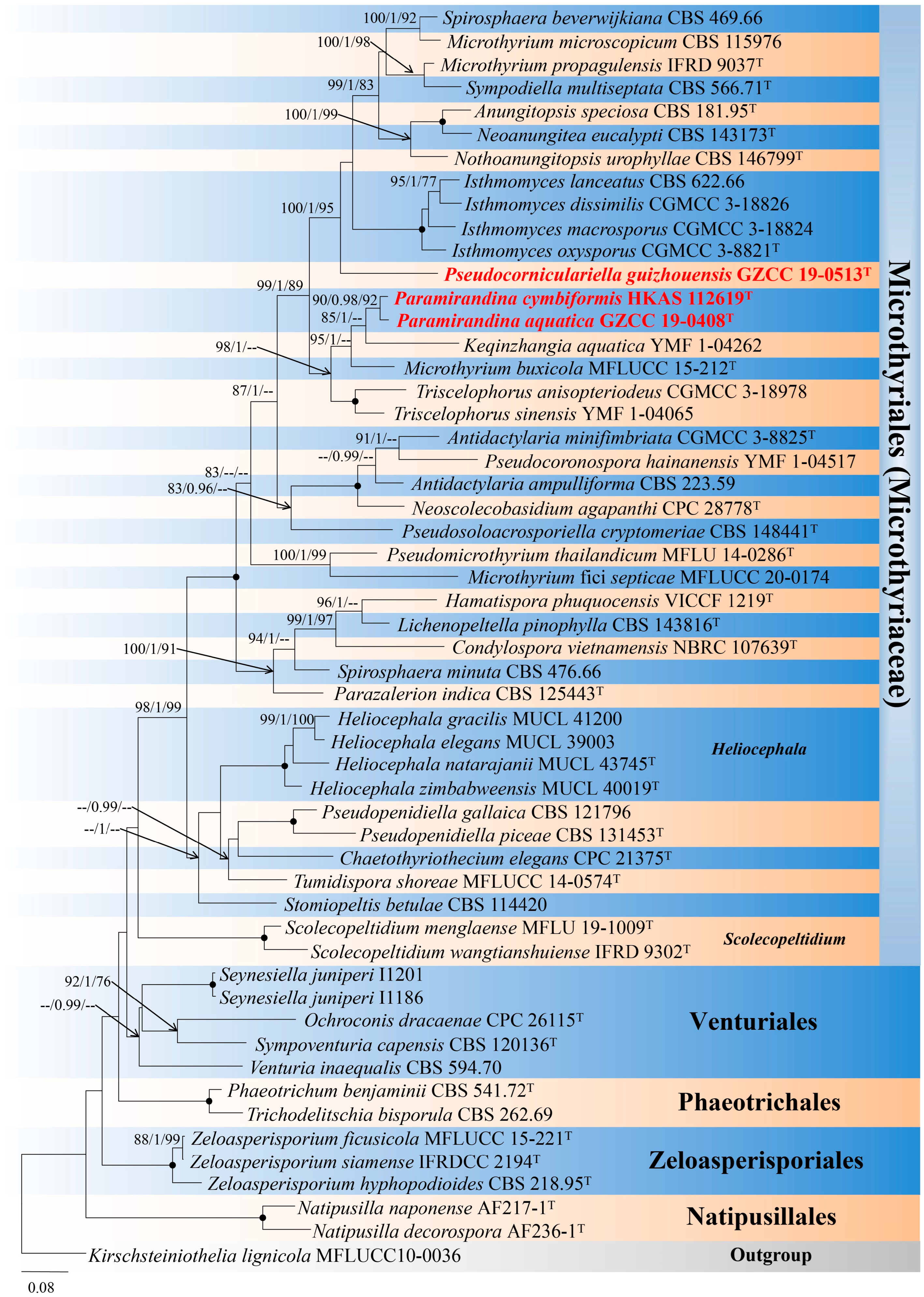
3. Phylogenetic Results

Phylogenetic relationships of three Microthyriales species were assessed in the combined analysis using ITS and LSU gene regions of 54 strains in Microthyriales and related orders Micropeltidales, Natipusillales, Phaeotrichales, Venturiales, and Zeloasperisporiales. The analyzed alignment consisted of combined ITS (1–516 bp) and LSU (517–1321 bp) sequence data, including gaps. Kirschsteiniothelia lignicola (MFLUCC10 0036) served as outgroup taxon. The best scoring RAxML tree is shown in Figure 1. The analyzed ML, MP, and bayesian trees were similar in topology and did not conflict significantly. Paramirandina and Pseudocorniculariella formed two distinct clades and nested within Microthyriaceae. Paramirandina aquatica (GZCC 19-0408) grouped with Pa. cymbiformis (HKAS 112619) with good support (90% MLBS/0.98 PP/92% MPBS), and they formed a sister clade to Keqinzhangia aquatica (YMF1-04262). Comparison of the LSU sequences of Pa. aquatica and Pa. cymbiformis vs. K. aquatica showed 93.29% (57 bp different in 850 bp) and 92.88% (57 bp different in 800 bp) sequence identity, respectively. Based on the molecular data, Pa. aquatica differs from Pa. cymbiformis by 11 bp in LSU (806 bp), 39 bp in tef1-α (1013 bp), and 48 bp in rpb2 (997 bp). Pseudocorniculariella guizhouensis (GZCC 19-0513) was resolved as a monophyletic clade with good statistical support (100% MLBS/1.0 PP/95% MPBS).

4. Taxonomy

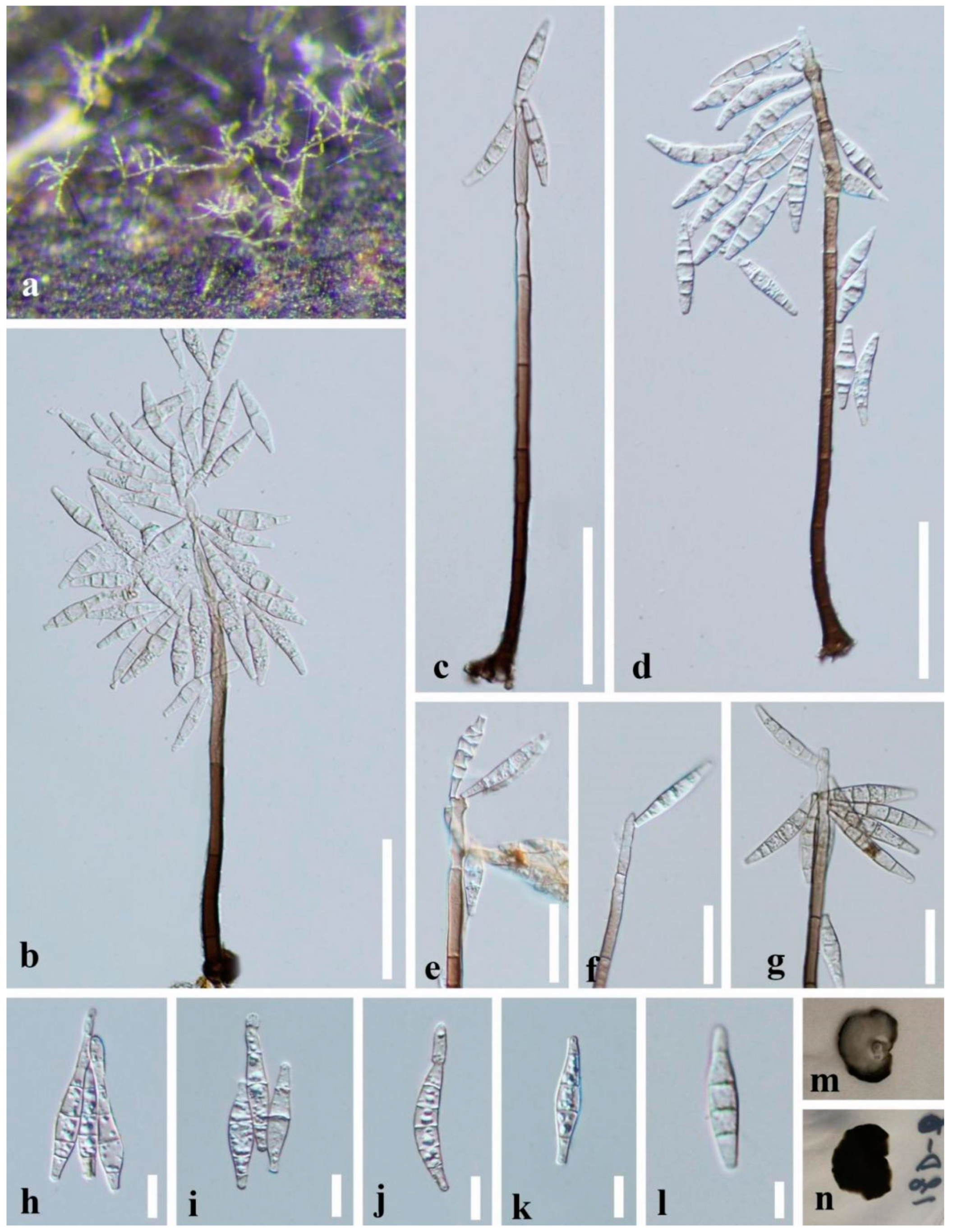
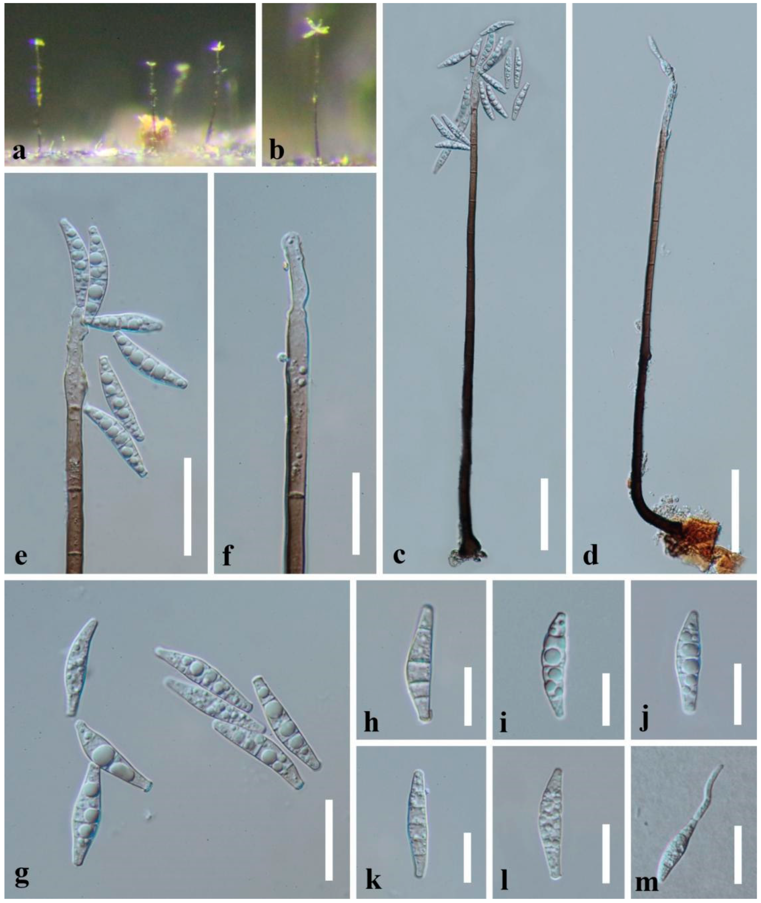
4.1. Paramirandina L.L. Liu & Z.Y. Liu, gen. nov.

Index Fungorum number: IF900034; Facesoffungi number: FoF13245
Etymology: named after its morphology similar to Mirandina.
Saprobic on decaying submerged wood in freshwater habitats. Asexual morph: Colonies on natural substrates effuse, hairy, scattered, yellowish brown to brown, with glistening conidial masses at the apex. Mycelium partly superficial, partly immersed, composed of septate, brown to hyaline, smooth-walled hyphae. Conidiophores macronematous, mononematous, single or in small groups, unbranched, erect, straight or slightly flexuous, cylindrical, smooth-walled, multi-septate, dark brown, becoming pale brown to subhyaline towards the apex, slightly tapering towards the apex. Conidiogenous cells polyblastic, integrated, terminal, cylindrical to lageniform, pale brown to subhyaline, often flexuous at the apex, sometimes elongating percurrently. Conidia holoblastic, solitary or gathered in chains, acropleurogenous, fusiform, cymbiform or narrowly lunate, hyaline, 2–6-septate, smooth-walled, truncate at the base. Sexual morph: undetermined.
Type species: —Paramirandina aquatica L.L. Liu & Z.Y. Liu
Notes: Paramirandina is similar to Heliocephala and Mirandina [41,42]. Heliocephala, typified by H. proliferans, is similar to Paramirandina by cylindrical, brown, erect conidiophores [41]. Paramirandina can be distinguished from Heliocephala in having integrated, polyblastic conidiogenous cells and cymbiform, fusiform or narrowly lunate conidia. Heliocephala has monoblastic, discrete conidiogenous cells and obclavate and rostrate conidia. Mirandina, typified by M. corticola, is characterized by brown, erect conidiophores, cylindrical, polyblastic conidiogenous cells, and hyaline, clavate, filiform or fusiform conidia, usually with short-cylindrical denticles in apical clusters [42]. However, the short-cylindrical denticles are absent in Paramirandina. Phylogeneticanalysis showed that Paramirandina belongs to Microthyriales, while Mirandina belongs to Helotiales [26]. Paramirandina shares the morphology with Pleurotheciella and Pleurothecium in having macronematous brown conidiophores, polyblastic conidiogenous cells and hyaline conidia. However, conidiogenous cells of Pleurotheciella and Pleurothecium are with minute or tooth-like denticles. Pleurotheciella and Pleurothecium are members of Pleurotheciaceae (Pleurotheciales, Sordariomycetes) [43]. In our phylogenetic analysis, Paramirandina is sister to Keqinzhangia aquatica (Figure 1). Paramirandina has cymbiform, fusiform or narrowly lunate conidia, while Keqinzhangia has cylindrical, obclavate, bacilliform, fusiform, sub-oblecythiform or cuneiform conidia [14]. Comparison of the LSU sequences of Pa. aquatica and K. aquatica showed 93.29% (793/850 bp) identity, while Pa. cymbiformis and K. aquatica showed 92.88% (743/800 bp) identity.
Paramirandina aquatica L.L. Liu & Z.Y. Liu, sp. nov., Figure 2.
Index Fungorum number: IF900038; Facesoffungi number: FoF13246
Etymology: referring to the aquatic habitat of the species
Holotype: GZAAS 20-0303
Saprobic on decaying submerged wood in freshwater habitats. Asexual morph: Colonies on natural substrates effuse, hairy, scattered, yellowish brown, with glistening conidial masses at the apex. Mycelium partly superficial, partly immersed, composed of septate, brown to hyaline, smooth-walled hyphae. Conidiophores macronematous, mononematous, solitary or in small groups, unbranched, erect, straight or slightly flexuous, cylindrical, smooth-walled, 6–10-septate, brown, becoming pale brown to subhyaline and tapering towards the apex, 138–200 × 4.5–8 μm ( X   ¯ = 171 × 6 μm, n = 15). Conidiogenous cells polyblastic, integrated, terminal, determinate, sympodial, cylindrical to lageniform, pale brown to subhyaline, often flexuous at the apex, sometimes elongating percurrently. Conidia holoblastic, solitary or gathered in chains, acropleurogenous, cymbiform, fusiform or narrowly lunate, obtuse at the apex, truncate at the base, sometimes slightly curved, hyaline, 2–5-septate, mostly 4-septate, 23–34 × 4–7.5 μm ( X   ¯ = 28 × 6 μm, n = 30), smooth, sometimes bearing a new conidium at the apex. Sexual morph: undetermined.
Culture characteristics: Conidia germinating on WA medium within 24 h and germ tube produced from one or both ends. Colonies on PDA medium slow growing, reaching about 10 mm diam. after two months at 25 °C in dark, circular, with dense, velvety, grayish white to brown mycelium on the surface; in reverse dark brown to black with entire margin.
Material examined: CHINA, Guizhou Province, Dushan District, deep ditch scenic spot, near 25°55′N, 107°37′E, at an altitude of 1205m, on decaying branch submerged in a stream, 5 July 2018, L.L. Liu, 18D-66 (GZAAS 20-0303, holotype), ex-type culture GZCC 19-0408; additional sequences, SSU: OQ025204; tef1-α: OQ032664; rpb2: OQ032662
Notes: Paramirandina aquatica is similar to Heliocephala variabilis [41] in conidiophores; similar to Mirandina inaequalis [44] and Keqinzhangia aquatica [14] in conidial shape; and similar to Pa. cymbiformis (HKAS 112619). However, Pa. aquatica differs from H. variabilis by cymbiform or narrowly lunate, fusiform conidia, and polyblastic conidiogenous cells. Paramirandina aquatica is distinguished from M. inaequalis in lacking the short-cylindrical denticles in the upper region of the conidiogenous cells. Keqinzhangia aquatica was described on culture. The conidial shape and size of Pa. aquatica on natural substrate differs from K. aquatica on culture. Conidia of Pa. aquatica are shorter than that of K. aquatica (23–34 μm vs. 12–76.5 μm). Paramirandina aquatica has mostly cymbiform, 4-septate conidia while K. aquatica has narrowly fusiform, 0–6(–7)-septate, conidia with acute ends. Paramirandina aquatica (GZCC 19-0408) and K. aquatica (YMF1-04262) showed 93.29% (793/850 bp) sequence identity of the LSU gene region. Paramirandina aquatica shares the similar morphology with Pa. cymbiformis but differs by shorter conidiophores (138–200 µm vs. 280–350 µm). Comparisons of the LSU, tef1-α, and rpb2 sequences of Pa. aquatica (GZCC 19-0408) and Pa. cymbiformis (HKAS 112619) showed 11 bp differences in LSU, 39 bp in tef1-α, and 48 bp in rpb2 gene regions, respectively.
Paramirandina cymbiformis J. Yang & Z.Y. Liu, sp. nov., Figure 3.
Index Fungorum number: IF900037; Facesoffungi number: FoF13247
Etymology: referring to the cymbiform conidia.
Holotype: HKAS 112619
Saprobic on decaying submerged wood in freshwater habitats. Asexual morph: Colonies on wood effuse, hairy, scattered, brown, with glistening conidial masses at the apex. Mycelium partly superficial, partly immersed, composed of septate, smooth-walled, brown to hyaline hyphae. Conidiophores macronematous, mononematous, erect, straight or slightly flexuous, solitary, cylindrical, smooth-walled, septate, unbranched, dark brown, becoming pale brown to subhyaline towards the apex, 280–350 × 5–9 µm ( X ¯ = 313 × 7 µm, n = 20). Conidiogenous cells polyblastic, integrated, terminal, determinate, sympodial, cylindrical, pale brown to subhyaline, often flexuous at the apex, elongating percurrently. Conidia acropleurogenous, aggregated in slimy masses, solitary, cymbiform or narrowly lunate, 3–6-septate, smooth-walled, hyaline, 24–30 × 5–6.5 µm ( X ¯ = 26.5 × 5.5 µm, n = 30), guttulate, thin-walled, sometimes slightly constricted at the septa. Sexual morph: Undetermined.
Material examined: CHINA, Guizhou Province, Chishui City, Sidonggou Waterfall, 25°27.38′ N, 107°39.85′ E, on decaying twig submerged in a freshwater stream, 11 July 2019, J. Yang, CS 53-1 (HKAS 112619, holotype; HKAS 125927, isotype); additional sequence, SSU: OQ025205; tef1-α: OQ032665; rpb2: OQ032663
Notes: Paramirandina cymbiformis share the similar morphology with Pa. aquatica except for the dimension of conidiophores. However, they are distinct species based on the molecular data.
Attempts to preserve the living culture were unsuccessful since few conidia germinated, with no growth after reaching 1–2 mm diam..

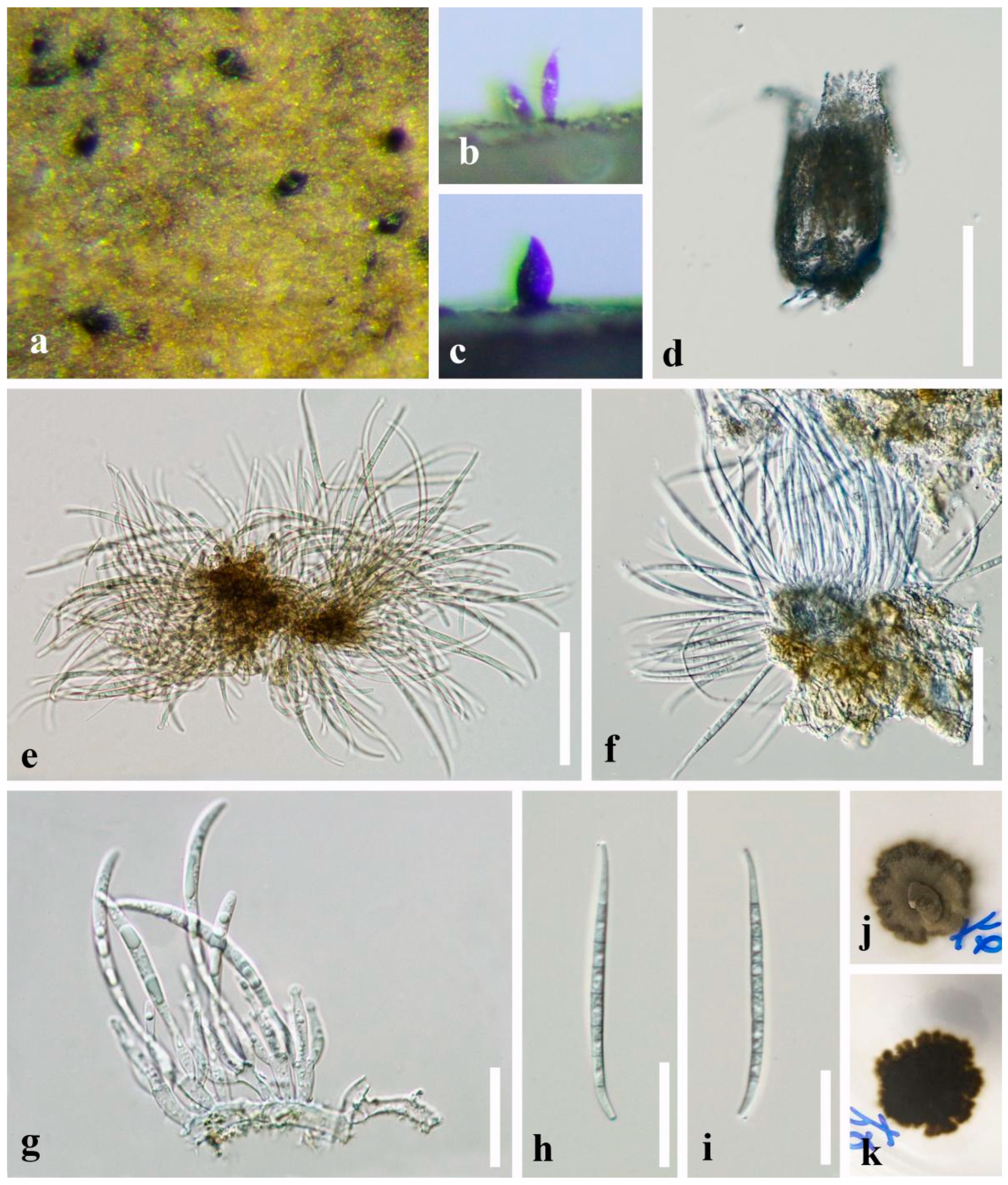
4.2. Pseudocorniculariella L.L. Liu & Z.Y. Liu, gen. nov.

Index Fungorum number: IF900035; Facesoffungi number: FoF13248
Etymology: referring to the morphology similar genus Corniculariella.
Saprobic on decaying submerged twigs in freshwater habitats. Asexual morph: Conidiomata effuse, sporodochial, synnematous or absent, solitary to gregarious, dark brown to black, stromatic, obpyriform, subcylindrical to subconical, slightly swollen at the base or level of the locule, narrower towards the apex, scattered over the substrate surface, minutely scabrous, reticular. Conidiomatal wall composed of closely interwoven septate hyphae, compacted towards exterior, dark brown to black cells of textura angularis, becoming thin-walled and hyaline toward the inner region. Conidiophores hyaline, cylindrical, branched, developed from the inner layer of the conidiomata, reduced to conidiogenous cells. Conidiogenous cells hyaline, enteroblastic, polyphialidic, subcylindrical or cylindrical to ampulliform, indeterminate, forming conidia at their tips, discrete or integrated, smooth, moderately thick-walled. Conidia solitary, hyaline, smooth, guttulate to granular, septate, slightly constricted at septa, thick-walled, filiform, acerose, tapering towards both ends, slightly curved, base truncate. Sexual morph: Undetermined.
Type species: —Pseudocorniculariella guizhouensis L.L. Liu & Z.Y. Liu, sp. nov.
Notes: Phylogenetic study based on ITS and LSU sequence data showed that Pseudocorniculariella formed a separate branch in Microthyriaceae (Microthyriales) close to Isthmomyces (Figure 1). Pseudocorniculariella is distinct from Isthmomyces in the formation of conidiogenous cells and conidia. Pseudocorniculariella has enteroblastic and polyphialidic conidiogenous cells and hyaline, filiform or falcate conidia. Isthmomyces has polyblastic conidiogenous cells, and two cellular isthmic-segment obclavate, clavate, pyriform conidia [13]. Pseudocorniculariella shares similar characteristics with Corniculariella (Dermateaceae, Medeolariales, Leotiomycetes) in having subconical conidiomata and hyaline, filiform conidia. However, it can be distinguished by stromatic conidiomata and polyphialidic conidiogenous cells. Phylogenetic analysis showed that Pseudocorniculariella belongs to Microthyriales, Dothideomycetes, while Corniculariella belongs to Medeolariales, Leotiomycetes [45]. Based on the morphology and phylogeny, Pseudocorniculariella is introduced as a new genus in Microthyriaceae. Additional collections and further molecular evidence are needed to confirm its taxonomy.
Pseudocorniculariella guizhouensis L.L. Liu & Z.Y. Liu, sp. nov., Figure 4.
Index Fungorum number: IF900036; Facesoffungi number: FoF13249
Etymology: referring to the collecting site in Guizhou Province, China.
Holotype: GZAAS 20-0408
Saprobic on decaying submerged twigs in freshwater habitats. Asexual morph: Conidiomata effuse, sporodochial, synnematous or absent, solitary to gregarious, dark brown to black, stromatic, obpyriform, subcylindrical to subconical, slightly swollen at the base or level of the locule, narrower towards the apex, scattered over the substrate surface, minutely scabrous, reticular, 65–90 µm diam., 135–175 µm high, uninoculated, reticular thin-walled, papillate. Conidiomatal wall composed of closely interwoven septate hyphae, compacted towards the exterior, dark brown cells of textura angularis, becoming thin-walled, up to 2 µm wide, and hyaline toward the inner region. Conidiophores formed from the inner wall of the conidiomata, reduced to conidiogenous cells. Conidiogenous cells polyphialidic, hyaline, definite, smooth, subcylindrical to ampulliform, 9.5–12 × 3.5–4.5 µm ( X ¯ = 10.8 × 4.2 μm, n = 20). Conidia solitary, hyaline, 6–8-septate, mostly 7-septate, slightly constricted at septa, smooth, guttulate to granular, filiform, acerose, base truncates, 55.5–76.5 × 2.5–4.0 μm ( X ¯ = 68.5 × 3.2 μm, n = 20). Sexual morph: Undetermined.
Culture characteristics: Conidia germinating on PDA medium within 24 h and germ tubes produced from both ends. Colonies growing on PDA medium slow growing, reaching 8–10 mm in three weeks at 25 °C in natural light, circular, with dense, olive-green mycelium in the middle, darker of the inner ring, with sparser, brown mycelium of the outer ring on the surface, in reverse dark brown to black with irregular margin.
Material examined: CHINA, Guizhou Province, Aha Lake, 26°32′ N, 106°40′ E, at an altitude of 1085 m, on decaying submerged twigs in the lake, 16 April 2018, L.L. Liu, 18A-14 (GZAAS 20-0408, holotype), ex-type culture GZCC 19-0513; additional sequence, tef1-α: OQ032666
Notes: Pseudocorniculariella guizhouensis resembles Corniculariella rhamni in possessing subconical conidiomata and hyaline, filiform, conidia [45]. However, Ps. guizhouensis possesses stromatic conidiomata and polyphialidic conidiogenous cells, while C. rhamni has monophialidic conidiomata and phialidic conidiogenous cells. Phylogenetic analyses based on ITS and LSU showed that Ps. guizhouensis (GZCC 19-0513) belongs to Microthyriales, Dothideomycetes, while Corniculariella is a member of Medeolariales, Leotiomycetes.

5. Discussion

Freshwater fungi are a heterogeneous group. With the increasing abundance of molecular data, the numbers have rapidly increased [46,47,48,49,50,51,52,53]. According to statistics, there are 3,870 freshwater fungal species [54]. They mainly consist of Ascomycota (Sordariomycetes, Dothideomycetes, Eurotiomycetes, Leotiomycetes) and other phyla, including Chytridiomycota, Basidiomycota and Rozellomycota species. The Ascomycota accounted for three-quarters of the total [54].
In recent years, the molecular phylogeny of freshwater fungi has been updated several times [48,49,54]. Nevertheless, Microthyriaceae (Microthyriales) was omitted, although a freshwater genus Hamatispora has been reported before then [10]. Recently, four new freshwater genera, Antidactylaria, Isthmomyces, Keqinzhangia, and Pseudocoronospora, were reported [10,11]. In this study, the combined ITS and LSU tree (Figure 1) showed that three new isolates formed two clades in Microthyriaceae. Based on the morphology and molecular evidence, we establish two new asexual genera and three new species, named Paramirandina and Pseudocorniculariella, with Pa. aquatica, Pa. cymbiformis and Ps. guizhouensis. Eight freshwater genera are known in Microthyriaceae, including the two new genera in this study. It is worth noting that both are asexual genera. In the previous studies about freshwater fungi, few aquatic asexual genera were included [55]. The new genus Paramirandina is phylogenetically close to the asexual genus Keqinzhangia (Figure 1). Paramirandina is well distinguishable from Keqinzhangia by relatively long conidiophores (more than 150 µm long vs. prostrate, not differentiated), conidiogenesis (holoblastic vs. holothallic) and the conidial shape (cymbiform or narrowly lunate vs. cylindrical, cylindrical-obclavate, obclavate, bacilliform) [12]. The sequence identity of the LSU gene region between two Paramirandina species and K. aquatica is relatively low (93.29% and 92.88%). Therefore, we introduce Paramirandina as a new genus.
Pseudocorniculariella is phylogenetically close to the asexual genus Isthmomyces (Figure 1). It is distinguished from Isthmomyces by the formation of conidiogenous cells and the morphology of conidia, and was identified as a new genus.
Microthyriaceae has ever been a poorly studied group. However, its taxonomic studies have greatly advanced with molecular data resulting in a rapid increase of genera numbers. Formerly, only seven genera were accepted in the family by Wu et al. [7], nine were accepted by Wijayawardene et al. [8,9], 11 were accepted by Hongsanan et al. [12], and 18 were accepted at present (this study). However, most genera contained fewer species, such as eight monotypic genera, Chaetothyriothecium, Hamatispora, Keqinzhangia, Paramicrothyrium, Pseudomicrothyrium, Pseudocoronospora, Tumidispora, and the new genus Pseudocorniculariella, and four genera with two or three species. Thus, more collections and further molecular evidence are needed to confirm the taxonomy of these genera.
Furthermore, the taxonomy of earlier proposed genera needs to be confirmed by molecular data. Heliocephala is the first hyphomycetous genus described in Microthyriales. There are eight species in the genus, five of which have available molecular DNA data without the type species H. proliferans [56,57]. Gonzalez et al. accepted Heliocephala in Microthyriaceae based on H. variabilis [57]. However, Calabon et al. [54] referred Heliocephala to Microthyriales incertae sedis. Thus, the taxonomy of Heliocephala needs to be reappraised with molecular DNA data of the type species and more collections.

Author Contributions

Conceptualization: L.L. and Z.L.; Methodology: L.L. and J.Y.; Formal analysis and investigation: L.L. and J.Y.; Resources: Z.L.; Writing—original draft preparation, L.L.; Writing—review and editing, L.L., J.Y., S.Z., X.G., Q.W. and M.Z.; Supervision, Z.L. and J.G.; Funding acquisition, L.L. All authors have read and agreed to the published version of the manuscript.

Funding

This research was funded by the National Natural Science Foundation of China (project IDs: 32260007) and the Foundation of Key Laboratory of Microbial Resources Collection and Preservation, Ministry of Agriculture and Rural Affairs (Project no. KLMRCP2021-08).

Institutional Review Board Statement

Not applicable.

Informed Consent Statement

Not applicable.

Data Availability Statement

The datasets generated for this study can be found in the NCBI database.

Acknowledgments

Lingling Liu thanks Jiankui Liu, University of Electronic Science and Technology of China, for his valuable correction and suggestion.

Conflicts of Interest

The authors declare no conflict of interest.

References

  1. Saccardo, P.A. Sylloge Pyrenomycetum. Sylloge Fungorum 1883, 2, 1–813. [Google Scholar]
  2. Hongsanan, S.; Hyde, K.D. Phylogenetic placement of Micropeltidaceae. Mycosphere 2017, 8, 1930–1942. [Google Scholar] [CrossRef]
  3. Hongsanan, S.; Hyde, K.D.; Phookamsak, R.; Wanasinghe, D.N.; McKenzie, E.H.C.; Sarma, V.V.; Boonmee, S.; Lücking, R.; Bhat, D.J.; Liu, N.G. Refined families of Dothideomycetes: Dothideomycetidae and Pleosporomycetidae. Mycosphere 2020, 11, 1553–2107. [Google Scholar] [CrossRef]
  4. Wu, H.X.; Hyde, K.D.; Chen, H. Studies on Microthyriaceae: Placement of Actinomyxa, Asteritea, Cirsosina, Polystomellina and Stegothyrium. Cryptogam Mycol. 2011, 32, 3–12. [Google Scholar] [CrossRef]
  5. Wu, H.X.; Li, Y.M.; Chen, H.; Hyde, K.D. Studies on Microthyriaceae: Some excluded genera. Mycotaxon 2010, 113, 147–156. [Google Scholar] [CrossRef]
  6. Wu, H.X.; Tian, Q.; Li, W.J.; Hyde, K.D. A reappraisal of Microthyriaceae. Phytotaxa 2014, 176, 201–212. [Google Scholar] [CrossRef] [Green Version]
  7. Wu, H.X.; Schoch, C.L.; Boonmee, S.; Bahkali, A.H.; Chomnunti, P.; Hyde, K.D. A reappraisal of Microthyriaceae. Fungal Divers. 2011, 51, 189–248. [Google Scholar] [CrossRef] [PubMed] [Green Version]
  8. Wijayawardene, N.N.; Hyde, K.D.; McKenzie, E.H.C.; Wang, Y. Notes for genera update Ascomycota: 6822–6917. Mycosphere 2018, 9, 1222–1234. [Google Scholar] [CrossRef]
  9. Wijayawardene, N.N.; Hyde, K.D.; Lumbsch, H.T.; Liu, J.K.; Maharachchikumbura, S.S.N.; Ekanayaka, A.H.; Tian, Q.; Phookamsak, R. Outline of Ascomycota: 2017. Fungal Divers. 2018, 88, 167–263. [Google Scholar] [CrossRef]
  10. Yen, L.T.H.; Yamaguchi, K.; Tsurumi, Y.; Hop, D.V.; Ando, K. Hamatispora, a new genus of aquatic fungi in Microthyriales isolated from fallen leaves in Vietnam. Mycoscience 2018, 59, 467–472. [Google Scholar] [CrossRef]
  11. Crous, P.W.; Wingfield, M.J.; Burgess, T.I.; Carnegie, A.J.; Hardy, G.E.S.; Smith, D.; Summerell, B.A.; Cano-Lira, J.F.; Guarro, J.; Hobraken, J.; et al. Fungal Planet description sheets: 625–715. Persoonia 2017, 39, 270–467. [Google Scholar] [CrossRef] [PubMed]
  12. Hongsanan, S.; Hyde, K.D.; Phookamsak, R.; Wanasinghe, D.N.; McKenzie, E.H.C.; Sarma, V.V.; Lucking, R.; Boonmee, S.; Bhat, J.D.; Liu, N.G. Refined families of Dothideomycetes: Orders and families incertae sedis in Dothideomycetes. Fungal Divers. 2020, 105, 17–318. [Google Scholar] [CrossRef]
  13. Qiao, M.; Zheng, H.; Guo, J.S.; Castañeda-Ruiz, R.F.; Xu, J.P.; Peng, J.; Zhang, K.Q.; Yu, Z.F. Two new asexual genera and six new asexual species in the family Microthyriaceae (Dothideomycetes, Ascomycota) from China. MycoKeys 2021, 85, 1–30. [Google Scholar] [CrossRef] [PubMed]
  14. Zheng, H.; Qiao, M.; Guo, J.; Castañeda-Ruiz, R.F.; Peng, J.; Yu, Z.F. Keqinzhangia aquatica gen. et sp. nov. and Pseudocoronospora hainanense gen. et sp. nov. isolated from freshwater in southern China. Antonie van Leeuwenhoek 2022, 115, 203–213. [Google Scholar] [CrossRef] [PubMed]
  15. Wijayawardene, N.N.; Hyde, K.D.; Al-Ani, L.K.T.; Tedersoo, L.; Haelewaters, D.; Rajeshkumar, K.C.; Zhao, R.L.; Aptroot, A.; Leontyev, D.V.; Saxena, R.K.; et al. Outline of fungi and fungus-like taxa. Mycosphere 2020, 11, 1060–1456. [Google Scholar] [CrossRef]
  16. Hyde, K.D.; Jones, E.B.G.; Liu, J.K.; Ariyawansa, H.; Boehm, E.; Boonmee, S.; Braun, U.; Chomnunti, P.; Crous, P.W.; Dai, D.Q. Families of Dothideomycetes. Fungal Divers. 2013, 63, 1–313. [Google Scholar] [CrossRef]
  17. Hyde, K.D.; Norphanphoun, C.; Abreu, V.P.; Bazzicalupo, A.; Kandawatte, T.C.; Clericuzio, M.; Dayarathne, M.; Dissanayake, A.J.; Ekanayaka, A.H.; He, M.Q.; et al. Fungal diversity notes 603–708: Taxonomic and phylogenetic notes on genera and species. Fungal Divers. 2017, 87, 1–235. [Google Scholar] [CrossRef]
  18. Yang, J.; Maharachchikumbura, S.S.N.; Liu, J.K.; Hyde, K.D.; Jones, E.B.G.; Al-Sadi, A.M.; Liu, Z.Y. Pseudostanjehughesia aquitropica gen. et sp. nov. and Sporidesmium sensu lato species from freshwater habitats. Mycol. Prog. 2018, 17, 591–616. [Google Scholar] [CrossRef]
  19. Liu, L.L.; Liu, N.G.; Yang, J.; Chen, Y.Y.; Gui, X.X.; Liu, Z.Y. Sporidesmium guizhouense sp. nov. (Sordariomycetes incertae sedis), a new species from a freshwater habitat in Guizhou Province, China. Phytotaxa 2019, 422, 144–156. [Google Scholar] [CrossRef]
  20. Hyde, K.D.; Dong, Y.; Phookamsak, R.; Jeewon, R.; Bhat, D.J.; Jones, E.B.G.; Liu, N.-G.; Abeywickrama, P.D.; Mapook, A.; Wei, D.; et al. Fungal diversity notes 1151–1276: Taxonomic and phylogenetic contributions on genera and species of fungal taxa. Fungal Divers. 2020, 100, 5–277. [Google Scholar] [CrossRef] [Green Version]
  21. Liu, L.L.; Liu, N.G.; Yang, J.; Chen, Y.Y.; Liu, Z.Y. Cucurbitinus gen. nov. (Halosphaeriaceae, Microascales), a new genus to accommodate Cucurbitinus constrictus comb. nov. and Cucurbitinus ibericus comb. nov. Phytotaxa 2020, 455, 119–136. [Google Scholar] [CrossRef]
  22. Yang, J.; Liu, L.L.; Jones, E.B.G.; Li, W.L.; .Hyde, K.D.; Liu, Z.Y. Morphological Variety in Distoseptispora and Introduction of Six Novel Species. J. Fungi. 2021, 7, 945. [Google Scholar] [CrossRef] [PubMed]
  23. Luo, Z.L.; Hyde, K.; Liu, J.K.; Bhat, D.J.; Bao, D.F.; Li, W.L.; Su, H.Y. Lignicolous freshwater fungi from China II: Novel Distoseptispora (Distoseptisporaceae) species from northwestern Yunnan Province and a suggested unified method for studying lignicolous freshwater fungi. Mycosphere 2018, 9, 444–461. [Google Scholar] [CrossRef]
  24. Indunil Chinthani, S.; Achala, R.R.; Diana, S.M.; Mark, S.C.; Eleni, G.; Hyang Burm, L.; Vedprakash, G.H.; Dhandevi, P.; Lakmali, S.D.; Subodini, N.W.; et al. Morphological approaches in studying fungi: Collection, examination, isolation, sporulation and preservation. Mycosphere 2020, 11, 2678–2754. [Google Scholar] [CrossRef]
  25. Jayasiri, S.C.; Hyde, K.D.; Ariyawansa, H.A.; Bhat, D.J.; Buyck, B.; Cai, L.; Dai, Y.; Abd-Elsalam, K.A.; Ertz, D.; Hidayat, I.; et al. The faces of fungi database: Fungal names linked with morphology, phylogeny and human impacts. Fungal Divers. 2015, 74, 3–18. [Google Scholar] [CrossRef]
  26. Index Fungorum. Available online: http://www.indexfungorum.org/names/names.asp (accessed on 12 January 2023).
  27. White, T.J.; Bruns, T.; Lee, S.; Taylor, J. Amplification and direct sequencing of fungal ribosomal RNA genes for phylogenetics. In PCR Protocols a Guide to Methods and Applications; Innis, M.A., Gelfand, D.H., Sninsky, J.J., White, T.J., Eds.; Academic Press: New York, NY, USA, 1990; pp. 315–322. [Google Scholar] [CrossRef]
  28. Vilgalys, R.; Hester, M. Rapid genetic identification and mapping of enzymatically amplified ribosomal DNA from several Cryptococcus species. J. Bacteriol. 1990, 172, 4238–4246. [Google Scholar] [CrossRef] [Green Version]
  29. Rehner, S.A.; Samuels, G.J. Taxonomy and phylogeny of Gliocladium analysed from nuclear large subunit ribosomal DNA sequences. Mycol. Res. 1994, 98, 625–634. [Google Scholar] [CrossRef]
  30. Rehner, S.A.; Buckley, E. A Beauveria phylogeny inferred from nuclear ITS and EF1-alpha sequences: Evidence for cryptic diversification and links to Cordyceps teleomorphs. Mycologia 2005, 97, 84–98. [Google Scholar] [CrossRef]
  31. Sung, G.H.; Sung, J.M.; Hywel-Jones, N.L.; Spatafora, J.W. A multi-gene phylogeny of Clavicipitaceae (Ascomycota, Fungi): Identification of localized incongruence using a combinational bootstrap approach. Mol. Phyl. Evol. 2007, 44, 1204–1223. [Google Scholar] [CrossRef]
  32. Liu, Y.J.; Whelen, S.; Hall, B.D. Phylogenetic relationships among ascomycetes: Evidence from an RNA Polymerase II subunit. Mol. Biol. Evol. 1999, 16, 1799–1808. [Google Scholar] [CrossRef] [Green Version]
  33. Katoh, K.; Standley, D.M. MAFFT multiple sequence alignment software version 7: Improvements in performance and usability. Mol. Biol. Evol. 2013, 30, 772–780. [Google Scholar] [CrossRef] [PubMed] [Green Version]
  34. Miller, M.A.; Pfeiffer, W.; Schwartz, T. Creating the CIPRES Science Gateway for inference of large phylogenetic trees. In Proceedings of the 2010 Gateway Computing Environments Workshop (GCE), New Orleans, LA, USA, 14 November 2010; pp. 1–8. [Google Scholar] [CrossRef] [Green Version]
  35. Stamatakis, A. RAxML Version 8: A tool for phylogenetic analysis and post-analysis of large phylogenies. Bioinformatics 2014, 30, 1312–1313. [Google Scholar] [CrossRef] [PubMed] [Green Version]
  36. Nylander, J. MrModeltest, version 2.3—Program for Selecting DNA Substitution Models Using PAUP*; Evolutionary Biology Centre: Uppsala, Sweden, 2008.
  37. Huelsenbeck, J.P.; Ronquist, F. MrBayes: Bayesian inference of phylogenetic trees. Bioinformatics 2001, 17, 754–755. [Google Scholar] [CrossRef] [PubMed] [Green Version]
  38. Larget, B.; Simon, D.L. Markov Chain Monte Carlo algorithms for the Bayesian analysis of phylogenetic trees. Mol. Biol. Evol. 1999, 16, 750–759. [Google Scholar] [CrossRef] [Green Version]
  39. Swofford, D.L. PAUP*, Phylogenetic Analysis Using Parsimony (*and Other Methods), Version 4; Sinauer Associates: Sunderland, UK, 2003.
  40. Hillis, D.M.; Bull, J.J. An empirical test of bootstrapping as a method for assessing confidence in phylogenetic analysis. Syst. Biol. 1993, 42, 182–192. [Google Scholar] [CrossRef]
  41. Rao, V.G.; Reddy, K.A.; de Hoog, G.S. Heliocephala, a new genus of dematiaceous Hyphomycetes. Persoonia 1984, 12, 239–242. [Google Scholar]
  42. Goos, R.D.; DeHoog, G.S. Taxonomy of the Dactylaria Complex, IV-VI. Mycologia 1986, 78, 978. [Google Scholar] [CrossRef]
  43. Réblová, M.; Seifert, K.A.; Fournier, J.; Stepánek, V. Phylogenetic classification of Pleurothecium and Pleurotheciella gen. nov. and its dactylaria-like anamorph (Sordariomycetes) based on nuclear ribosomal and protein-coding genes. Mycologia 2012, 104, 1299–1314. [Google Scholar] [CrossRef]
  44. Ma, Y.R.; Xia, J.W.; Zhang, X.G.; Castaeda-Ruiz, R.F. New species of Phaeomonilia and Mirandina from dead branches in China. Mycotaxon 2015, 130, 775–781. [Google Scholar] [CrossRef]
  45. Li, W.J.; McKenzie, E.H.C.; Liu, J.K.; Bhat, D.J.; Dai, D.Q.; Camporesi, E.; Tian, Q.; Maharachchikumbura, S.S.N.; Luo, Z.L.; Shang, Q.J.; et al. Taxonomy and phylogeny of hyaline-spored coelomycetes. Fungal Divers. 2020, 100, 279–801. [Google Scholar] [CrossRef]
  46. Shearer, C.A. The freshwater ascomycetes. Nova Hedwig 1993, 56, 1–33. [Google Scholar]
  47. Hyde, K.D.; Fryar, S.; Tian, Q.; Bahkali, A.H.; Xu, J.C. Lignicolous freshwater fungi along a north-south latitudinal gradient in the Asian/Australian region; can we predict the impact of global warming on biodiversity and function? Fungal Ecol. 2016, 19, 190–200. [Google Scholar] [CrossRef]
  48. Luo, Z.L.; Hyde, K.D.; Liu, J.K.; Maharachchikumbura, S.S.N.; Jeewon, R.; Bao, D.F.; Bhat, D.J.; Lin, C.G.; Li, W.L.; Yang, J.; et al. Freshwater Sordariomycetes. Fungal Divers. 2019, 99, 451–660. [Google Scholar] [CrossRef] [Green Version]
  49. Dong, W.; Wang, B.; Hyde, K.D.; McKenzie, E.H.C.; Raja, H.A.; Tanaka, K.; Abdel-Wahab, M.A.; Abdel-Aziz, F.A.; Doilom, M.; Phookamsak, R.; et al. Freshwater Dothideomycetes. Fungal Divers. 2020, 105, 319–575. [Google Scholar] [CrossRef]
  50. Yang, J.; Liu, J.K.; Hyde, K.D.; Jones, E.B.G.; Liu, Z.Y. New species in Dictyosporium, new combinations in Dictyocheirospora and an updated backbone tree for Dictyosporiaceae. MycoKeys 2018, 36, 83–105. [Google Scholar] [CrossRef]
  51. Yang, J.; Liu, N.G.; Liu, J.K.; Hyde, K.D.; Jones, E.B.G.; Liu, Z.Y. Phylogenetic placement of Cryptophiale, Cryptophialoidea, Nawawia, Neonawawia gen. nov. and Phialosporostilbe. Mycosphere 2018, 9, 1132–1150. [Google Scholar] [CrossRef]
  52. Yu, X.D.; Dong, W.; Bhat, D.J.; Boonmee, S.; Zhang, D.I.; Zhang, H. Cylindroconidiis aquaticus gen. et sp. nov., a new lineage of aquatic hyphomycetes in Sclerococcaceae (Eurotiomycetes). Phytotaxa 2018, 372, 79–87. [Google Scholar] [CrossRef]
  53. Wang, G.N.; Yu, X.D.; Dong, W.; Bhat, D.J.; Boonmee, S.; Zhang, D.; Zhang, H. Freshwater hyphomycetes in Eurotiomycetes: A new species of Minimelanolocus and a new collection of Thysanorea papuana (Herpotrichiellaceae). Mycol Prog. 2019, 18, 511–522. [Google Scholar] [CrossRef]
  54. Calabon, M.S.; Hyde, K.D.; Jones, E.B.G.; Luo, Z.L.; Dong, W.; Hurdeal, V.G.; Gentekaki, E.; Rossi, W.; Leonardi, M.; Thiyagaraja, V.; et al. Freshwater fungal numbers. Fungal Divers. 2022, 003, 3–235. [Google Scholar] [CrossRef]
  55. Wijayawardene, N.N.; Crous, P.W.; Kirk, P.M.; Hawksworth, D.L.; Boonmee, S.; Braun, U.; Dai, D.Q.; D’souza, M.; Diederich, P.; Dissanayake, A.; et al. Naming and outline of Dothideomycetes-2014 including proposals for the protection or suppression of generic names. Fungal Divers. 2014, 69, 1–55. [Google Scholar] [CrossRef]
  56. Heredia-Abarca, G.; Castañeda-Ruiz, R.F.; Arias-Mota, R.M.; Becerra-Hernandez, C.I.; Gómez, S.; Bogale, M.; Untereiner, W.A. A new species of Heliocephala from México with an assessment of the systematic positions of the anamorph genera Heliocephala and Holubovaniella. Mycologia 2011, 103, 631–640. [Google Scholar] [CrossRef] [PubMed]
  57. Gonzalez, I.I.; Garcia, D.; Guarro, J.; Gene, J. Heliocephala variabilis and Pseudopenidiella vietnamensis: Two new Hyphomycetous species in the Microthyriaceae (Dothideomycetes) from Vietnam. Microorganisms 2020, 8, 478. [Google Scholar] [CrossRef] [PubMed] [Green Version]
Figure 1. Maximum likelihood majority rule consensus tree for Microthyriales and related orders using ITS and LSU sequence data. Bootstrap support values for maximum likelihood (ML) and maximum parsimony (MP) greater than 75% and Bayesian posterior probabilities greater than 0.95 are indicated above branches as ML BS/PP/MP BS. The scale bar represents the expected number of changes per site. The tree is rooted with Kirschsteiniothelia lignicola (MFLUCC 10-0036). Ex-type strains are indicated with T. The new taxa are in red bold. Branches with 100% ML BS, 1.0PP and 100% MP BS were dotted with black dots. Orders are indicated as colored blocks.
Jof 09 00178 g001
Figure 2. Paramirandina aquatica (GZAAS 20-0303, holotype). (a) Colony on wood. (bd) Conidiophores and conidia. (eg) Conidiogenous cells and conidia. (hl) Conidia. (m,n) Culture, m from above, n from below. Scale bars: (bd) = 50 μm, (eg) = 25 μm, (hl) = 10 μm.
Jof 09 00178 g002
Figure 3. Paramirandina cymbiformis (HKAS 112619, holotype) (a,b) Colony on wood. (c,d) Conidiophores with conidia. (e) Conidiogenous cell with conidia. (f) Conidiogenous cell. (gl) Conidia. (m) Germinated conidium. Scale bars: (c,d) = 50 µm, (e) = 30 µm, (f,g,m) = 20 µm, (hl) = 15 µm.
Jof 09 00178 g003
Figure 4. Pseudocorniculariella guizhouensis (GZAAS20-0408, holotype) (ac) Conidiomata on wood. (d) Pycnidium. (eg) Conidiogenous cells and conidia. (h,i) Conidia. (j,k) Culture, j from above, k from below. Scale bars: (d) =100 μm, (e,f) =50 μm, (gi) =25 μm.
Jof 09 00178 g004
Table 1. Taxa used in the phylogenetic analyses and their GenBank accession numbers. T denotes ex-type strains. Newly generated sequences are in bold.
TaxonVoucher/Strain NumberGenBank Accession Number
LSUITS
Antidactylaria ampulliformaCBS 223.59MH869386MH857845
Antidactylaria minifimbriataCGMCC 3-18825TMK577808MK569506
Anungitopsis speciosaCBS 181.95TEU035401EU035401
Chaetothyriothecium elegansCPC 21375TKF268420-
Condylospora vietnamensisNBRC 107639TLC146725LC146723
Hamatispora phuquocensisVICCF 1219TLC064073LC064074
Heliocephala elegansMUCL 39003HQ333478HQ333478
Heliocephala gracilisMUCL 41200HQ333479HQ333479
Heliocephala natarajaniiMUCL 43745THQ333480HQ333480
Heliocephala zimbabweensisMUCL 40019THQ333481HQ333481
Isthmomyces dissimilisCGMCC 3 18826MK577811MF740794
Isthmomyces lanceatusCBS 622.66MH870563MH858897
Isthmomyces macrosporusCGMCC 3-18824MK577812MF740796
Isthmomyces oxysporusCGMCC 3-18821TMK577810MF740793
Keqinzhangia aquaticaYMF 1-04262MK577809MK569507
Kirschsteiniothelia lignicolaMFLUCC 10-0036HQ441568HQ441567
Lichenopeltella pinophyllaCBS 143816TMG844152-
Microthyrium buxicolaMFLUCC 15-0212TKT306551-
Microthyrium fici-septicaeMFLUCC 20-0174TMW063252-
Microthyrium microscopicumCBS 115976GU301846-
Microthyrium propagulensisIFRD 9037TKU948989-
Natipusilla decorosporaAF236-1THM196369-
Natipusilla naponenseAF217-1THM196371-
Neoanungitea eucalyptiCBS 143173TMG386031MG386031
Neoscolecobasidium agapanthiCPC 28778TNG_059748NR_152546
Nothoanungitopsis urophyllaeCBS 146799TMW883825MW883433
Ochroconis dracaenaeCPC 26115TKX228334KX228283
Paramirandina aquaticaGZCC 19-0408TOQ025201OQ025199
Paramirandina cymbiformisHKAS 112619TOQ025202-
Parazalerion indicaCBS 125443TMH874977MH863483
Phaeotrichum benjaminiiCBS 541.72TMH872266MH860561
Pseudocorniculariella guizhouensisGZCC 19-0513TOQ025203OQ025200
Pseudocoronospora hainanensisYMF 1-04517MK577807MK569505
Pseudomicrothyrium thailandicumMFLU 14-0286TMT741680-
Pseudopenidiella gallaicaCBS 121796LT984843LT984842
Pseudopenidiella piceaeCBS 131453TJX069852JX069868
Pseudosoloacrosporiella cryptomeriaeCBS 148441TNG_081320NR_175206
Scolecopeltidium menglaenseMFLU 19-1009TMW003710MW003724
Scolecopeltidium wangtianshuienseIFRD 9302TNG_067860NR_166263
Seynesiella juniperiI1201MW405232MW405223
Seynesiella juniperiI1186MW405230MW405222
Spirosphaera beverwijkianaCBS 469.66HQ696657HQ696657
Spirosphaera minutaCBS 476.66HQ696659HQ696659
Stomiopeltis betulaeCBS 114420GU214701GU214701
Sympodiella multiseptataCBS 566.71TMH872028MH860264
Sympoventuria capensisCBS 120136TKF156104DQ885906
Trichodelitschia bisporulaCBS 262.69MH871039MH859305
Triscelophorus anisopteriodeusCGMCC 3-18978MK577818MK569511
Triscelophorus sinensisYMF 1-04065MK577820MK569513
Tumidispora shoreaeMFLUCC 14-0574TKT314074-
Venturia inaequalisCBS 594.70GU301879KF156040
Zeloasperisporium ficusicolaMFLUCC 15-0221TKT387733-
Zeloasperisporium hyphopodioidesCBS 218.95TEU035442EU035442
Zeloasperisporium siamenseIFRDCC 2194TJQ036228-
Disclaimer/Publisher’s Note: The statements, opinions and data contained in all publications are solely those of the individual author(s) and contributor(s) and not of MDPI and/or the editor(s). MDPI and/or the editor(s) disclaim responsibility for any injury to people or property resulting from any ideas, methods, instructions or products referred to in the content.

