Article

First Isolation and Identification of Neopestalotiopsis clavispora Causing Postharvest Rot of Rosa sterilis and Its Control with Methyl Jasmonate and Calcium Chloride

1
College of Biological Sciences and Agriculture, Qiannan Normal University for Nationalities, Duyun 558000, China
2
State Key Laboratory Breeding Base of Green Pesticide and Agricultural Bioengineering, Key Laboratory of Green Pesticide and Agricultural Bioengineering, Ministry of Education, Center for R&D of Fine Chemicals of Guizhou University, Guiyang 550025, China
*
Author to whom correspondence should be addressed.
Horticulturae 2022, 8(3), 190; https://doi.org/10.3390/horticulturae8030190
Submission received: 29 January 2022 / Revised: 16 February 2022 / Accepted: 18 February 2022 / Published: 22 February 2022
(This article belongs to the Special Issue Postharvest Physiology and Disease of Fruits)

Abstract

:
Postharvest rot is a major issue in fruit. However, the cause of postharvest rot on R. sterilis fruit has not been clarified, and there are few studies on the disease control. In this study, the fungus causing postharvest rot is isolated from the symptomatic R. sterilis fruit, and identified by morphological characteristic, pathogenicity test and molecular identification. Moreover, the effects of methyl jasmonate (MeJA) or calcium chloride (CaCl2) alone and their combination on disease resistance to fruit rot were assessed by the determination of defense-related enzyme activity and other indicators. N. clavispora was identified as the main fungus causing the postharvest rot of R. sterilis fruit. The infected fruits were treated with MeJA and CaCl2, and these partially controlled the disease, were additive in effectiveness when used together, increased retention of vitamin C content and fruit firmness, and both enhanced and improved the retention of PAL, POD and PPO activities. The treatment of 500 μL/L MeJA and 3% CaCl2 resulted in the high inhibition of the disease. To our knowledge, this is the first report of N. clavispora causing R. sterilis fruit rot, and the combined treatment is a promising method for controlling postharvest rot on R. sterilis fruit.

1. Introduction

Rosa sterilis S. D. Shi (R. sterilis) is a deciduous shrub with perennial rootstock, which is a wild fruit tree and grows at the altitude of 1000 m above the sea level on valleys, slopes, along roads and with bushes [1]. It has been reported that R. sterilis has a very close genetic relationship to Rosa roxburghii (R. roxburghii) [2,3]. Similar to R. roxburghii, R. sterilis has also been widely cultivated in Guizhou, China [4]. R. sterilis fruit is a nutritional product and a rich source of vitamins, amino acids, minerals, trace elements, functional components, volatile aroma compounds, among others [5,6,7]. However, postharvest rot on R. sterilis fruit predominated in most orchards, and it is considered as the most serious cause of a loss of production and commercial value at postharvest stage. Therefore, it is necessary to clarify the cause of R. sterilis fruit rot and develop a reliable method to control and reduce the occurrence of the disease during fruit storage.
Disease investigation, pathogen identification and disease control are commonly used methods in the protection of fruit products worldwide [8,9,10,11]. Numerous studies have indicated that the main pathogens causing postharvest fruit rot include Botrytis cinerea [8,12], Alternaria alternata [13], Aspergillus tubingensis [14], Alternaria tenuissima, Fusarium moniliforme, among others. Many pathogens, mostly fungi, cause the postharvest decay of perishable fruits [15], for example, the families of Botryosphaeriaceae (Diplodia mutila and Diplodia seriata (D. seriata)), Bulgariaceae (Phacidiopycnis washingtonensis) and Phacidium lacerum were considered as the fungal pathogens causing apple rot [16]. Colletotrichum fructicola was identified as a pathogen of pear rot [17]. Additionally, Pestalotiopsis bicilita, D. seriata, Diaporthe eres and Colletotrichum viniferum were reported as causing fruit rot in grapes [10,18], while Fusarium solani and Phomopsis amygdali were considered as causing fruit rot in peaches [19,20]. Botryosphaeria dothidea, Lasiodiplodia theobromae, Neofusicoccum parvum and Fusarium proliferatum were reported as causing kiwifruit postharvest rot [21,22]. In addition, as an important plant pathogenic species, Neopestalotiopsis clavispora (N. clavispora) was reported as a causative pathogen on postharvest fruit rot and trunk diseases; it caused canker and dieback on blueberries in Chile [23] and root and crown rot on strawberries in Spain [24]. In the field, initial symptoms of fruit rot disease are mostly observed at the splits and the wounds of the fruits. However, there are no reports on R. sterilis fruit rot caused by N. clavispora, and the pathogens that cause the postharvest decay of R. sterilis in Guizhou, China are not known, and determining them was one objective of our research.
In recent years, many chemical and biological agents have been widely used for controlling fruit diseases. As a naturally occurring plant growth regulator, methyl jasmonate (MeJA) was used as an activator for defense responses against pathogenic infections [11,25,26,27]. The possible reason is that the exogenous application of MeJA can effectively enhance the activities of antioxidant protective enzymes and defense-related enzymes, increase phenolic content, alter volatile patterns in the fruit and, finally, enhance the innate disease resistance of postharvest fruit against pathogens, extend fruit shelf-life, and improve fruit quality [28]. Additionally, due to its ability to inhibit the spread of postharvest diseases, inorganic salt, such as calcium chloride (CaCl2), has been widely used in the field of disease control. Postharvest fruit handling with CaCl2 have been proposed as a safe and effective way to control fruit rot and prolong storage life [29,30]. However, to the best of our knowledge, the pathogenic role of fungi causing postharvest rot on R. sterilis fruit is largely unknown. Moreover, there is still little information about the synergistic inhibitory effect of MeJA and CaCl2 on the control of the disease.
The aims of this study are to (i) isolate and identify the main fungus causing postharvest decay on R. sterilis fruit via morphological observation, molecular identification and pathogenicity test; (ⅱ) evaluate the inhibitory effects of MeJA and CaCl2, used separately or in combination on R. sterilis fruit against with postharvest rot. The results of the present work will provide a reference for future research on R. sterilis fruit postharvest rot, such as disease prediction, effective disease control and resistance breeding.

2. Materials and Methods

2.1. Sample Collection

In early October 2021, the ripe R. sterilis fruits (as shown in Figure 1a) used for the experiment were collected from 3 commercial orchards (no chemical pesticides were used in these orchards during the first 2 months of fruit harvest) in the Puding county, Guizhou province, China, and packed into 6 plastic bags, each containing about 150 pieces. In addition, the symptomatic fruits (as shown in Figure 1b) were obtained from the storehouse in these orchards, which were placed into 3 plastic bags. All the plastic bags were packed into a portable refrigerator (4 °C) and transported to the microbiology lab of College of Biological Sciences and Agriculture of Qiannan Normal University for Nationalities on the same day. The geographical coordinates of Puding county are 105.74285 E longitude and 26.30141 N latitude, and the sea level is about 1205 m. The average temperature and precipitation were about 15.0 °C and 1300 mm in 2021.

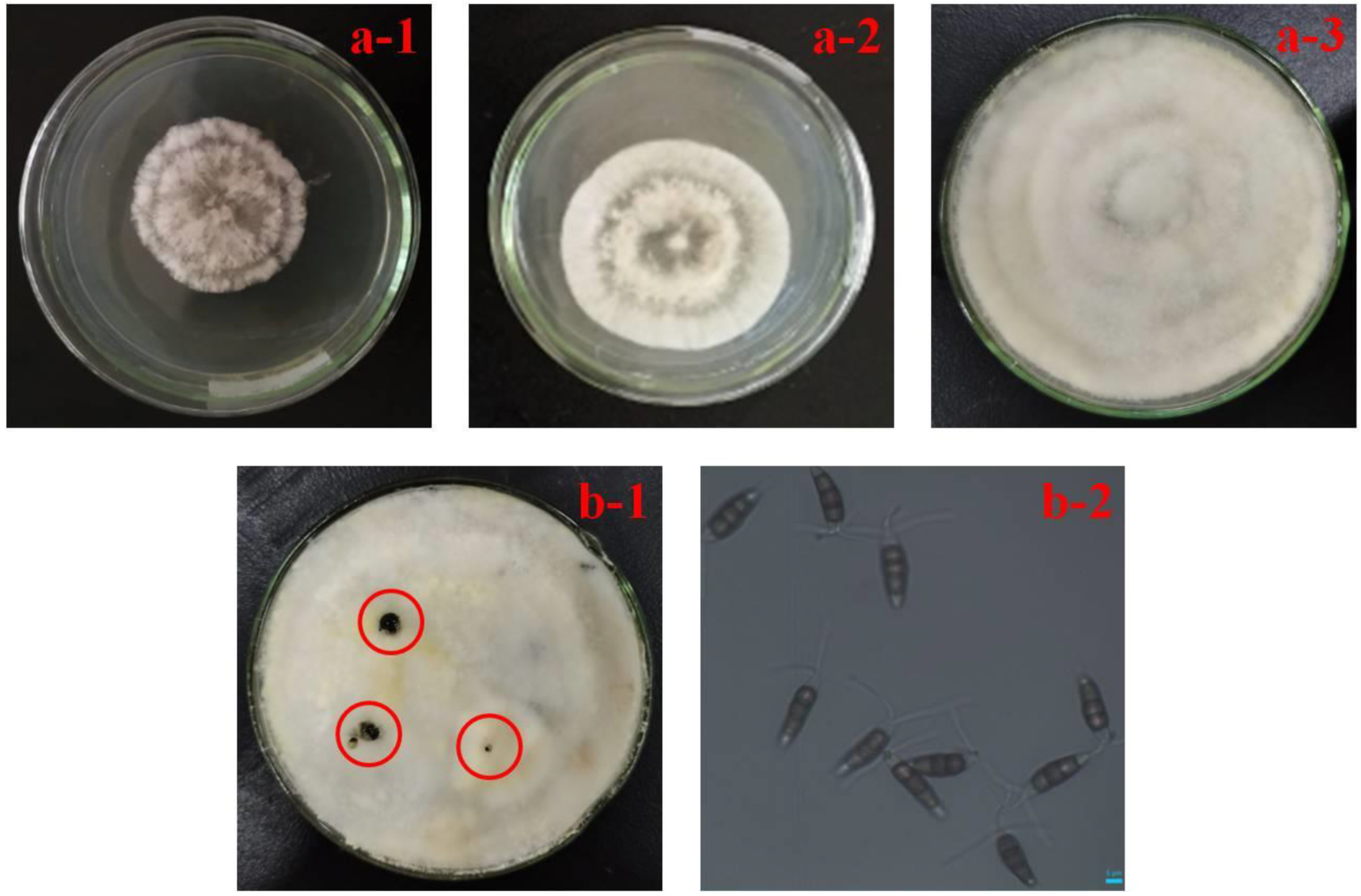
2.2. Pathogen Isolation and Morphological Characterization

The pathogens were isolated from the symptomatic R. sterilis fruits. The tissue at the junction of disease and health was removed from the rotten fruit and spread on potato dextrose agar (PDA, 200 g potato, 20 g dextrose, 20 g agar, 1 L H2O, pH 7.0), and then incubated at 28 °C. To obtain a pure culture, after 3 days of initial incubation, the mycelia at the growing edge of each colony was transferred to a new PDA, and then incubated at 28 °C for 10 days [31]. A total of 30 symptomatic fruits (divide into 3 groups with 10 samples in each group) were selected for the experiment and compared.
The appearance, texture, and color of colony and conidia of the pathogen were observed by naked eye and an optical microscope. Then, preliminary identification was performed based on the morphological features of the colony and conidia.

2.3. Preparation of Spore Suspension

The spore suspension was prepared by flooding a 10 day old PDA plates with sterile water containing 0.05% (v/v) Tween 80. The suspension was filtered through sterile cheesecloth to remove the adhering mycelia. The spore concentration of the fungus was measured by hemocytometer and adjusted to 1 × 10⁶ spores/mL with sterile water.

2.4. Pathogenicity Test

A pathogenicity test was conducted on the sterilized surface of matured R. sterilis fruits. In detail, a total of 40 fresh fruits were sterilized with 75% ethanol for 2 min, rinsed in sterile water and air dried, and then they were wounded with a 2 mm deep hole with a sterile needle. After that, 20 μL of spore suspension (1 × 10⁶ spores/mL) was injected into the hole of 30 wounded fruits from 3 sources (10 fruits from each orchard), and the remaining 10 fruits (from 3 different orchards) were treated with sterile water as a control. Finally, all the treated fruits were incubated in a constant temperature incubator with a plastic bag at 28 °C, 80% humidity. The symptoms on the treated samples were observed and recorded after 1 day of inoculation. Rotten tissue at the lesion margins of the infected fruit was transferred to PDA plates, and the isolated fungus was re-identified by morphological and microscopic analysis.

2.5. DNA Extraction, PCR Amplification and ITS Sequence Analysis

For DNA extraction, the isolated fungal strain was grown on a PDA plate for 7 days at 28 °C. An approximately 20 mg of the mycelia of strain were harvested and ground in a frozen and sterile mortar, and a fine powder was obtained using liquid nitrogen. Genomic DNA was extracted from the fine powder using a plant DNA extraction kit (Tsingke, Biotechnology Co., Ltd., Beijing, China). The internal transcribed spacer (ITS) region of the nuclear rDNA was amplified by the universal primers ITS1 and ITS4. PCR reaction of the ITS gene region was conducted using a thermal cycler, where each reaction was a 50 μL reaction solution containing 45 μL of a PCR master mix (TSE101, Tsingke, Biotechnology Co., Ltd., Beijing, China), 2 μL of each primer set, and 1 μL of genomic DNA. Thermal cycling parameters were 98 °C for 120 s, 35 cycles of 30 s at 98 °C, 30 s at 56 °C, 60 s at 72 °C, and a final extension for 300 s at 72 °C. The PCR products were verified by 1% AGE (agarose gel electrophoresis). Both strands were custom sequenced (Tsingke, Biotechnology Co., Ltd., Kunming, China) and clustal W was applied to generate consensus sequences. All sequence data were submitted to Genbank. Homologous sequences were analyzed against GenBank sequences by using the nucleotide BLAST search tool on the website of http://www.ncbi.nlm.nih.gov/blast/ (4 January 2022), and phylogenetic analysis was performed by using MEGA 6 software with the neighbor-joining method, and a bootstrap test of 1000 replications.

2.6. Fruits and Treatment

Fruits with uniform size (the diameter was about 20 mm), color and shape, and without physical damaged were selected. All fruits were first sanitized in a 2% (m/v) sodium hypochlorite solution for 5 min, then rinsed with sterile water, and finally dried prior to wounding.
The fruits were wounded with a sterile needle to make a uniform hole with 3 × 2 mm in diameter and depth at the equatorial zone of fruit. The wounded fruits were randomly divided into four groups, three treated groups and one control group. For treated groups, a portion of 20 μL of 500 μL/L MeJA, 3% CaCl2 and 500 μL/L MeJA-3% CaCl2 solution was pipetted into the wound hole, respectively. The control group was pipetted with distilled water, instead of the chemical solutions. After drying for 30 min, 20 μL of N. clavispora spore suspension was pipetted into the hole. All the treated fruits were stored at 28 °C and 80% relative humidity for 7 days.
For physiological and biochemical analysis, approximately 1 cm of healthy fruit tissue surrounding the wound holes was obtained and cut into small pieces at days 0, 1, 2, 3, 5, and 7. The excised samples were frozen in liquid nitrogen, ground into powder, and stored at refrigerator (−80 °C) until determination.

2.7. Inhibitory Effect of MeJA or CaCl2 Alone and Their Combination on Artificially Inoculated Fruit

2.7.1. Determination of Disease Incidence

The number of infected fruit (showing visible mycelium in the inoculated hole) was recorded and the disease incidence was expressed as the percentage of infected fruit. The measurement was performed on the 1st, 3rd, 5th and 7th day after inoculation. Each treatment was replicated 3 times with a total of 20 fruits per replicate.

2.7.2. Assay of Defense-Related Enzyme Activity

Phenylalanine ammonia-lyase (PAL) activity was assayed according to the method of a previous study with slight modifications [12]. In detail, the fruit flesh was weighed (1.0 g) and homogenized in 9.0 mL of 50 mM sodium borate buffer extract (pH 8.8, pre-cooled). The homogenate was centrifuged at 10,000 rpm for 10 min at 5 °C, and the supernatant was used as enzyme source. The enzyme reaction was carried out according to the instruction of test kits (Nanjing Jiancheng Bioengineering Institute, Nanjing, China), and then the absorbance at 290 nm was measured by a spectrophotometry (TU-1901, Beijing Purkinje General Instrument Co., Ltd., Beijing, China). One unit (U) of PAL activity was defined as the quantity of PAL causing increase of 0.1 OD290 per minute. The activity was expressed as U/g (FW).
Peroxidase (POD) activity was measured as described in Xie et al. with some modifications [32]. The fruit flesh (1.0 g) was weighed and crushed with a pestle in an ice-cold mortar with 9.0 mL of 0.1 M phosphate buffer. The homogenate was centrifuged at 3500 rpm for 10 min at 4 °C, and the supernatant was used as enzyme source. The reaction mixture for the POD assay was prepared according to the instruction of test kits (Nanjing Jiancheng Bioengineering Institute, Nanjing, China), and the absorbance at 420 nm was recorded by a spectrophotometry (TU-1901, Beijing Purkinje General Instrument Co., Ltd., Beijing, China). One unit (U) of enzyme activity was defined as the quantity of POD causing an increase of 0.001 OD420 per minute. The activity of POD was expressed as U/g (FW).
Polyphenoloxidase (PPO) activity was determined following the method of Xie et al. [32]. The fruit flesh (1.0 g) was weighed and ground with a pestle in an ice-cold mortar with 5.0 mL of sodium acetate buffer (100 mM, pH 5.5). The homogenate was centrifuged at 8000 rpm for 10 min at 4 °C, and the supernatant was used as the enzyme source. The reaction mixture for the PPO assay consisted of 4.0 mL of distilled water, 1.0 mL of 0.1 M catechol and 1.0 mL of enzyme source. The absorbance at 420 nm was measured by spectrophotometry (TU-1901, Beijing Purkinje General Instrument Co., Ltd., Beijing, China) for the PPO activity assay. One unit (U) was defined as the quantity of POD, causing an increase of 0.01 OD420 per minute, and the activity of PPO was expressed as U/g (FW).

2.7.3. Determination of Hardness and Vitamin C Content

Hardness was measured by a hardness detector (GY-4, Ningbo kecheng insturment co. LTD, Ningbo, China) with a 3.5 mm diameter head.
The 2,6-dichloroindophenol titrimetric method was used to measure the vitamin C content of R. sterilis fruit, and the result was expressed as milligrams of vitamin C per 100 g of fresh sample.

2.8. Statistical Analysis

Statistical analysis of experimental data was conducted using SPSS 22.0 software. Analysis of variance was applied followed by Duncan’s multiple range tests to separate means. A value of p < 0.05 was considered significant.

3. Results

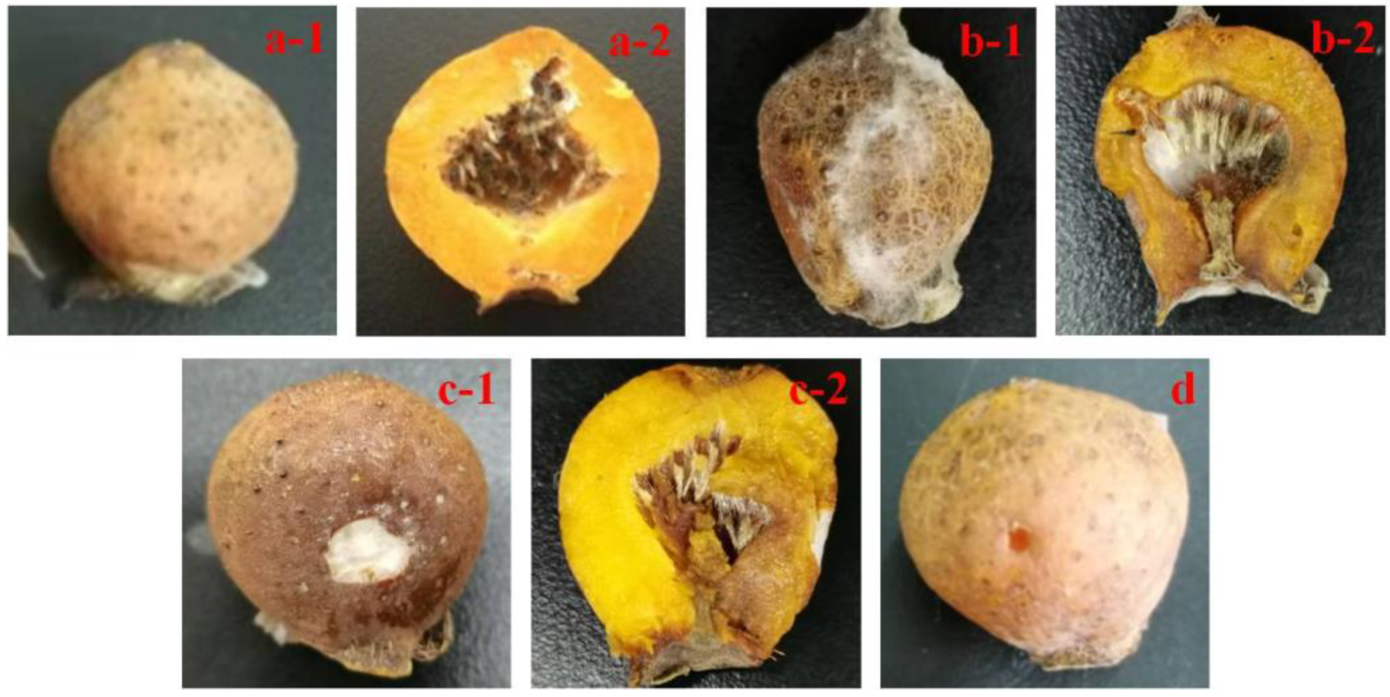
3.1. Typical Symptoms of Rot Fruit and Morphological Identification of Pathogen

Postharvest rot symptoms were observed on both the infected fruits collected from orchards and the stored fruits, and the typical symptoms were as follows: the surface of fruit was covered with an amount of gray-white mycelium, which had small black granular conidia discs, and soft occurred on the outer surface of fruit, then extended to the whole fruit (Figure 1(b1)). Water-soaked flesh tissue could be seen in the sunken area when the collapsed portion was cut (Figure 1(b2)). From the symptomatic fruit, fungal strains with similar features were consistently isolated and cultured on PDA plates for 10 days. In the early stage of growth (within 2 days), the mycelium grew slowly; the colony was white, cottony, and circular growth appearance (Figure 2(a1)). After 3 days of inoculation, the mycelium grew luxuriant; the wheel stripe was observed, and entire colonies formed concentric rings (Figure 2(a2)). On the 7th day of incubation, the Petri dish (d = 5 cm) was filled with mycelium due to the rapid growth (Figure 2(a3)). In the later stage of growth (on the 10th day), small black spots and molds were observed on the surface of the mycelium layer, which developed acervuli that contained scattered conidia (Figure 2(b1)). In addition, there were some granular acervuli buried under the mycelium layer. Conidia were slightly curved, fusiform to clavate, five-celled with four constricted septa; the two dimensions were 17.21 ± 1.39 × 6.11 ± 0.64 μm (n = 20). Three median cells were brown or darker, and the apical and basal cells were hyaline and white, one of which had 2–4 appendages (Figure 2(b2)). It was found that all cultured strains had similar morphological characteristics, thus they were considered to be the same pathogen. The morphological characteristics of the isolated fungus were similar to those described in the literature for the species of N. clavispora or P. clavispora [24,33]. The isolate could be characterized as N. clavispora by colony and conidia morphology.

3.2. Pathogenicity

The inoculated fruits were kept at 28 °C in plastic bags. At 7 days after inoculation, the white mycelium developed within the wounds, and discoloration extended outward from the inoculated sites. Slight rot began to develop on the inoculated fruits. At 14 days after inoculation, the area of decay covered more than half of the fruit surface and abundant white mycelia and black spores were observed on the rotten area (Figure 1(c1)). The symptoms on the inoculated fruits (Figure 1(c2)) phenotypically matched those observed on the naturally occurring fruits (Figure 1(b2)) from the appearance of the rotten tissue. The control fruits (inoculated with sterile water) showed discoloration at the edges of the hole, but no disease symptoms developed (Figure 1d). The test results of spore suspensions from different orchards were consistent. The pathogen was re-isolated from the inoculated fruits, and re-identified by morphological features. The resulting colony phenotypically matched the colony obtained from the originally diseased fruit, therefore completing Koch’s postulates. Based on the observed pathological symptom, morphology features, and pathogenicity, the pathogen was further identified as N. clavispora.

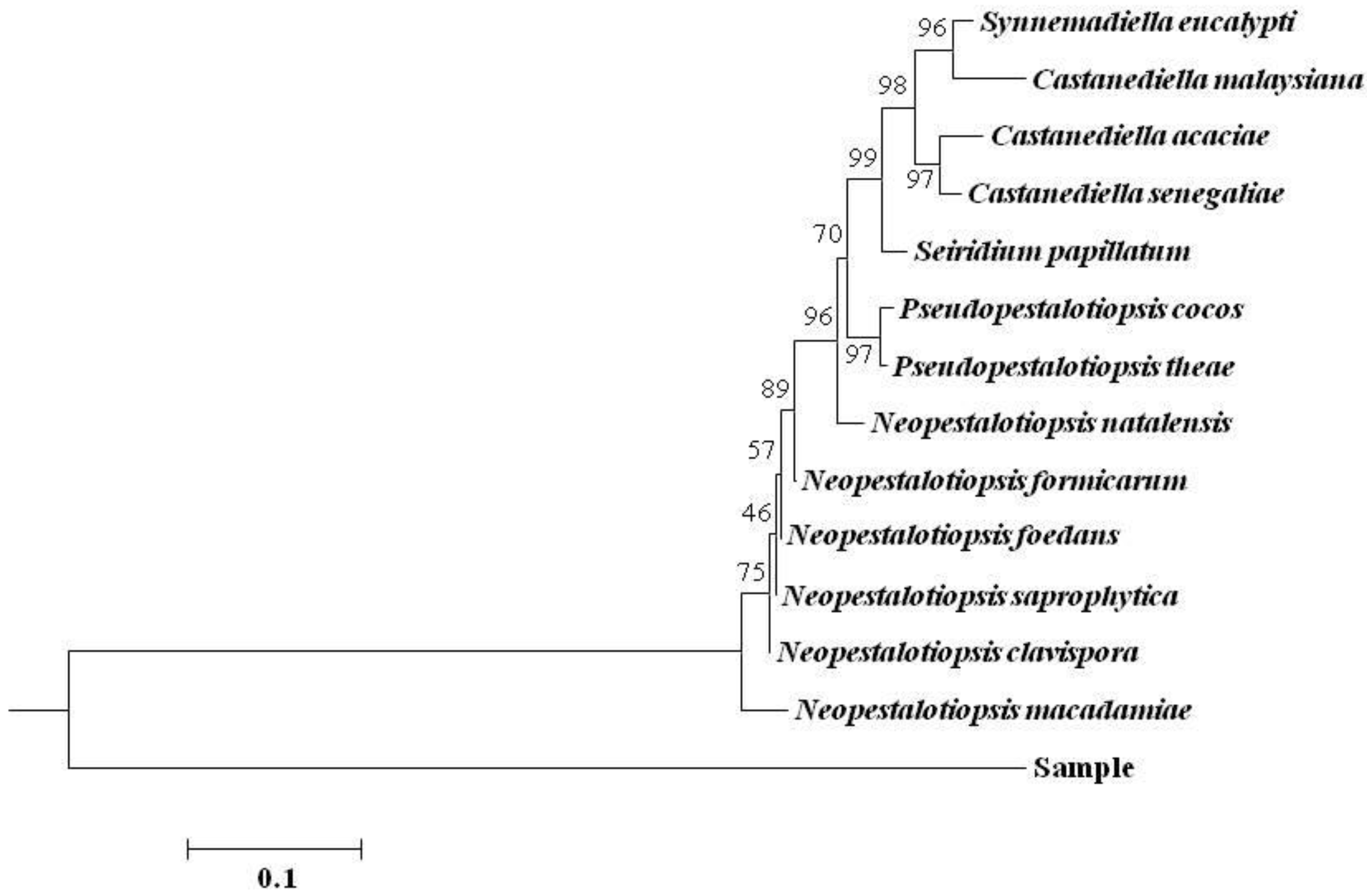
3.3. rDNA-ITS Sequence Analysis of Pathogen

The isolated N. clavispora was further confirmed by molecular identification. The gene region of ITS was PCR amplified and sequenced. The rDNA-ITS sequence of the isolated strain was amplified with ITS-1 and ITS-4 as universal primers. The obtained PCR products were detected by 1% AGE. The results showed a single band, no more than specific amplification, and the size of ITS sequence was about 600 bp (as shown in Figure S1).
The sequencing result (Doc. S1) shows that about 600 nucleotide sequences of the rDNA-ITS sequence of the fungal strain were effectively amplified. The sequencing results were submitted to GenBank and subjected to BLAST analysis. The results show that the ITS sequence of the fungal strain was 99–100% homologous to the ITS sequence of N. clavispora. The ITS sequence with higher homology was selected and compared with the ITS sequence of the isolated strain to establish a phylogenetic tree, and the result is shown in Figure 3. According to the phylogenetic tree, the isolated strain and N. clavispora belong to the same branch of the phylogenetic tree; the relationship was closest, and the homology was over 99%.
The molecular analysis confirmed the morphological identification for the fungal pathogen. Combined with the results of morphological identification, the pathogen was identified as N. clavispora.

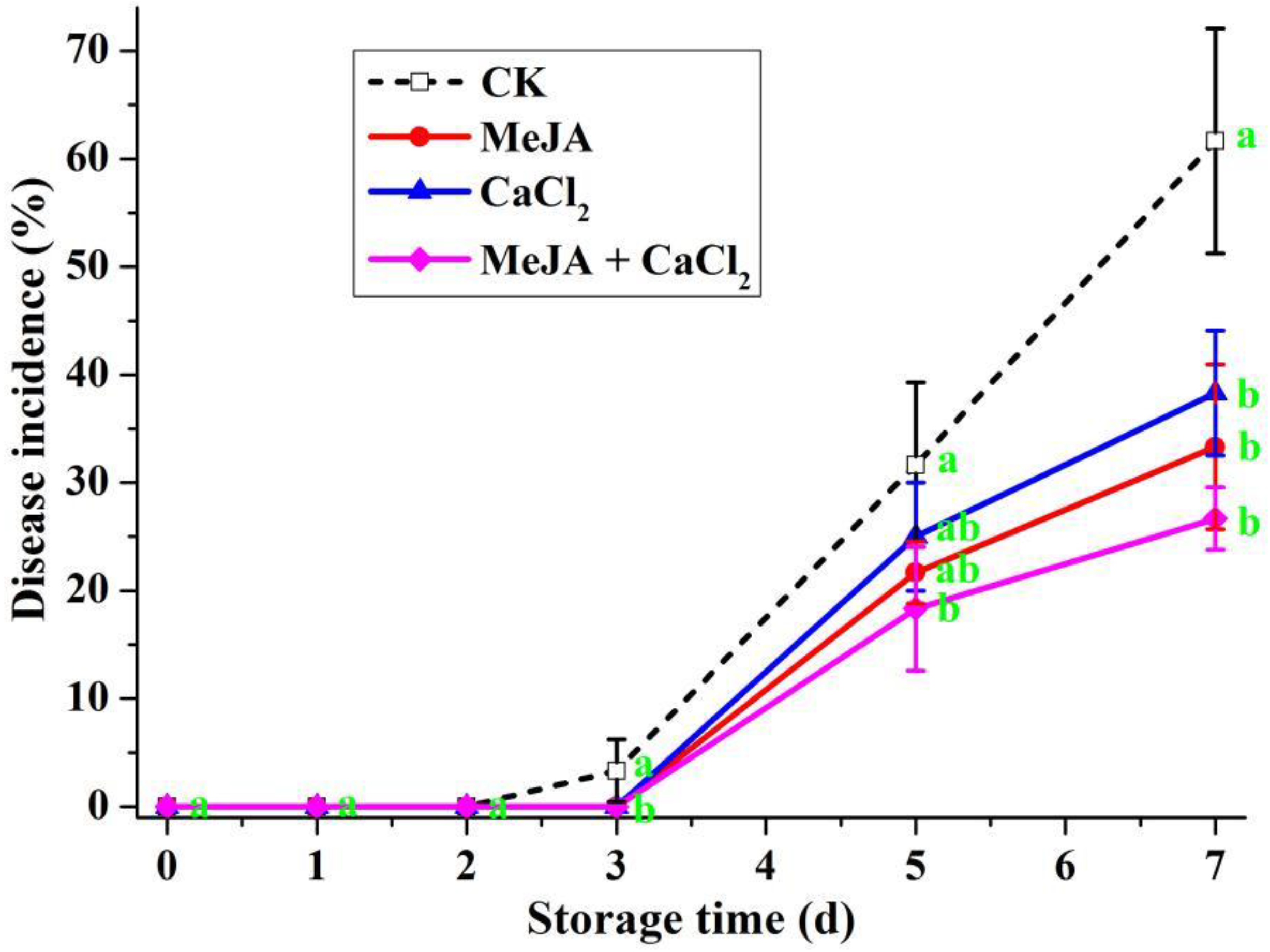
3.4. Effects of Different Treatments in Controlling Decay on Artificially Inoculated R. sterilis Fruit

The effects of different treatments on the induction of R. sterilis fruit resistance based on the disease incidence are shown in Figure 4. Disease incidence at 7 days after inoculation for control, MeJA, CaCl2 and MeJA + CaCl2 treatments was 61.67, 33.33, 38.33 and 26.67%, respectively. The control had significantly more decay than the other treatments, which did not differ significantly from each other. The combined treatment further reduced fruit decay in comparison with the treatment of MeJA or CaCl2 alone, but no significant difference of incidence was observed among different treatment groups. On the 5th day after inoculation, the disease incidence of the control group (31.67%) was significantly higher than the MeJA–CaCl2 treated group (18.33%) and no significant difference of disease incidence was found among different treated groups or the control group, MeJA alone and CaCl2 alone. The differences in disease incidence among different groups were very small during the first three days of inoculation, although the incidence in all treatment groups was significantly lower than that in the control group (3.33%).

3.5. Effect of Different Treatments on the Activity of Defense-Related Enzymes

The activities of all resistance-related enzymes are shown in Figure 5. As shown in Figure 5A, PAL activity exhibited a similar trend in both control and treatment groups; in detail, it can be seen that it first increased and then decreased, but the activity increased to its peak value at different incubation times. PAL activity in MeJA-treated fruits increased on the 1st day of inoculation, and decreased rapidly during the 1st to 3rd days, then became smooth on the 3rd to 5th days, and finally decreased again after 5 days. On the 3rd day of inoculation, the PAL activities in the three groups (control, CaCl2, and MeJA–CaCl2 treatment) peaked simultaneously; the activity in the MeJA–CaCl2 treatment group (56.23 U/g FW) was significantly higher than in the control (47.78 U/g FW), and there was no significant difference between the CaCl2 treatment and control groups, while PAL activity in the MeJA treatment group (40.20 U/g FW) was significantly lower than in the control. On the 5th day, PAL activity in MeJA–CaCl2 treated fruits (50.90 U/g FW) was significantly higher than the control, MeJA and CaCl2 treatment groups. At the end of observation period (on the 7th day of inoculation), PAL activities were still significantly higher in the treatment groups than in the control (27.34 U/g FW), and the PAL activity in the MeJA–CaCl2 treatment group (38.55 U/g FW) was significantly higher than those of the MeJA and CaCl2 treatment groups.
As can be seen from Figure 5B, POD activities followed different trends in the control and treatment groups during the monitored time. POD activity in the MeJA–CaCl2 treated fruits showed a slight upward trend on the 1st day and dramatically increased on the 2nd day of inoculation, and then decreased gradually within the remainder of storage. POD activity in the infected fruits treated with CaCl2 slightly increased within the first 2 days, then increased dramatically and reached the highest value on the 3rd day, and finally decreased until the end of the monitored period, while the enzyme activities followed the same trend in both control and MeJA treatment groups, i.e., first increasing and then decreasing. The POD activities of different groups showed significant difference on the 2nd day, and the value was 8.59, 12.22, 5.16, and 15.29 U/g FW for the control, MeJA-treated, CaCl2-treated, and combination-treated group, respectively. There was no difference in the POD activities among different groups on the 3rd day of inoculation, while the differences among these four groups were significant on the 5th day. POD activity in all groups exhibited a downtrend at the later stage of the measurement period (after 3 days of inoculation), and the activities of different groups decreased to almost the same level (corresponding to no significant difference) at the end of the observation period (on the 7th day of inoculation).
The PPO activities of different fruits are shown in Figure 5C. The PPO activities followed the same trend in the four groups (increasing and then decreasing), but they did not take the same amount of time to reach their highest value. After inoculation, the PPO activity in the control fruits started to rise and peaked on the 3rd day (9.54 U/g FW);–similarly, the PPO activity in fruits treated with MeJA-CaCl2 was also at a maximum during this time (17.50 U/g FW), while PPO activities in MeJA-treated and CaCl2-treated fruits reached their maximum value on the 5th day. The activities in the treatment groups were always higher than that in the control group. On the 3rd day of inoculation, the PPO activity of MeJA–CaCl2 treatment group was significantly higher than those of the other three groups. On the and 5th day, the PPO activities for all treatments were significantly higher than that of the control (3.72 U/g FW). On the 7th day of inoculation (the end time of the experiment), the activity in control fruits (1.59 U/g FW) was significantly lower than those in the MeJA and MeJA–CaCl2 treatment fruits, while no significant difference between the control and CaCl2 treatment groups was observed. The PPO activity in the MeJA–CaCl2 treatment fruits (7.53 U/g FW) was significantly higher than those in the control and CaCl2 treatment fruits, while there was no significant difference between the fruits treated with MeJA alone and in combination with CaCl2.

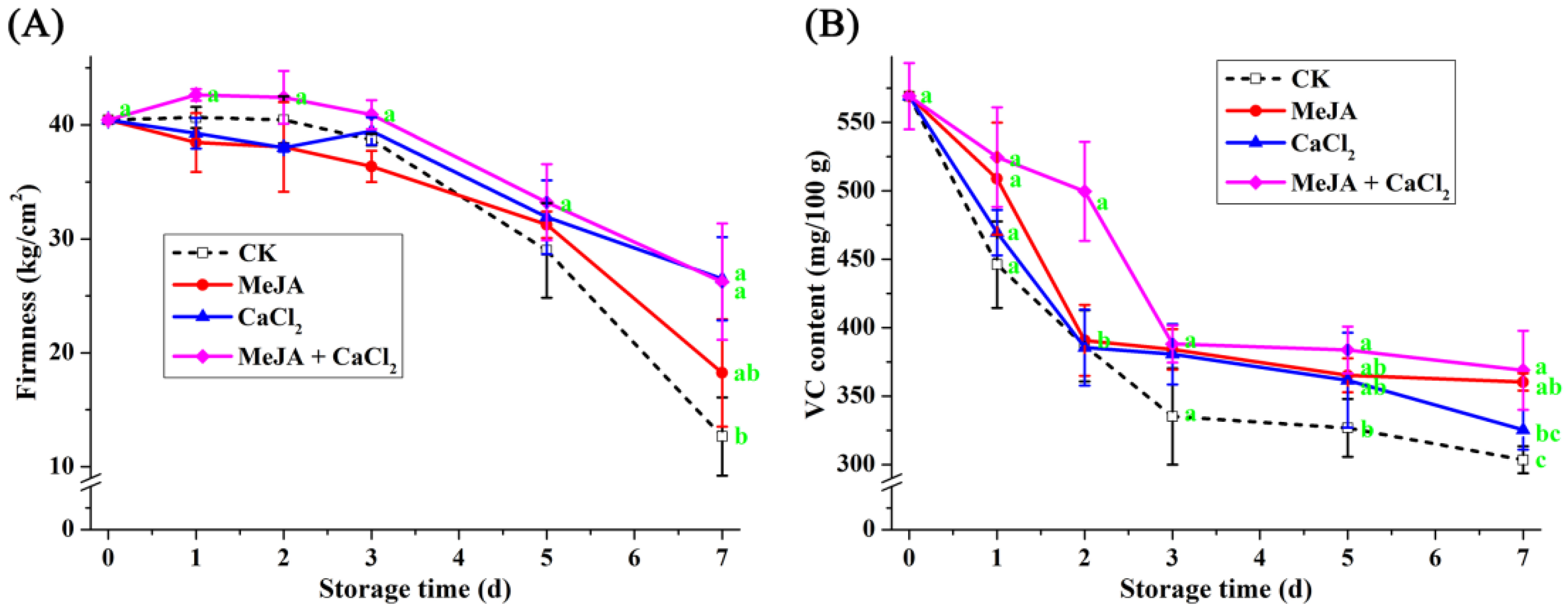
3.6. Effect of Different Treatments on Hardness and Vitamin C Content

As shown in Figure 6A, fruit hardness in both control and treatment groups decreased gradually with the prolongation of incubating time; the decrease in hardness may be due to the hydrolysis of pectin and carbohydrates and the occurrence of fruit rot. The degree of decrease in hardness was less in the MeJA–CaCl2 and CaCl2 treated fruits. On the 7th day of inoculation, the fruits treated with CaCl2 alone or in combination with MeJA maintained a significantly high hardness (26.51 kg/cm2 or 26.24 kg/cm2) compared with the control (18.24 kg/cm2). MeJA showed no significant effect on the hardness of R. sterilis fruits throughout the whole storage period.
The vitamin C contents of different fruits are given in Figure 6B. The vitamin C content during storage was retained significantly better after the MeJA, CaCl2 or MeJA+CaCl2 treatments than the control. On the 7th day of inoculation, vitamin C contents in R. sterilis fruits were well maintained by different treatments.

4. Discussion

In this paper, a systematic study of postharvest rot occurring on R. sterilis fruit during the postharvest storage period was carried out. N. clavispora was firstly identified as the main pathogen causing postharvest rot on R. sterilis fruit based on the results of morphological identification, molecular analysis and pathogenicity test. N. clavispora is widely distributed as opportunistic plant pathogen [24,34], and our experiments showed that the wounded R. sterilis fruit can quickly become ruined by the action of the fungus.
It is well known that MeJA, as a naturally growth regulator, plays important roles in regulating a great diversity of biochemical and physiological processes [26]. Previous studies have showed that MeJA has been widely used to control the postharvest diseases of various fruits, including Chinese bayberry [25,26], kiwifruit [11] and cherry tomato [27], possibly by inhibiting pathogen growth or/and inducing disease resistance. In this study, we found that the treatment with 500 μL/L MeJA in combination with 3% CaCl2 resulted in the better control of N. clavispora on R. sterilis fruit than the use of MeJA or CaCl2 alone due to the fact that it can significantly reduce disease incidence and increase the induction effect of R. sterilis fruit infected with N. clavispora. PAL acts as a marker of plant-induced disease resistance due to being the first key enzyme in the phenylpropanoid pathway, which is highly associated with the biosynthesis of lignins, phenolic compounds and phytoalexins to prevent pathogen invasion and slow disease development during pathogenic infection [12,35]. In this study, PAL activities were generally higher in treatment groups than in the control during the whole infection period (especially in the initial period), indicating that the treated fruits showed higher resistance. Many researches suggest that there is a direct relationship between the increase in POD activity and the natural resistance of fruit [36]. This is why a rapid increase in POD activity was observed after 2 days of inoculation. POD plays a key role in the accumulation of lignin since it can catalyze the biosynthesis of lignin [35,37]. The higher POD activities in fruits treated with MeJA alone and in combination with CaCl2 than in the control may be related to MeJA-induced resistance. The activation of PPO is associated with triggering and accelerating the infection process in the treated fruits [35]. In general, the activities of PPO were higher in the inoculated fruits (especially in the MeJA–CaCl2 and MeJA treatment groups) compared to the corresponding healthy fruits. These results suggest that the effect of MeJA in reducing the decay of R. sterilis fruit is mainly due to inducing disease resistance in the fruit. In addition, our data have shown that R. sterilis fruit treated with CaCl2 alone or CaCl2 with MeJA exhibited significantly higher level of hardness compared with those treated with MeJA alone and control, which may be attributed to the key role of calcium ions in reducing fruit softening by strengthening the cell walls [36]. These results indicate that MeJA in combination with CaCl2 also had an additive effect in inhibiting the decrease in the hardness of the fruit. Vitamin C, an important organic acid in fruits and plants, has great potential in suppressing fungal disease [38,39]. The vitamin C contents of the treated fruits always remained at high levels during the infection period, indicating that the fruits in the treatment groups showed higher resistance to fruit rot disease. Therefore, the combination of MeJA and CaCl2 has a potential application in postharvest treatment for reducing rot in R. sterilis fruit during postharvest storage and distribution.

5. Conclusions

To the best of our knowledge, N. clavispora was identified as the main pathogen causing postharvest rot on R. sterilis fruit in China for the first time. In addition, MeJA in combination with CaCl2 treatment can reinforce the resistance of postharvest rot on R. sterilis fruit during N. clavispora infection. The possible mechanism is that the MeJA–CaCl2 treatment induced disease resistance against N. clavispora in the infected fruits by priming of defense responses, such as increasing the activities of PAL, PPO and POD in the phenylpropanoid pathway and maintaining a high level of vitamin C content. Further research is needed to understand the pathogenicity and infection process of the N. clavispora related to fruit rot, which can be carried out as follows: first, to further confirm that the postharvest rot is also caused by other pathogens by the isolation and identification of strains from the symptomatic fruits collected from more different regions. Second, to elucidate the mode of action of different treatments on the disease of R. sterilis fruit. Third, to explore the possible mechanisms involved in postharvest disease control by the determination of other indicators, such as the activities of cinnamate-4-hydroxylase and cinnamyl-alcohol dehydrogenase, total phenol and flavonoid contents.

Supplementary Materials

The following supporting information can be downloaded at: https://www.mdpi.com/article/10.3390/horticulturae8030190/s1, Figure S1: Gel electrophoresis of pathogen PCR-amplification results; Doc. S1: The full-length rDNA-ITS sequence of the strain to be tested as follows.

Author Contributions

Conceptualization, T.S.; methodology, T.S.; software, T.S.; validation, T.S.; formal analysis, T.S.; investigation, T.S.; resources, T.S.; data curation, T.S. and T.P.; writing—original draft preparation, T.P.; writing—review and editing, T.S., T.P., and M.G.; visualization, T.S.; supervision, T.P.; project administration, T.S. and T.P.; funding acquisition, T.S. and T.P. All authors have read and agreed to the published version of the manuscript.

Funding

This research was funded by the Natural Science Research Project of Education Department of Guizhou Province, grant number [2018] 432, China Postdoctoral Science Foundation, grant number 2019M663570 and Education Quality Improvement Project of Qiannan Normal University for Nationalities, grant number 2021xjg018.

Institutional Review Board Statement

Not applicable.

Informed Consent Statement

Not application.

Data Availability Statement

Not applicable.

Acknowledgments

The authors thank all those who provided assistance and advice to this work and manuscript.

Conflicts of Interest

The authors declare no conflict of interest.

References

  1. Li, J.L.; Quan, W.X.; Li, C.C.; Shi, D.H.; Hu, J.W.; Jin, J.; Ruan, W. Effects of ecological factors on content of flavonoids in Rosa sterilis from different Karst areas of Guizhou, SW China. Pak. J. Bot. 2018, 50, 1125–1133. [Google Scholar]
  2. Wen, X.P.; Deng, X.X. Characterization of genotypes and genetic relationship of cili (Rosa roxburghii) and its relatives using RAPD markers. Chin. J. Agr. Biotech. 2003, 11, 605–611. [Google Scholar] [CrossRef]
  3. Wen, X.P.; Pang, X.M.; Deng, X.X. Characterization of genetic relationships of Rosa roxburghii Tratt and its relatives using morphological traits, RAPD and AFLP markers. J. Hortic. Sci. Biotech. 2004, 79, 189–196. [Google Scholar] [CrossRef]
  4. Liang, G.-Y.; Gray, A.I.; Waterman, P.G. Pentacyclic triterpenes from the fruits of Rosa sterilis. J. Nat. Prod. 1989, 52, 162–166. [Google Scholar] [CrossRef]
  5. He, J.Y.; Zhang, Y.H.; Ma, N.; Zhang, X.L.; Liu, M.H.; Fu, W.M. Comparative analysis of multiple ingredients in Rosa roxburghii and R. sterilis fruits and their antioxidant activities. J. Funct. Food. 2016, 27, 29–41. [Google Scholar] [CrossRef]
  6. Liu, M.H.; Zhang, Q.; Zhang, Y.H.; Lu, X.Y.; Fu, W.M.; He, J.Y. Chemical analysis of dietary constituents in Rosa roxburghii and Rosa sterilis fruits. Molecules 2016, 21, 1024. [Google Scholar] [CrossRef]
  7. Luo, X.L.; Dan, H.L.; Li, N.; Li, Y.H.; Zhang, Y.J.; Zhao, P. A new catechin derivative from the fruits of Rosa sterilis SD. Shi. Nat. Prod. Res. 2017, 31, 2239–2244. [Google Scholar] [CrossRef]
  8. Shi, J.-F.; Sun, C.-Q. Isolation, identification, and biocontrol of antagonistic bacterium against Botrytis cinerea after tomato harvest. Braz. J. Microbiol. 2017, 48, 706–714. [Google Scholar] [CrossRef]
  9. Zhang, S.W.; Zheng, Q.; Xu, B.L.; Liu, J. Identification of the fungal pathogens of postharvest disease on peach fruits and the control mechanisms of Bacillus subtilis JK-14. Toxins 2019, 11, 322. [Google Scholar] [CrossRef] [Green Version]
  10. Lorenzini, M.; Zapparoli, G. Identification of Pestalotiopsis bicilita, Diplodia seriata and Diaporthe eres causing fruit rot in withered grapes in Italy. Eur. J. Plant Pathol. 2018, 151, 1089–1093. [Google Scholar] [CrossRef]
  11. Pan, L.Y.; Zhao, X.Y.; Chen, M.; Fu, Y.Q.; Xiang, M.L.; Chen, J.Y. Effect of exogenous methyl jasmonate treatment on disease resistance of postharvest kiwifruit. Food Chem. 2020, 305, 125483. [Google Scholar] [CrossRef] [PubMed]
  12. He, J.H.; Ma, L.J.; Wang, D.J.; Zhang, M.Y.; Zhou, H.L. Ferulic acid treatment reinforces the resistance of postharvest apple fruit during gray mold infection. J. Plant Pathol. 2019, 101, 503–511. [Google Scholar] [CrossRef]
  13. Pan, T.T.; Sun, D.-W.; Pu, H.B.; Wei, Q.Y.; Xiao, W.; Wang, Q.J. Detection of A. alternata from pear juice using surface-enhanced Raman spectroscopy based silver nanodots array. J. Food Eng. 2017, 215, 147–155.10. [Google Scholar] [CrossRef]
  14. Guo, M.J.; Wang, Q.T.; Cheng, Y.H.; Hou, C.L. Identification of Aspergillus tubingensis causing pomegranate fruit rot in China. Australas. Plant Path. 2021, 50, 233–240. [Google Scholar] [CrossRef]
  15. Palou, L.; Smilanick, J.L. Postharvest Pathology of Fresh Horticultural Produce; CRC Press: New York, NY, USA, 2020; pp. 375–406. [Google Scholar]
  16. Diaz, G.A.; Latorre, B.A.; Ferrada, E.; Lolas, M. Identification and characterization of Diplodia mutila, D. seriata, Phacidiopycnis washingtonensis and Phacidium lacerum obtained from apple (Malus x domestica) fruit rot in Maule Region, Chile. Eur. J. Plant Pathol. 2019, 153, 1259–1273. [Google Scholar] [CrossRef]
  17. Jiang, J.J.; Zhai, H.Y.; Li, H.N.; Wang, Z.H.; Chen, Y.S.; Hong, N.; Wang, G.; Chofong, G.N.; Xu, W. Identification and characterization of Colletotrichum fructicola causing black spots on young fruits related to bitter rot of pear (Pyrus bretschneideri Rehd.) in China. Crop. Prot. 2014, 58, 41–48. [Google Scholar] [CrossRef]
  18. Oo, M.M.; Oh, S.K. Identification and characterization of new record of grape ripe rot disease caused by Colletotrichum viniferum in Korea. Mycobiology 2017, 45, 421–425. [Google Scholar] [CrossRef] [Green Version]
  19. Zhu, J.Z.; Li, C.X.; Zhang, C.J.; Wang, Y.; Li, X.G.; Zhong, J. Fusarium solani causing fruit rot of peach (Prunus persica) in Hunan, China. Crop. Prot. 2019, 122, 171–174. [Google Scholar] [CrossRef]
  20. Michailides, T.J.; Thomidis, T. First report of Phomopsis amygdali causing fruit rot on peaches in Greece. Plant Dis. 2006, 90, 1551. [Google Scholar] [CrossRef]
  21. Li, L.; Pan, H.; Chen, M.Y.; Zhang, S.J.; Zhong, C.H. Isolation and identification of pathogenic fungi causing postharvest fruit rot of kiwifruit (Actinidia chinensis) in China. J. Phytopathol. 2017, 165, 782–790. [Google Scholar] [CrossRef]
  22. Zhou, Y.; Gong, G.S.; Cui, Y.L.; Zhang, D.X.; Chang, X.L.; Hu, R.P.; Liu, N.; Sun, X. Identification of Botryosphaeriaceae species causing kiwifruit rot in sichuan province, China. Plant Dis. 2015, 99, 699–708. [Google Scholar] [CrossRef] [PubMed]
  23. Espinoza, J.G.; Briceno, E.X.; Keith, L.M.; Latorre, B.A. Canker and twig dieback of blueberry caused by Pestalotiopsis spp. and a Truncatella sp in Chile. Plant Dis. 2008, 92, 1407–1414. [Google Scholar] [CrossRef] [PubMed] [Green Version]
  24. Chamorro, M.; Aguado, A.; De los Santos, B. First report of root and crown rot caused by Pestalotiopsis clavispora (Neopestalotiopsis clavispora) on strawberry in Spain. Plant Dis. 2016, 100, 1495. [Google Scholar] [CrossRef]
  25. Wang, K.T.; Jin, P.; Han, L.; Shang, H.T.; Tang, S.S.; Rui, H.J.; Duan, Y.; Kong, F.; Kai, X.; Zheng, Y. Methyl jasmonate induces resistance against Penicillium citrinum in Chinese bayberry by priming of defense responses. Postharvest Biol. Tec. 2014, 98, 90–97. [Google Scholar] [CrossRef]
  26. Wang, K.T.; Jin, P.; Shang, H.T.; Zheng, Y.H. Effect of methyl jasmonate in combination with ethanol treatment on postharvest decay and antioxidant capacity in Chinese bayberries. J. Agric. Food Chem. 2010, 58, 9597–9604. [Google Scholar] [CrossRef]
  27. Chen, J.P.; Zou, X.; Liu, Q.; Wang, F.; Feng, W.; Wan, N. Combination effect of chitosan and methyl jasmonate on controlling Alternaria alternata and enhancing activity of cherry tomato fruit defense mechanisms. Crop. Prot. 2014, 56, 31–36. [Google Scholar] [CrossRef]
  28. Wang, S.-Y.; Shi, X.-C.; Liu, F.-Q.; Laborda, P. Effects of exogenous methyl jasmonate on quality and preservation of postharvest fruits: A review. Food Chem. 2021, 353, 129482. [Google Scholar] [CrossRef]
  29. Gupta, N.; Jawandha, S.K.; Gill, P.S. Effect of calcium on cold storage and post-storage quality of peach. J. Food Sci. Technol.-Mysore 2011, 48, 225–229. [Google Scholar] [CrossRef] [Green Version]
  30. Li, L.; Ban, Z.J.; Li, X.H.; Xue, T. Effect of 1-methylcyclopropene and calcium chloride treatments on quality maintenance of ’Lingwu Long’ Jujube fruit. J. Food Sci. Technol.-Mysore 2014, 51, 700–707. [Google Scholar] [CrossRef] [Green Version]
  31. Zhao, J.; Liu, W.C.; Liu, D.W.; Lu, C.G.; Zhang, D.P.; Wu, H.L.; Dong, D.; Meng, L. Identification and evaluation of Aspergillus tubingensis as a potential biocontrol agent against grey mould on tomato. J. Gen. Plant Pathol. 2018, 84, 148–159. [Google Scholar] [CrossRef]
  32. Xie, G.F.; Wang, L.R.; Fan, K.X.; Liu, N.; Liu, Y.L.; Zhao, Z.B. Postharvest 1-methylcyclopropene treatments maintain the quality of Rosa sterilis D. shi during storage. Food Sci. Technol. 2020, 40, 89–94. [Google Scholar] [CrossRef] [Green Version]
  33. Biju, C.N.; Peeran, M.F.; Gowri, R. Identification and characterization of Neopestalotiopsis clavispora associated with leaf blight of small cardamom (Elettaria cardamomum Maton). J. Phytopathol. 2018, 166, 532–546. [Google Scholar] [CrossRef]
  34. Jayawardena, R.S.; Zhang, W.H.; Liu, M.H.; Maharachchikumbura, S.S.N.; Zhou, Y.; Huang, J.B.; Nilthong, S.; Wang, Z.; Li, X.; Yan, J.; et al. Identification and characterization of Pestalotiopsis-like fungi related to grapevine diseases in China. Fungal Biol. 2015, 119, 348–361. [Google Scholar] [CrossRef] [PubMed]
  35. Ngadze, E.; Icishahayo, D.; Coutinho, T.A.; Van der Waals, J.E. Role of polyphenol oxidase, peroxidase, phenylalanine ammonia lyase, chlorogenic acid, and total soluble phenols in resistance of potatoes to soft rot. Plant Dis. 2012, 96, 186–192. [Google Scholar] [CrossRef] [PubMed] [Green Version]
  36. Feng, W.; Zheng, X.D.; Chen, J.P. Combined inhibitory effect against postharvest storage rots and their effects on postharvest quality parameters in cherry tomatoes by cassia oil and calcium chloride. J. Food Protect. 2013, 76, 1873–1878. [Google Scholar] [CrossRef]
  37. Cesarino, I.; Araujo, P.; Domingues Junior, A.P.; Mazzafera, P. An overview of lignin metabolism and its effect on biomass recalcitrance. Braz. J. Bot. 2012, 35, 303–311. [Google Scholar] [CrossRef]
  38. El-Samawaty, A.M.A. Inducing resistance in a flax cultivar susceptible to rust disease caused by Melampsora lini. J. Pure Appl. Microbio. 2013, 7, 1831–1836. [Google Scholar]
  39. El-Samawaty, A.M.A.; Yassin, M.A.; Moslem, M.A.; Omar, M.R. Role of some antioxidants in suppressing of flax powdery mildew. Fresen. Environ. Bull. 2016, 25, 1059–1067. [Google Scholar]
Figure 1. Postharvest rot caused by N. clavispora on R. sterilis fruit. (a) Fresh fruit (1: outside; 2: inside). (b) Symptoms of fruit rot by natural inoculation (1: outside; 2: inside). (c) Symptoms on wound inoculated fruit after 14 days (1: outside; 2: inside). (d) Control.
Horticulturae 08 00190 g001
Figure 2. Morphological characteristics of N. clavispora. (a) Colony on PDA (1/2/3-two/three/seven-day-old); (b1) acervuli and (b2) conidia.
Horticulturae 08 00190 g002
Figure 3. Phylogenetic tree constructed using the neighbor-joining method based on rDNA-ITS sequences.
Horticulturae 08 00190 g003
Figure 4. Effect of different treatments on the disease incidence of R. sterilis fruit inoculated with N. clavispora within 7 days of storage at 28 °C. Data are expressed as mean ± SD of triplicate assays.
Horticulturae 08 00190 g004
Figure 5. Activities of resistance-related enzymes in the control and treated R. sterilis fruits inoculated with N. clavispora. (A) PAL; (B) POD; and (C) PPO.
Horticulturae 08 00190 g005
Figure 6. Hardness and vitamin C content of the control and treated R. sterilis fruits inoculated with N. clavispora. (A) Hardness; and (B) Vitamin C content.
Horticulturae 08 00190 g006
Publisher’s Note: MDPI stays neutral with regard to jurisdictional claims in published maps and institutional affiliations.

