
In Vitro Germination and Propagation of Dyckia brevifolia, An Ornamental and Endangered Bromeliad


Laboratory of Floriculture and Landscape Architecture, Department of Crop Science, School of Plant Sciences, Agricultural University of Athens, 75 Iera Odos Street, GR-11855 Athens, Greece
*
Author to whom correspondence should be addressed.
Horticulturae 2022, 8(5), 390; https://doi.org/10.3390/horticulturae8050390
Submission received: 30 March 2022
/
Revised: 18 April 2022
/
Accepted: 25 April 2022
/
Published: 28 April 2022
(This article belongs to the Special Issue Seed Germination and Micropropagation of Ornamental Plants)
Abstract
:Dyckia brevifolia is an endangered plant used for ornamental purposes. As no references to the in vitro propagation of the species exist, the present study aims at investigating the possibility of an efficient micropropagation protocol. Seeds collected from mother plants were germinated at high percentages (84–86%) at a range of 15–25 °C, without any pre-treatment, and demonstrated their highest germination speed index (191.51) at 25 °C. In vitro-grown seedlings were used as the starting material for micropropagation on solid, or liquid, MS medium, supplemented with a variety of concentrations of cytokinins (BA, KIN or 2IP). Shoots and leaves were used as starting explants. Liquid media supplemented with BA or 2IP at 1.0 mg L−1 led to high multiplication rate and 2.7, or 2.3, lateral shoots were regenerated while on 2IP a high percentage (77.5%) of rooting occurred at the same time. Rooted microshoots were acclimatised ex vitro at 100% and acclimatised plants were transplanted in pots where they grew with a survival rate of 100% after two months. The in vitro propagation protocol presented in this study could enhance the large-scale propagation use of D. brevifolia as an ornamental plant and, simultaneously, contribute to the ex-situ conservation of the species.
1. Introduction
Bromeliads are widely used in floriculture. Their attractive rosettes, range of colours of flowers, and graceful foliage, make them ideal ornamental plants. Still, information on their reproductive strategies remains scant [1,2]. Certain members of Bromeliaceae are often viewed as a prime example of adaptive radiation [3] as they can be found in a wide range of environments, from mesic to xeric, from terrestrial to epiphytic, and from sea level areas to high elevation ones. Their key adaptations include leaf succulence, spiny leaves, which gives them herbivore protection, and a crassulacean acid metabolism (CAM), which allows them to inhabit rocky, semi-arid environments [4,5]. However, bromeliads are relatively difficult to propagate, with one plant producing no more than a few individual plantlets characterised by slow growth [6]. Thus, in the recent past, the development of plant tissue culture-based techniques has been gaining ground since such techniques have proved valuable tools in developing new strategies for the bromeliads’ mass propagation [7].
Dyckia is a genus that includes 170 species (either terrestrial or saxicolous, tankless perennial plants) and is one of the most species-rich genera in the Pitcairnioideae subfamily [8]. Many of the Dyckia members are of high ornamental interest [9]. According to recent studies, the Dyckia genus is one of the three Bromeliaceae genera which have formed a well-supported xerophytic clade [4,10,11], and includes species which are native to and thrive in environments with poor soil, limited water supply, and high sunlight exposure [12]. Dyckia brevifolia Baker (aka ‘sawblade’) is a species endemic to Southern Brazil [13]. It has yellow flowers on a 30-cm-long spike. It forms about 30 leaves, with fierce spiny margins (20 cm long), grouped in a dense rosette. Leaves are erect in the centre and recurve outwards, growing to a height of 40–50 cm [14]. D. brevifolia is a heliophyte that tolerates full sunlight, but can also adapt to a river’s flow, whether such adaptation entails its submergence during floods or its dehydration during periods of low tide [15]. What is more, the morphoanatomical aspects of D. brevifolia carry both xeromorphic and hydromorphic characteristics which, in a rheophytic environment, give rise to adaptations during periods of both low and high water [16]. At present, the species, a member of the Brazilian consolidated ornamentals [17], can be found in an area no broader than a mere two hectares [1] and is faced with extinction as a result of the human activities [13] taking place in the Itajaí-Açu River Basin for construction of a hydroelectric plant [18]. It is similar to Dyckia distachya, another rare bromeliad included in the official list of Brazilian species threatened with extinction [19,20]. The special characteristics described above add such a high ornamental value to the species that a study of seed germination, in tandem with the development of in vitro propagation methods, could enhance its exploitation as an ornamental plant and lead to the establishment of strategies for its conservation in situ and ex situ alike.
In Greece, D. brevifolia is used as a landscape or potted plant, suitable for landscape architecture. It can also be used as a xerophyte. In vitro propagation of the bromeliad can take place using explants derived from the leaves’ basal region since application of plant growth regulators can activate the vascular elements present in the explants [6]. This type of explant has been successfully used in other bromeliad in vitro systems [21,22,23,24]. For instance, in the case of Dyckia macedoi [9], in vitro morphogenesis was carried out starting from leaf explants and, in the case of Dyckia distachya, in vitro morphogenesis began with flower stalk segments [7]. In the cases of Dyckia sulphurea Koch [25] and Dyckia maritima Baker [26], the role of starting material towards the establishment of a successful micropropagation technique was played by shoot explants. Pompelli [27] as well as Pompelli and Guerra [28] used seeds as starting material for in vitro propagation of Dyckia distachya Hassler and so did Silva et al. [29] for Dyckia agudensis. The use of explants of seedling origin for in vitro cultures initiation does encourage a high proliferation rate [30], but the fact that Bromeliad seeds lose their viability quite rapidly [31] must be taken into account. Propagation by seed enhances genetic diversity which, in turn, contributes to the selection of genotypes of exceptional commercial value [32,33,34], has little environmental impact [35], and is followed by efficient clonal propagation strategies necessary in meeting the increased demands of the ornamental plant market. Proper molecular methods should be employed, in tandem with the study of the genetic variability in natural populations, for the assessment of genetic diversity and the relationship between individuals aiming at the preservation of natural variation. Cases of ex vitro seed germination of D. brevifolia have indeed been reported [36,37]. However, to the best of our knowledge, there are no studies researching in vitro propagation of D. brevifolia.
All Dyckia species show promising potential for the ornamental plant market [37]. Moreover, an efficient protocol for seed and clonal propagation could facilitate the introduction of suitable clones of D. brevifolia into nursery production and supporting floriculture use. The use of seedlings as starting plant material has proved quite effective for other species [30,38], enhancing those species’ high genetic diversity and making it possible to select clones suitable for the floricultural industry as well as for breeding programs. The present study examines the in vitro germinability of D. brevifolia seeds and the use of in vitro-grown seedlings as starting material in order to investigate the effect of explant type, cytokinins, and medium phase of in vitro morphogenesis and micro-shoot rooting.
2. Materials and Methods
2.1. Plant Material
Seeds were collected fully mature, at Gryllis Water Lilies Nurseries (Marathon, Attica, Greece, 38°08′02.7″ N 23°56′33.7″ E) from potted mother plants. They were transferred to the Laboratory of Floriculture and Landscape Architecture, Agricultural University of Athens, Athens. They were stored dry, in the dark, in 9-cm, unsealed, plastic Petri dishes between filter paper, at T = 25 °C, and a relative humidity of 30%. After three months of collection, in-vitro germination and propagation took place (Figure 1).
2.2. In Vitro Germination
Before initiating germination treatments, seeds were surface-sterilized with 20% (v/v) commercial bleach (4.6% w/v sodium hypochlorite) for 10 min and rinsed three times for three min per time with sterile distilled water. Seeds were sown in 9-cm plastic Petri dishes containing Hf, half-strength Murashige and Skoog (MS) medium [39]. Petri dishes were incubated at 5, 10, 15, 20, 25, 30, and 35 °C. In compliance with the rules of the International Seed Testing Association [40], germination was defined as the appearance of a radicle that would be at least 2 mm long and would be recorded every 1 d. T50 was defined as time taken by cumulative germination to reach 50% of its maximum [41]. One hundred seeds were used per treatment (five Petri dishes per treatment/20 seeds per Petri dish). The germination speed index (GSI) was calculated using the formula proposed by Maguire [42]:
in which: G1, G2 and Gn = number of normal seedlings, computed during the first, second and last counts; and N1, N2, Nn = number of sowing days during the first, second and last counts.
GSI = G1/N1 + G2/N2 + … + Gn/Nn
2.3. Micropropagation
2.3.1. Establishment of Initial Cultures
Ten days after the completion of seed germination, young seedlings (plantlets) were transferred to Hf medium (8 g L−1 agar) and to MS medium supplemented with 6-benzyladenine (BA) (0.5 or 1.0 mg L−1) for in vitro cultures. Forty (40) days later, the plantlets were sub-cultured on solid medium supplemented with BA, kinetin (KIN), and 6-γ-γ-(dimethylallylamino)-purine (2IP) at 0.5 and 1.0 mg L−1, or Hf medium. The root system of young plantlets was excised before culturing on a new MS medium in both previous stages.
2.3.2. Effect of Explant Type and Medium Type on Shoot Multiplication
During the multiplication stage, lateral shoots and leaves excised from the base of plantlet stems were cultured on solid or liquid, Hf MS medium; or supplemented with BA, KIN and 2IP at 1.0 mg L−1. Explants were sub-cultured on the same medium as that on which each had originated. A total of four subcultures followed.
2.3.3. In-Vitro Culture Conditions and Data Collection
In vitro cultures of the initial and establishment phases were carried out in 60 mL glass vessels (three explants per vessel); and covered with plastic wrap (Sanitas; Sarantis S.A., Athens, Greece). In vitro liquid, or solid, cultures of the multiplication phase were conducted in Magenta GA-7 vessels (77 mm × 77 mm × 77 mm, Sigma-Aldrich) (four explants per vessel). All cultures were maintained at 25 °C, with a 16 h photoperiod at 37.5 μmol m−2 s−1 provided by cool-white, fluorescent lamps. All solid media were solidified with 8 g L−1 agar and the pH of all media was adjusted to 5.7–5.8 before addition of the agar and autoclaving (121 °C for 20 min). Data on the initial cultures were collected after 40 days of culture. In compliance with previous studies [30], data on the establishment and multiplication phase were collected after 60 days of culture. Data were also collected on shoot proliferation percentages, shoot numbers per explant, shoot lengths, and number of leaves per shoot. To obtain the proliferation potential of the cultures, the “multiplication index” (MI) of each culture was calculated by multiplying the percentage of explants that produced shoots by the mean number of shoots per responding explant. Rooting percentages and root numbers and lengths were recorded during the rooting experiments.
2.3.4. Ex Vitro Acclimatisation
Rooting and shooting took place at the same time. Rooted microshoots 2.0–2.5 cm long, on various media, separated from shoot-clusters and thoroughly rinsed under running tap water in order to remove the medium before being transferred to 500 mL containers (eight plantlets per container), on peat (pH 5.5–6.5, Klasmann-Delimann Gmbh, Geeste, Germany) and perlite (particles diameter 1–5 mm, Perloflor, Isocon S.A., Athens, Greece) substrate 1:1 (v/v). All containers were covered with transparent plastic wrap (Sanitas) to maintain humidity. Pots were then placed for one week in a growth chamber (25 °C and 16-h cool white fluorescent light 37.5 μmol.m−2 s−1/8-h dark photoperiod). Next, pots were uncovered for one week more. Afterwards, pots were transferred to a heated glasshouse (37°58′58.0″ N, 23°42′19.2″ E) and placed on a greenhouse bench for another 7 days. At the end of that period, data on acclimatisation were recorded. The plants were transplanted singly in 500 mL plastic pots with peat: perlite (1/1, v/v) and fertilized monthly with 2.0 g L−1 complete water-soluble fertilizer (Nutrileaf 60, 20-20-20; Miller Chemical and Fertilizer Corp., Hanover, PA, USA). The final survival rate was checked two months later.
2.4. Statistical Analysis
Our statistical analysis used the completely randomized design (CRD) method. The significance of the results was tested by one- or two-way analysis of variance (ANOVA). Data on percentages were arcsine-transformed prior to statistical analysis. The treatment means were compared via use of the Student’s t-test at p ≤ 0.05 (JMP 14.0 software, SAS Institute Inc., Cary, NC, USA, 2013). As shown on the data tables, the number of replicates per treatment differed between experiments. Experiments on the multiplication phase involved four subcultures. Data for the purposes of our statistical analysis were pooled.
3. Results
3.1. Seed Germination
The disinfection method of seeds resulted in 100% of success. The germination percentages were high at 15, 20, and 25 °C, i.e., 86.00, 89.00, and 84.00%, respectively (Table 1; Figure 2 and Figure 3), with no statistical differences. Germination at 25 °C was completed in 3 days. At 30 °C the germination percentage was 35.00%. Cardinal germination temperatures were at 15 and 30 °C (86 and 35% germination, respectively). T50 was completed in 2 days at 25 °C. Seeds for that treatment had a higher germination speed index (191.51) (Table 1).
3.2. Micropropagation
3.2.1. Initial Culture and Establishment
D. brevifolia plantlets deriving from in vitro grown seedlings were transferred on MS medium supplemented with BA at 0.5 or 1.0 mg L−1 or Hf medium to continue their growth (initial culture). There was no difference between the treatments, and the 40-day plantlets were 0.7 cm in height, and showed vigorous growth, of five to six leaves. In the subsequent establishment stage, the survival of plantlets was 95–100%. The plantlets were more vigorous, with more leaves than previously, i.e., during the initial phase (Figure 4A). They were similar in height (0.6–0.8 cm) in most of the media tested, with the exception of the medium containing 0.5 mg L−1 BA on which the growth of shoots was the shortest one (0.5 cm). The number of leaves was higher (8.4 leaves) on a Hf medium and presented no difference from the MS medium supplemented with KIN or 2IP at 1.0 mg L−1 (Table 2).
3.2.2. Multiplication Stage
Shoot Explants
The multiplication stage comprised a total of four subcultures so as to ascertain whether growth would be optimized in: (a) solid MS containing BA, KIN, or 2IP, at 1.0 mg L−1, or Hf; (b) liquid MS containing BA, KIN, or 2IP, at 1.0 mg L−1, or Hf. The two-way analysis showed that the liquid media were superior to the solid ones in terms of growth (height of plantlets), leaf number/main shoot, number of lateral shoots and MI (Table 3). The plantlets showed sturdy growth and were vigorous with lateral shoots (Figure 4B,C,D). The type of cytokinin used also played a significant, positive role. The maximum MI (1.93 and 1.97) was recorded on liquid media supplemented with 2IP or BA at 1.0 mg L−1 (Table 3, Figure 4B). The percentage of lateral shoot formation was higher on solid and liquid media containing BA (83 and 73%, respectively) and on liquid media with 2IP (84%) (Figure 4D). The higher lateral shoot number (2.7) was observed in the Hf liquid medium with BA (Table 3, Figure 4C). More leaves were formed on Hf MS, and the higher growth was estimated when explants were cultured on MS containing KIN or 2IP (2.3 and 2.2 cm, respectively).
Leaf Explants
Bud proliferation started at the base of both shoot and leaf explants 2 weeks after culture initiation (Figure 4B,E). Regarding leaf explants, shoots originated directly from protuberances which had formed at the leaf blade’s cut without any intermediate callus phase. Two-way analysis revealed that solid media were more effective than liquid media, as lateral shoot formation was higher (Table 4). On the other hand, explants cultured in liquid media, gave rise to plantlets of greater growth. Multiple shoots formed in all media, but the lateral shoot formation was higher on solid MS supplemented with BA, KIN or 2IP (41.0, 35.0 and 44.5%, respectively). Lateral shoot number stood at 7.4 and the MI was higher on liquid media containing BA, registering 1.9 (Table 4).
3.2.3. In Vitro Rooting and Ex Vitro Acclimatisation
Shoot-origin plantlets produced on establishment media, rooted spontaneously at a high percentage (95%) on Hf medium, or supplemented with 0.5–1.0 mg L−1 KIN or 2IP (Table 5). Rooted plantlets produced 2.1 and 2.3 roots per plantlet of 1.2 and 1.1 cm length on Hf and MS containing 0.5 mg L−1 KIN, respectively. During the multiplication phase, rooting was continuous. Shoot- and leaf-origin plantlets rooted at a higher percentage (94.0 and 74.5%, respectively) on a solid MS medium (Table 6, Figure 4F). The percentage of rooting was high on liquid HF or MS medium supplemented with 1 mg L−1 2IP (74.5 and 77.5%, respectively) for shoot-origin shoots. Leaf-origin shoots rooted at a lower percentage (Table 6). Rooted microshoots were acclimatised ex vitro at 100% (Figure 4G). Acclimatised plants were transplanted in pots. Their survival rate while growing was 100% after two months (Figure 4H). Lateral sprouts were formed at the base of some plantlets during the acclimatisation phase (Figure 5).
4. Discussion
The small size of Bromeliaceae seeds and their diminished reproduction capacity [43,44] has resulted in scientists’ turning to alternative propagation by means of tissue culture-based techniques, starting with young tissues of in vitro grown seedlings. With regard to the temperature range for seed germination of the D. brevifolia, the present study is the first one to investigate and define cardinal temperatures as ranging from 15 °C to 30 °C. At temperatures of 20–25 °C, D. brevifolia exhibited maximum germination in a brief period of time. The present study’s finding has been confirmed by Paula et al. [36] who recently evaluated the effect of liquid nitrogen during cryopreservation of D. brevifolia seeds and determined a germination percentage of 95% at 25 °C. At the same time, Moresco et al. [37] have also cited a high germination rate. Such references lead to the thought that fast germination of D. brevifolia may prove an adaptive strategy of taking advantage of the short period of rainfall and water availability that D. brevifolia has at its disposal in its natural habitat. That strategy has been reported as successful for Haloxylon recurvum Bunge ex. Boiss, for H. salicornicum Bunge ex. Boiss [45], and for Limonium axillare (Forssk.) Boiss [46]. The study also indicates that D. brevifolia seeds have no dormancy period as reported for D. distachya, another Dyckia species [28]. With regard to germination percentages of two other Dyckia species, it has been observed that D. distachya seeds germinate at higher percentages when the temperature range is 20–30 °C [47], in the presence of light. Similarly, seeds of Dyckia tuberosa have shown a higher germination percentage at a range of 30–35 °C [48], with cardinal temperatures for germination ranging from 10 °C to 40 °C. The lower optimum temperature for germination of D. brevifolia may be explained by the major impact of the environment on seed germinability during seed production [49]. Regarding the optimum temperatures for germination of Mediterranean species overall, the usual range is between 15–20 °C [50,51,52]. The range of germination of D. brevifolia, which is similar to that of other Mediterranean species, could enhance its use as an ornamental plant, as is the case with D. encholirioides, another endangered species, which has no limitations when germinated in vitro, in contrast to its germination in a natural environment where there are certain obstacles that could reduce its germination rate [53,54]. It could also lead to the introduction of new varieties/types into the Mediterranean Basin countries: plants grown from seeds are characterised by high quality that could lead to the production of disease- and/or virus-free plants enjoying a prolonged lifespan [55]. In vitro propagated plants should be indexed as being free of viruses and virus-like diseases through enzyme-linked immunosorbent assay (ELISA) and molecular methods [56]. Moreover, seed propagation is the basis for production of commercially attractive agronomic and horticultural plants [57]. Thus, using seeds as the starting material could meet the increasing demand for ornamental succulent plants [58].
Many areas around the planet face strong anthropogenic pressure [59]. Inevitably as well as regrettably, several species will be included in the official lists of threatened species. It is quite the paradox that, although certain species are faced with extinction in their natural habitat, those very species are widely used in ornamental horticulture and by landscape designers. Cases in point, Origanum dictamnus, a plant endemic to Crete, Greece, [60] or Neoregelia cruenta, an endemic bromeliad found exclusively on the sandy parts of the coastal plain of Rio de Janeiro [22,61]. In vitro techniques are of paramount importance for conservation purposes. In view of that, an efficient micropropagation protocol for D. brevifolia could enhance as well as accelerate efforts to that direction. In the present study, initial culture and establishment phases were successful for explants derived from young seedlings, providing an effective method of production of in vitro-grown plantlets. An effective plant tissue culture protocol is based on the selection of the appropriate explant type [62]. The present study has shown that shoot-origin explants can lead to a successful, direct-shoot regeneration on solid or liquid media supplemented with BA at 1.0 mg L−1. Of particular interest is the ability of the explants to produce lateral shoots with a high rate of spontaneous rooting on liquid media with 2IP (1.0 mg L−1) at the same time. Regarding leaf origin-explants, shoots originating directly from protuberances located at the cut end of the leaf blade, without any intermediate callus phase, produced multiple shoots both in liquid and solid media. Bud and protuberance formation at the base of leaf explants prior to bud development has been described in the case of Dyckia macedoi [9].
The relevant literature indicates that successful propagation systems of other bromeliads do exist. Those systems are based on tissue culture through liquid media on which the number of leaf-derived buds could reach double the number of buds as produced on solid media with the same composition [63]. Similar base leaf regeneration in bromeliads was also observed on the base of leaf sheaths removed from greenhouse-grown seedlings of Puya tuberosa [64]. In the present study, the shoot production that took place by leaf-derived explants on a liquid MS medium supplemented with 1.0 mg L−1 BA was exceptionally high (7.4 shoots/explant). On the other hand, the percentage of lateral shoot formation was a mere 26.0%, since the task of excising a specific leaf explant that could be used as a suitable explant successfully proved particularly daunting.
The ultimate survival of acclimatised plantlets is crucial for a successful in vitro propagation protocol. In the present study, the percentage of survival after two months reached a full 100%, an acclimatisation rate which is quite high and observed also in the case of D. distachya [27], and of D. agudensis [29]. The present experimentation resulted in an efficient plant propagation method which could be used either for commercial propagation of selected clones or for in vitro and ex situ conservation programs.
5. Conclusions
In conclusion, the present study investigated a fully reliable procedure for propagation on D. brevifolia starting from a small quantity of plant material. In quite a short period, three-month-old seeds of D. brevifolia germinated profusely, at 15 to 25 °C. Cardinal temperatures for germination were defined at 15 °C and 30 °C. Regarding micropropagation, seedling-origin shoot explants responded more eagerly than single leaves to in vitro culture. Nevertheless, both exhibited high shoot multiplication on liquid MS medium supplemented with 1.0 mg L−1 2IP, with simultaneous rooting. Microshoots rooted abundantly on Hf, half-strength MS medium and were successfully established at ex vitro conditions.
In many countries, ornamental bromeliads fetch a high market price as they are sought after by the fields of floriculture and landscape architecture. To our knowledge, this is the first report on in vitro propagation of D. brevifolia. This experimental procedure leads to the production of a high number of individuals, independently of the natural vegetative cycle of D. brevifolia in the wild. This type of propagation may facilitate both the needs for increased demand that floriculture or ornamental horticulture face and in producing new specimens for in vitro and ex situ conservation purposes. Starting with young tissue taken from in vitro germinated seeds is essential for the preservation of the genetic diversity that threatened species are in need of. Future studies on genetic stability could evaluate the use of regenerated plants for reintroduction in those plants’ natural habitats.
Author Contributions
Conceptualization, K.B.; methodology, K.B. validation, K.B.; formal analysis, K.B.; investigation, K.B.; resources, K.B. and K.-P.P.; data curation, K.B. and K.-P.P.; writing—original draft preparation, K.B.; writing—review and editing, K.B.; visualization, K.B.; supervision, K.B.; project administration, K.B.; funding acquisition, K.B. All authors have read and agreed to the published version of the manuscript.
Funding
This research received no external funding.
Institutional Review Board Statement
Not applicable.
Informed Consent Statement
Not applicable.
Acknowledgments
The authors would like to thank the plant nursery Gryllis Water Lilies (POBOX Ζ2-04, Vranas—Marathon Attica, Greece) for donating the seeds of the species investigated.
Conflicts of Interest
The authors declare no conflict of interest.
References
- Martinelli, G. The Bromeliads of the Atlantic Forest. Sci. Am. 2000, 282, 86–93. [Google Scholar] [CrossRef] [PubMed]
- Zanella, C.M.; Janke, A.; Palma-Silva, C.; Kaltchuk-Santos, E.; Pinheiro, F.G.; Paggi, G.M.; Soares, L.E.S.; Goetze, M.; Büttow, M.V.; Bered, F. Genetics, evolution and conservation of Bromeliaceae. Genet. Mol. Biol. 2012, 35, 1020–1026. [Google Scholar] [CrossRef] [PubMed] [Green Version]
- Benzing, D.H. Bromeliaceae: Profile of an Adaptive Radiation; Cambridge University Press: Cambridge, UK, 2000. [Google Scholar] [CrossRef]
- Givnish, T.J.; Barfuss, M.H.J.; Ee, B.V.; Riina, R.; Schulte, K.; Horres, R.; Gonsiska, P.A.; Jabaily, R.S.; Crayn, D.M.; Smith, J.A.C.; et al. Phylogeny, adaptive radiation, and historical biogeography in Bromeliaceae: Insights from an eightlocus plastid phylogeny. Am. J. Bot. 2011, 98, 872–895. [Google Scholar] [CrossRef] [PubMed] [Green Version]
- Silvestro, D.; Zizka, G.; Schulte, K. Disentangling the effects of key innovations on the diversification of Bromelioideae (Bromeliaceae). Evolution 2014, 68, 163–175. [Google Scholar] [CrossRef]
- Hosoki, T.; Asahira, T. In Vitro propagation οf bromeliads in liquid culture. HortScience 1980, 15, 603–604. [Google Scholar]
- Daquinta, M.; Almeida, A.P.; Guerra, M.P. In Vitro morphogenesis of immature flower and buds of flower stalk in Dyckia distachya. J. Bromel. Soc. 1998, 49, 72–76. Available online: https://isb.emnuvens.com.br/iheringia/article/view/162 (accessed on 4 March 2022).
- Krapp, F.; de Barros Pinangé, D.S.; Benko-Iseppon, A.M.; Leme, E.M.; Weising, K. Phylogeny and evolution of Dyckia (Bromeliaceae) inferred from chloroplast and nuclear sequences. Plant Syst. Evol. 2014, 300, 1591–1614. [Google Scholar] [CrossRef]
- Mercier, H.; Kerbauy, G.B. Micropropagation of Dyckia macedoi–an endangered endemic Brazilian bromeliad. Bot. Gard. Microprop. News 1993, 1, 70–72. [Google Scholar]
- Givnish, T.J.; Millam, K.C.; Berry, P.E.; Sytsma, K.J. Phylogeny, adaptive radiation, and historical biogeography of Bromeliaceae inferred from ndhF sequence data. Aliso 2007, 23, 3–26. [Google Scholar] [CrossRef]
- Rex, M.; Schulte, K.; Zizka, G.; Peters, J.; Vásquez, R.; Ibisch, P.L.; Weising, K. Phylogenetic analysis of Fosterella L.B. Sm. (Pitcairnioideae, Bromeliaceae) based on four chloroplast DNA regions. Mol. Phylogenet. Evol. 2009, 51, 472–485. [Google Scholar] [CrossRef]
- Givnish, T.J.; Barfuss, M.H.J.; Van Ee, B.; Riina, R.; Schulte, K.; Horres, R.; Gonsiska, P.A.; Jabaily, R.S.; Crayn, D.M.; Smith, J.A.C.; et al. Adaptive radiation, correlated and contingent evolution, and net species diversification in Bromeliaceae. Mol. Phylogenetics Evol. 2014, 71, 55–78. [Google Scholar] [CrossRef]
- Santos-Silva, F.; Saraiva, D.P.; Monteiro, R.F.; Pita, P.; Mantovani, A.; Forzza, R.C. Invasion of the South American dry diagonal: What can the leaf anatomy of Pitcairnioideae (Bromeliaceae) tell us about it? Flora 2013, 208, 508–521. [Google Scholar] [CrossRef]
- Synge, P.M. Dictionary of Gardening; Clarendon Press: Oxford, UK; Cumberlege: London, UK, 1981; p. 725. [Google Scholar]
- Reitz, R. Bromeliaceas e a malaria—bromelia endémica. In Flora Ilustrada Catarinense. Parte I. Fasciculo Bromelia; Reitz, R., Ed.; Herbário Barbosa Rodrigues: Itajaí, Brazil, 1983; p. 518. [Google Scholar]
- Lobo, G.M.; de Souza, T.V.; Voltolini, C.H.; Reis, A.; Santos, M. Leaf epidermis of the rheophyte Dyckia brevifolia Baker (Bromeliaceae). Sci. World J. 2013, 2013, 307593. [Google Scholar] [CrossRef] [Green Version]
- da Silva Sousa, R.P.; Costa, W.S.; Matos, P.e.S.; Carvalho, A.S.; Martins, F.D.; Torres, K.R. Ornamental potential of species from the ferruginous Campo rupestre of the Carajás National Forest, Brazilian Amazon. Comun. Sci. 2012, 12, e3260. [Google Scholar] [CrossRef]
- Rogalski, J.M.; Reis, A.; Rogalski, M.; Montagna, T.; Dos Reis, M.S. Mating System and Genetic Structure Across All Known Populations of Dyckia brevifolia: A Clonal, Endemic, and Endangered Rheophyte Bromeliad. J. Hered. 2017, 108, 299–307. [Google Scholar] [CrossRef] [Green Version]
- Ibama. Reconhecer Como Espécies da Flora Brasileira Ameaçadas de Extinção Aquelas Constantes do Anexo i a Esta Instrução Normativa 2014, n. 443, de 17 de Dezembro de 2014. Available online: http://www.ibama.gov.br/sophia/cnia/legislacao/MMA/PT0443-171214.pdf (accessed on 4 March 2022).
- Strehl, T. Periodically submersed bromeliads. Bromélia 1994, 3, 19–21. [Google Scholar]
- Mercier, H.; Kerbauy, G.B. Micropropagation of ornamental bromeliads (Bromeliaceae). In Biotechnology in Agriculture and Forestry; Bajaj, Y.P.S., Ed.; Springer: Berlin, Germany, 1997; pp. 43–57. [Google Scholar]
- Carneiro, L.; Araújo, R.; Brito, G.; Fonseca, M.H.P.B.; Costa, A.; Crocomo, O.J.; Mansur, E. In Vitro regeneration from leaf explants of Neoregelia cruenta (R. Graham) L.B. Smith, an endemic bromeliad from Eastern Brazil. Plant Cell Tissue Organ Cult. 1998, 55, 79–83. [Google Scholar] [CrossRef]
- Alves, G.M.; Guerra, M.P. Micropropagation for mass propagation and conservation of Vriesea friburgensis var. paludosa from microbuds. J. Bromel. Soc. 2001, 51, 202–212. [Google Scholar]
- Pompelli, M.F.; Fernandes, D.; Guerra, M.P. Somatic embryogenesis in Dyckia distachia Hassler (Bromeliaceae)—An endangered bromeliad from South Brazil. Propag. Ornam. Plants 2005, 5, 192–198. [Google Scholar]
- Murashige, T. Plant propagation through tissue culture. Annu. Rev. Plant Phys. 1974, 25, 135–165. Available online: http://dx.doi.org/10.1146/annurev.pp.25.060174.001031 (accessed on 4 March 2022). [CrossRef]
- Silva, A.L.L.; Franco, E.T.H.; Dornelles, E.B.; Gesing, J.P.A. Micropropagation of Dyckia maritima Baker-Bromeliaceae. Iheringia Sér. Botânica 2008, 63, 135–138. [Google Scholar]
- Pompelli, M.F. Morfogênese In Vitro, Métodos Demicropropagação e Conservação de Germoplasma de Dyckia distachya Hassler. Master’s Thesis, Universidade Federal de Santa Catarina, Florianópolis, Brazil, 2002; 93p. [Google Scholar]
- Pompelli, M.F.; Guerra, M.P. Micropropagation enables the mass propagation and conservation of Dyckia distachya Hassler. Crop Breed. Appl. Biotechnol. 2005, 5, 117–126. [Google Scholar] [CrossRef]
- Silva, A.L.L.; Dornelles, E.B.; Bisognin, D.A.; Franco, E.T.H.; Horbach, M.A. Micropropagation of Dyckia agudensis Irgang & Sobral—An extinction threatened bromeliad. Iheringia Sér. Botânica 2007, 62, 39–43. [Google Scholar]
- Papafotiou, M.; Bertsouklis, K.F.; Trigka, M. Micropropagation of Arbutus unedo, A. andrachne, and their natural hybrid, A. × andrachnoides from seedling explants. J. Hortic. Sci. Biotechnol. 2013, 6, 768–775. [Google Scholar] [CrossRef]
- Zotz, G. A longer story than expected: Seeds of several species (Tillandsioideae) remain viable for up to two years. J. Bromel. Soc. 2013, 63, 83–86. Available online: https://go.gale.com/ps/i.do?p=AONE&u=anon~898338be&id=GALE|A610341224&v=2.1&it=r&sid=bookmark-AONE&asid=b882c762 (accessed on 4 March 2022).
- Sarasan, V.; Kite, G.C.; Sileshi, G.W.; Stevenson, P.C. Applications of phytochemical and in vitro techniques for reducing over-harvesting of medicinal and pesticidal plants and generating income for the rural poor. Plant Cell Rep. 2011, 30, 1163–1172. [Google Scholar] [CrossRef]
- Generoso, A.L.; Carvalho, V.S.; Walter, R.; Campbell, G.; Araújo, L.S.; Santana, J.G.S.; Cunha, M. Mature-embryo culture in the cryopreservation of passion fruit (Passifora edulis Sims) seeds. Sci. Hortic. 2019, 256, 108638. [Google Scholar] [CrossRef]
- Silva, S.S.S.; Souza, E.H.; Souza, F.V.D.; Max, D.A.S.; Rossi, M.L.; Costa, M.A.P.C. Post-seminal development and cryopreservation of endemic or endangered bromeliads. An. Acad. Bras. Cienc. 2021, 93, e20191133. [Google Scholar] [CrossRef]
- Silva, L.F.; de Souza, D.C.; Resente, L.V.; Gonçalves, W.M. Manejo de recursos genéticos vegetais. An. da Acad. Pernambucana de Ciência Agronômica 2018, 15, 109–126. Available online: http://www.journals.ufrpe.br/index.php/apca/article/view/1824 (accessed on 4 March 2022).
- de Paula, J.C.B.; Men, G.B.; Biz, G.; Júnior, W.A.R.; de Faria, R.T. Cryopreservation of seeds from endangered Brazilian bromeliads-Dyckia brevifolia Baker and D. delicata Larocca & Sobral. Rev. Bras. de Ciências Agrárias 2020, 15, 1–8. [Google Scholar] [CrossRef]
- Moresco, V.P.; Omura, M.S.; de Paula, J.C.B.; Furlan, F.F.; Takahashi, L.S.A. Physiological potential of Dyckia spp. bromeliad seeds under different temperatures. Ciências Agrárias 2021, 42, 2639–2650. [Google Scholar] [CrossRef]
- Vlachou, G.; Papafotiou, M.; Bertsouklis, K.F. Studies on Seed Germination and Micropropagation of Clinopodium nepeta: A medicinal and aromatic plant. HortScience 2019, 54, 1558–1564. [Google Scholar] [CrossRef]
- Murashige, T.; Skoog, F. A revised medium for rapid growth and bioassays with tobacco tissue cultures. Physiol. Plant. 1962, 15, 473–497. [Google Scholar] [CrossRef]
- International Seed Testing Association. International rules for seed testing. Seed Sci. Technol. 1999, 27, 333. [Google Scholar]
- Soltani, A.; Galeshi, S.; Zeinali, E.; Latifi, N. Genetic variation for and interrelationships among seed vigor traits in wheat from the Caspian Sea coasts of Iran. Seed Sci. Technol. 2001, 29, 653–662. [Google Scholar]
- Maguire, J.D. Speed of germination-aid in selection and evaluation for seedling emergence and vigor. Crop. Sci. 1962, 2, 176–177. [Google Scholar] [CrossRef]
- Kowalski, V.K.; Tardivo, R.S.; Oliveira, F.M.C.; Mourão, K.S.M. Morphology and anatomy of seedlings of Bromeliaceae from the perspective of ecophysiological types. Flora 2021, 285, 151959. [Google Scholar] [CrossRef]
- Malda, G.; Suzán, H.; Backhaus, R.A. In vitro culture as a potential method for the conservation of endangered plants possessing crassulacean acid metabolism. Sci. Hort. 1999, 81, 71–87. [Google Scholar] [CrossRef]
- Sharma, T.P.; Sen, D.N. A new report on abnormally fast germinating seeds of Haloxylon spp.: An ecological adaptation to saline habitat. Curr. Sci. 1989, 58, 382–385. [Google Scholar] [CrossRef] [Green Version]
- Mahmoud, A.; El Sheikh, A.M.; Baset, S.A. Germination of two halophytes: Halopeplis perfoliata and Limonium axillare from Saudi Arabia. J. Arid. Environ. 1983, 6, 87–98. [Google Scholar] [CrossRef]
- Wiesbauer, M.B.; Hmeljevski, K.V.; Zimmermann, T.G.; dos Reis, M.S.; Reis, A.; de Souza, S.L. Reintrodução de Dyckia distachya Hassler nas áreas de Influência das Hidrelétricas de Itá e Machadinho. V Congresso de Inovação Tecnológica em Energia Elétrica. 2009, Volume 20011, p. 202006. Available online: https://www.cgti.org.br/publicacoes/wp-content/uploads/2016/03/Reintroduc%CC%A7a%CC%83o-de-Dyckia-distachya-Hassler-nas-a%CC%81reas-de- Influe%CC%82ncia-das-Hidrele%CC%81tricas-de-Ita%CC%81-e-Machadinho.pdf (accessed on 4 March 2022).
- Vieira, D.C.; Socolowski, F.; Takaki, M. Germinação de sementes de Dyckia tuberosa (Vell.) Beer (Bromeliaceae) sob diferentes temperaturas em luz e escuro. Rev. Bras. Bot. 2007, 30, 183–188. [Google Scholar] [CrossRef] [Green Version]
- Penfield, S.; MacGregor, D.R. Effects of environmental variation during seed production on seed dormancy and germination. J. Exp. Bot. 2017, 68, 819–825. [Google Scholar] [CrossRef] [PubMed] [Green Version]
- Thanos, C.A.; Doussi, M.A. Ecophysiology of seed germination in endemic labiates of Crete. Isr. J. Plant Sci. 1995, 43, 227–237. [Google Scholar] [CrossRef]
- Kadis, C.; Georghiou, K. Seed dispersal and germination behavior of three threatened endemic labiates of Cyprus. Plant Spec. Biol. 2010, 25, 77–84. [Google Scholar] [CrossRef]
- Bertsouklis, K.; Papafotiou, M. Seed germination of Arbutus unedo, A. andrachne and their natural hybrid A. andrachnoides in relation to temperature and period of storage. HortScience 2013, 48, 347–351. [Google Scholar] [CrossRef] [Green Version]
- Pompelli, M. Germinação de Dyckia encholirioides var encholirioides (Bromeliaceae, Pitcairnioideae). Rev. Floresta E Ambiente 2004, 13, 1–9. [Google Scholar]
- Pompelli, M.F.; Fernandes, D.; Guerra, M.P. Germination of Dyckia encholirioides (Gaudichaud) Mez var. encholirioides under saline conditions. Seed Sci. Technol. 2006, 34, 759–763. [Google Scholar] [CrossRef]
- Adams, C.R.; Early, M.P. Principles of Horticulture; Elsevier Butterworth-Heinemann Publication: Burlington, MA, USA, 2004. [Google Scholar]
- Cruz-Cruz, C.A.; González-Arnao, M.T.; Engelmann, F. Biotechnology and Conservation of Plant Biodiversity. Resources 2013, 2, 73–95. [Google Scholar] [CrossRef]
- Hartmann, H.T.; Kester, D.E.; Geneve, R.L. Hartmann & Kester’s Plant Propagation Principles and Practices; (No. 631.53 H2555p Ej.1 025385); Prentice Hall: Hoboken, NJ, USA, 2011; 2555p. [Google Scholar]
- Cabahug, R.A.M.; Nam, S.Y.; Lim, K.B.; Jeon, J.K.; Hwang, Y.J. Propagation techniques for ornamental succulents. Korean Flower Assoc. 2018, 26, 90–101. [Google Scholar] [CrossRef]
- Le Roux, J.J.; Hui, C.; Castillo, M.L.; Iriondo, J.M.; Keet, J.H.; Khapugin, A.A.; Médail, F.; Rejmánek, M.; Theron, G.; Yannelli, F.A.; et al. Recent Anthropogenic Plant Extinctions Differ in Biodiversity Hotspots and Coldspots. Curr. Biol. 2019, 29, 2912–2918. [Google Scholar] [CrossRef]
- Kougioumoutzis, K.; Kokkoris, I.P.; Panitsa, M.; Strid, A.; Dimopoulos, P. Extinction Risk Assessment of the Greek Endemic Flora. Biology 2021, 10, 195. [Google Scholar] [CrossRef]
- Leme, E.M.C. Considerations on a new Neoregelia from Brazil. J. Bromel. Soc. 1983, 33, 118–120. [Google Scholar]
- Tsay, H.S.; Lee, C.Y.; Agrawal, D.C.; Basker, S. Influence of ventilation closure, gelling agent and explant type on shoot bud proliferation and hyperhydricity in Scrophularia yoshimurae—A medicinal plant. Vitr. Cell. Dev. Biol. Plant 2006, 42, 445–449. Available online: https://www.jstor.org/stable/20461600 (accessed on 4 March 2022). [CrossRef]
- Guerra, M.P.; Vesco, L.L.D. Strategies for the Micropropagation of Bromeliads. In Protocols for In Vitro Propagation of Ornamental Plants. Methods in Molecular Biology (Methods and Protocols); Jain, S., Ochatt, S., Eds.; Humana Press: Totowa, NJ, USA, 2010; Volume 589. [Google Scholar] [CrossRef]
- Varadarajan, G.S.; Varadarajan, U.; Locy, R.D. Application of tissue culture techniques to maintain a rare species, Puya tuberosa. J. Bromel. Soc. 1993, 43, 112–118. [Google Scholar]
Figure 1.
Schematic representation of Dyckia brevifolia in vitro propagation procedures starting from in vitro germinated seeds.
Figure 1.
Schematic representation of Dyckia brevifolia in vitro propagation procedures starting from in vitro germinated seeds.

Figure 2.
Germinated seeds on MS, Hf, of Dyckia brevifolia at 25 °C after 3 (A) and 20 (B) days of culture in Petri dishes. Bars represent 1.0 cm of length.
Figure 2.
Germinated seeds on MS, Hf, of Dyckia brevifolia at 25 °C after 3 (A) and 20 (B) days of culture in Petri dishes. Bars represent 1.0 cm of length.

Figure 3.
Germination time course curves of Dyckia brevifolia seeds as affected by temperature. Five replicates of 20 seeds per treatment were used.
Figure 3.
Germination time course curves of Dyckia brevifolia seeds as affected by temperature. Five replicates of 20 seeds per treatment were used.

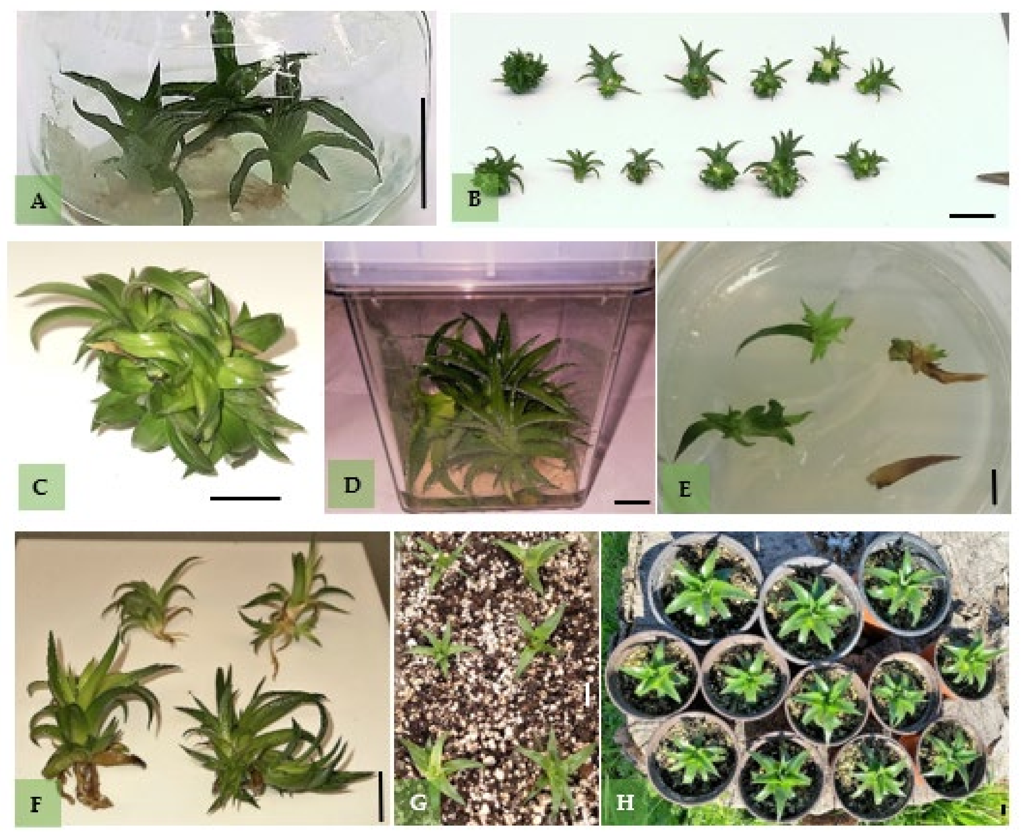
Figure 4.
In vitro regeneration of Dyckia brevifolia: (A) plantlets growing and root formation during initial transfer of young seedlings after forty days on solid MS-Hf medium; (Β) first shoots forming on shoot explants after two weeks of culture on solid MS medium containing BA 1 mg L−1; (C) multiple shoot formation on a shoot explant after six weeks on a solid MS medium with 1 mg L−1 BA; (D) multiple shoot and spontaneous root formation on a shoot explant after six weeks of culture on a liquid MS medium with 1 mg L−1 2IP; (E) first shoots forming on leaf- explants after two weeks of culture on a solid MS medium containing BA 1 mg L−1; (F) multiple shoot and spontaneous root formation on a shoot explant after six weeks of culture on a solid MS medium with 1 mg L−1 2IP; (G) plantlets during ex vitro acclimatisation; (H) two-month-old plants showing vigorous growth. Bars represent a length of 1 cm.

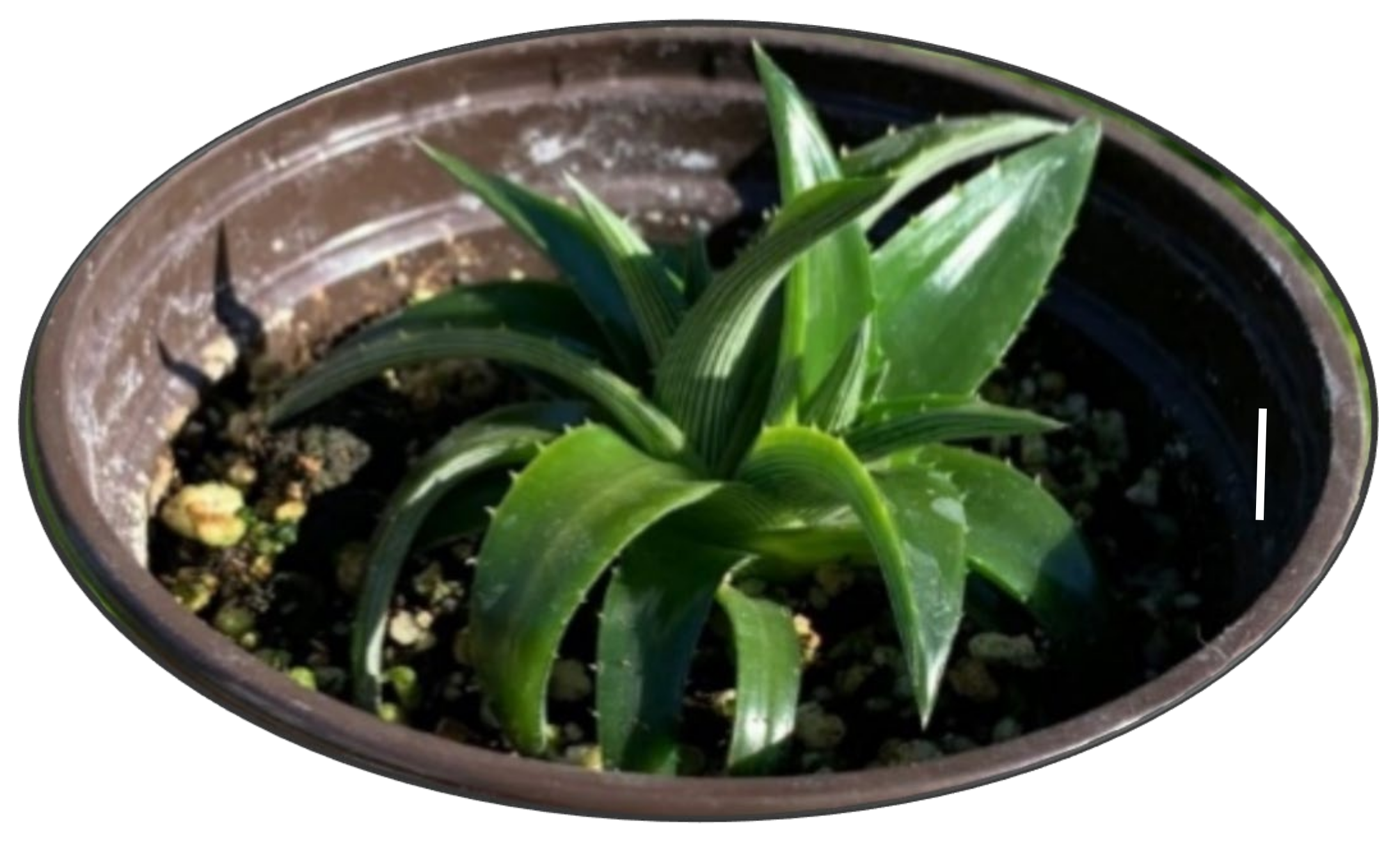
Figure 5.
Lateral sprouts formed at the base of one of the plantlets during acclimatisation. Bar represents length of 1 cm.
Figure 5.
Lateral sprouts formed at the base of one of the plantlets during acclimatisation. Bar represents length of 1 cm.


Table 1.
In vitro germination of Dyckia brevifolia seeds, T50 and germination speed index (GSI) at temperatures shown, after six months of storage at room temperature.
Table 1.
In vitro germination of Dyckia brevifolia seeds, T50 and germination speed index (GSI) at temperatures shown, after six months of storage at room temperature.
| Temperature (°C) | Germination (%) ± SD * | T50 (Days) | GSI |
|---|---|---|---|
| 5 | 0 | - | - |
| 10 | 0 | - | - |
| 15 | 86.00 ± 8.21 a | 15 | 25.24 c |
| 20 | 89.00 ± 5.47 a | 5 | 135.33 ab |
| 25 | 84.00 ± 11.81 a | 2 | 191.51 a |
| 30 | 35.00 ± 16.62 b | 4 | 84.17 b |
| 35 | 0 | - | - |
* Different letters in the same column indicate significant differences. Mean (±SE) separation in columns by Student’s t-test at p ≤ 0.05, n = 5, 20 seeds/Petri dish (total 100 seeds per treatment).
Table 2.
Effect of cytokinin type and concentration on shoot proliferation from shoot explants excised from shoots produced on MS medium (Hf or containing BA at 0.5 or 1.0 mg).
Table 2.
Effect of cytokinin type and concentration on shoot proliferation from shoot explants excised from shoots produced on MS medium (Hf or containing BA at 0.5 or 1.0 mg).
| Cytokinin | Concentration (mg L−1) | Survival (%) | Growth (cm) | Number of Leaves |
|---|---|---|---|---|
| Hf † | - | 95.0 a | 0.7 ab | 8.4 a |
| BA | 0.5 | 100.0 a | 0.5 c | 6.5 c |
| 1.0 | 97.5 a | 0.6 abc | 6.4 c | |
| KIN | 0.5 | 97.5 a | 0.8 a | 6.8 bc |
| 1.0 | 95.0 a | 0.6 abc | 7.3 ab | |
| 2IP | 0.5 | 100.0 a | 0.7 abc | 6.1 c |
| 1.0 | 100.0 a | 0.7 abc | 7.6 ab | |
| Fone-way ANOVA | NS | *** | *** | |
| Fcyt | NS | |||
| Fconc | NS | |||
| Fcyt×conc | NS | * | * | |
Different letters in the same column indicate significant differences. NS, *, *** Nonsignificant or significant at p ≤ 0.05, p ≤ 0.001, respectively, n = 40–50. † Hf (hormone free) treatment was excluded for 2-way ANOVA.
Table 3.
Effect of cytokinin type and concentration on shoot proliferation from shoot explants, excised from plantlets on solid and liquid media containing BA, KIN, or 2IP, at 1.0 mg L−1, or Hf.
Table 3.
Effect of cytokinin type and concentration on shoot proliferation from shoot explants, excised from plantlets on solid and liquid media containing BA, KIN, or 2IP, at 1.0 mg L−1, or Hf.
| MS | Cytokinin | Stem Growth (cm) | Number of Leaves/Main Shoot | Formation of Lateral Shoots (%) | Lateral Shoot Number | MI | Number of Leaves/Lateral Shoot |
|---|---|---|---|---|---|---|---|
| Solid | Hf † | 0.9 b | 8.5 cd | 0.0 c | 0.0 c | - | 0.0 c |
| BA | 0.8 b | 6.2 e | 83.0 a | 1.8 ab | 1.50 ab | 4.6 b | |
| KIN | 0.9 b | 7.7 de | 0.0 c | 0.0 c | - | 0.0 c | |
| 2IP | 0.7 b | 6.1 e | 54.0 b | 2.0 ab | 1.08 b | 4.9 b | |
| Liquid | Hf † | 1.7 ab | 12.1 a | 8.0 b | 1.5 bc | 0.11 b | 4.9 b |
| BA | 1.9 ab | 10.2 bc | 73.0 a | 2.7 a | 1.97 a | 4.9 b | |
| KIN | 2.3 a | 11.6 ab | 63.0 ab | 1.9 ab | 1.19 b | 6.1 a | |
| 2IP | 2.2 a | 10.5 bc | 84.0 a | 2.3 ab | 1.93 a | 6.0 a | |
| Fone-way ANOVA | *** | *** | ** | *** | * | *** | |
| Fmed | *** | *** | *** | ** | *** | ||
| Fcyt | NS | ** | *** | ** | *** | ||
| Fcyt×med | NS | NS | * | NS | NS | NS | |
Different letters in the same column indicate significant differences. NS, *, **, *** Non-significant or significant at p ≤ 0.05, p ≤ 0.01, p ≤ 0.001, respectively, n= 50–60, † Hf = hormone-free.
Table 4.
Shoot proliferation from leaf-origin explants excised from shoots produced either on Hf MS medium or on MS containing BA at 0.5 or 1.0 mg L−1, on solid and liquid media containing BA, KIN or 2IP at 1.0 mg L−1 or Hf.
Table 4.
Shoot proliferation from leaf-origin explants excised from shoots produced either on Hf MS medium or on MS containing BA at 0.5 or 1.0 mg L−1, on solid and liquid media containing BA, KIN or 2IP at 1.0 mg L−1 or Hf.
| MS | Cytokinin | Formation of Lateral Shoots (%) | Growth (cm) | Lateral Shoot Number | MI | Number of Leaves/Shoot |
|---|---|---|---|---|---|---|
| Solid | Hf † | 20.0 b | 1.0 ab | 1.4 c | 0.28 c | 8.8 a |
| BA | 41.0 a | 0.5 c | 1.4 c | 0.57 c | 7.0 a | |
| KIN | 35.0 a | 0.8 bc | 1.5 c | 0.53 c | 8.0 a | |
| 2IP | 44.5 a | 0.9 b | 1.5 c | 0.67 bc | 8.9 a | |
| Liquid | Hf† | 17.0 b | 1.2 ab | 1.0 c | 0.17 c | 7.4 a |
| BA | 26.0 ab | 1.0 ab | 7.4 a | 1.90 a | 6.6 a | |
| KIN | 22.5 b | 1.3 a | 2.6 b | 0.59 bc | 7.6 a | |
| 2IP | 27.5 b | 1.0 ab | 3.3 b | 0.90 b | 6.3 a | |
| Fone-way ANOVA | *** | ** | *** | ** | NS | |
| Fmed | *** | * | ||||
| Fcyt | NS | NS | ||||
| Fcyt×med | NS | NS | * | * | * | |
Different letters in the same column indicate significant differences. NS, *, **, *** Non-significant or significant at p ≤ 0.05, p ≤ 0.01, p ≤ 0.001, respectively, n = 50–60. † Hf = hormone-free.
Table 5.
In vitro rooting of shoots during first subculture on MS supplemented BA, KIN or 2IP at 0.5 or 1.0 mg L−1, or Hf.
Table 5.
In vitro rooting of shoots during first subculture on MS supplemented BA, KIN or 2IP at 0.5 or 1.0 mg L−1, or Hf.
| Cytokinin | Concentration (mg L−1) | Rooting (%) | Root Number | Root Length (cm) |
|---|---|---|---|---|
| Hf † | - | 95.0 a | 2.1 a | 1.2 a |
| BA | 0.5 | 20.0 b | 1.0 b | 0.5 c |
| 1.0 | 13.0 b | 1.0 b | 0.5 c | |
| KIN | 0.5 | 93.0 a | 2.3 a | 1.1 ab |
| 1.0 | 81.0 a | 1.6 ab | 0.9 bc | |
| 2IP | 0.5 | 100.0 a | 1.3 b | 0.9 bc |
| 1.0 | 79.0 ab | 1.6 ab | 0.8 c | |
| Fone-way ANOVA | ** | ** | ** | |
| Fcyt | NS | |||
| Fconc | ** | |||
| Fcyt×conc | * | * | NS | |
Different letters in the same column indicate significant differences, † Hf = hormone-free. NS, *, ** Non-significant or significant at p ≤ 0.05, p ≤ 0.01, respectively, n = 50–60.
Table 6.
In vitro rooting of shoots derived from shoots or leaves as affected by the type of MS (solid or liquid) and of cytokinin during multiplication stage.
Table 6.
In vitro rooting of shoots derived from shoots or leaves as affected by the type of MS (solid or liquid) and of cytokinin during multiplication stage.
| Shoot-Origin | Leaf-Origin | ||||||
|---|---|---|---|---|---|---|---|
| MS | Cytokinin | Rooting (%) | Root Number | Root Length (cm) | Rooting (%) | Root Number | Root Length (cm) |
| Solid | Hf † | 94.0 a | 2.2 b | 1.1 c | 73.5 a | 2.0 a | 1.2 a |
| BA | 0.0 c | 0.0 c | 0.0 c | 50.0 ab | 1.0 a | 0.5 a | |
| KIN | 47.0 b | 1.4 b | 1.0 c | 18.0 b | 1.7 a | 1.0 a | |
| 2IP | 43.0 b | 1.8 b | 0.9 c | 52.5 ab | 2.0 a | 0.9 a | |
| Liquid | Hf † | 74.5 ab | 4.2 a | 1.7 b | 62.5 ab | 2.4 a | 1.4 a |
| BA | 0.0 c | 0.0 c | 0.0 c | 0.0 b | 0.0 b | 0.0 b | |
| KIN | 18.3 b | 3.9 a | 2.3 a | 0.0 b | 0.0 b | 0.0 b | |
| 2IP | 77.5 ab | 3.8 a | 2.4 a | 0.0 b | 0.0 b | 0.0 b | |
| Fone-way ANOVA | *** | *** | *** | * | NS | NS | |
| Fmed | *** | ||||||
| Fcyt | NS | ||||||
| Fcyt × med | *** | NS | * | ** | *‘ | * | |
Different letters in the same column indicate significant differences. NS, *, **, *** Non-significant or significant at p ≤ 0.05, p ≤ 0.01, p ≤ 0.001, respectively, n = 50–60. † Hf = hormone-free.
Publisher’s Note: MDPI stays neutral with regard to jurisdictional claims in published maps and institutional affiliations. |
© 2022 by the authors. Licensee MDPI, Basel, Switzerland. This article is an open access article distributed under the terms and conditions of the Creative Commons Attribution (CC BY) license (https://creativecommons.org/licenses/by/4.0/).
Share and Cite
MDPI and ACS Style
Bertsouklis, K.; Panagaki, K.-P. In Vitro Germination and Propagation of Dyckia brevifolia, An Ornamental and Endangered Bromeliad. Horticulturae 2022, 8, 390. https://doi.org/10.3390/horticulturae8050390
AMA Style
Bertsouklis K, Panagaki K-P. In Vitro Germination and Propagation of Dyckia brevifolia, An Ornamental and Endangered Bromeliad. Horticulturae. 2022; 8(5):390. https://doi.org/10.3390/horticulturae8050390
Chicago/Turabian StyleBertsouklis, Konstantinos, and Konstantina-Panagiota Panagaki. 2022. "In Vitro Germination and Propagation of Dyckia brevifolia, An Ornamental and Endangered Bromeliad" Horticulturae 8, no. 5: 390. https://doi.org/10.3390/horticulturae8050390
Note that from the first issue of 2016, this journal uses article numbers instead of page numbers. See further details here.