Revealing Ancient Faces
The Reconstruction of a Neandertal
[authors]
POST-MORTEM facial reconstruction is a technique that uses anatomical knowledge of the human skull to flesh out the face of a deceased individual. Forensic artists work with law enforcement to identify victims of crime when skeletonized remains are found. Archaeologists use the same technique to learn what a person who lived and died long before photographs may have looked like.
For many people, skulls are an object of fear, a vivid reminder of inevitable death and decay. Skulls represent the universality of our mortality, and, without flesh, one may not look much different from another. It takes observational skill and specialized training to know whether a skeleton is male or female and what the age and lifestyle of that person may have been. Our detailed understanding of the human body, amassed over centuries, has allowed us to develop new techniques that tell us more from bones than we have ever been able to learn before. Through the work of Dr. Janet Monge, Associate Curator and Keeper of the Physical Anthropology Section, and Pennsylvania Academy of Fine Arts (PAFA) artist and fourth-year student Kathleen Gallo, visitors to the Museum’s Sphinx Gallery will be able to view the face of a man who may have died 60,000 years ago.
The “Old Man” of Shanidar
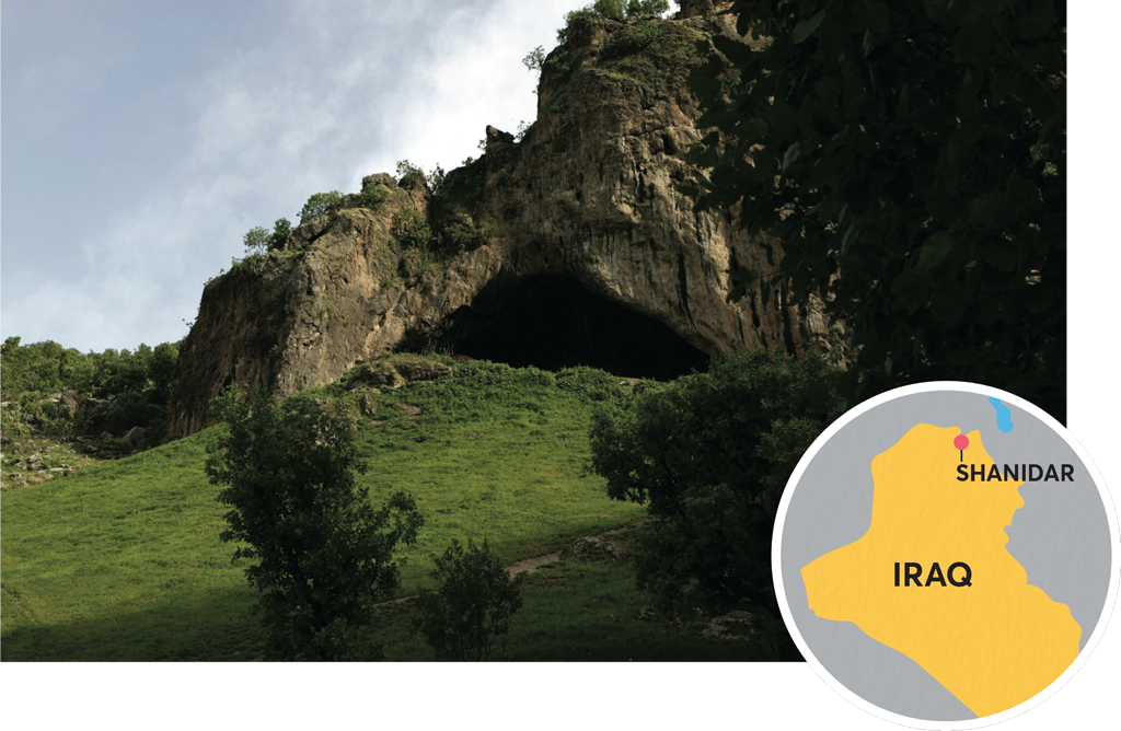
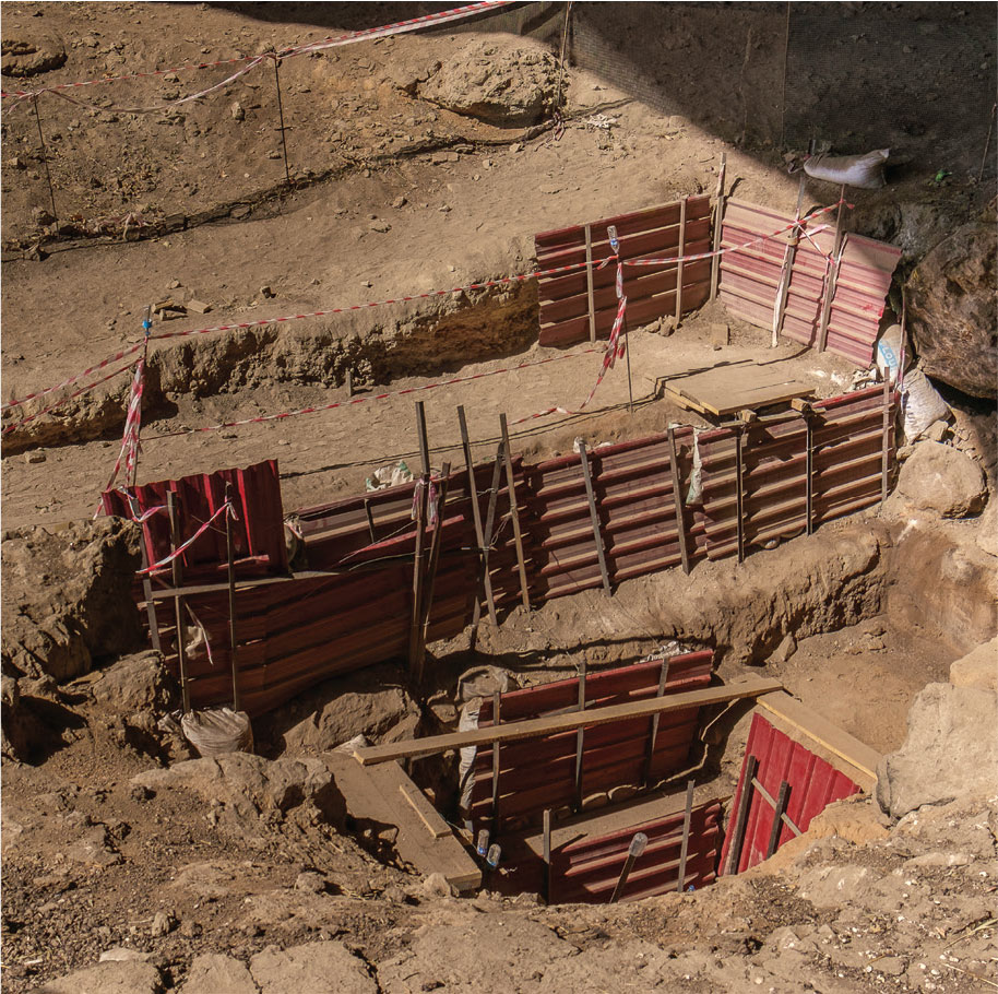
Shanidar Cave is located in what is today Kurdistan, in northern Iraq. It was here in the 1950s that archaeologist Dr. Ralph Solecki excavated the bodies of eight adults and two children, thought to have been buried during occupations in the cave between 45,000 and 60,000 years ago. Among some of the remains, Solecki identified evidence that changed our thinking about Neandertals and their culture.
When the first Neandertal bones were discovered in Germany in 1856, it was assumed that these thick-boned, heavy-browed hominins were more apelike than modern humans, lacking our cognitive abilities and unable to develop what might be thought of as culture. At the burial site at Shanidar, however, Solecki’s intensive study of the skeletal remains revealed signs of social relationships and ritual, exposing a cultural complexity previously thought to belong only to modern humans. Forensic evidence showed that Neandertals took care of each other, helping to heal injuries and tending to the sick. The discovery of pollen around the remains of some of the individuals also suggested that Neandertals mourned their dead and ritualized their burial with flowers.
Solecki’s excavation uncovered a man known as Shanidar 1, who had lost part or all of the vision in his left eye due to a traumatic injury. Studies have also shown that debris and earwax impacted in his right ear would have made him deaf on that side. His right arm was amputated below the elbow and he suffered from arthritis. Yet he lived to what we could consider an advanced age for a Neandertal: while it is difficult
to assess the exact age of an individual from skeletal remains, it is believed that he lived to be at least 50 years old. After being treated for his injuries, he must have been cared for by his community and provided with food and shelter despite being physically and sensory disabled. This required a social structure that was not previously thought to exist in Neandertal society. The position of his body indicated that he died in a rockfall inside the cave; a collection of smaller stones on the fossilized remains suggested to Solecki that his companions covered the body with these stones afterwards. While Solecki’s work has been heavily critiqued, many scholars now believe—through the study of other Neandertal burial sites—that Neandertals did in fact intentionally bury their dead.
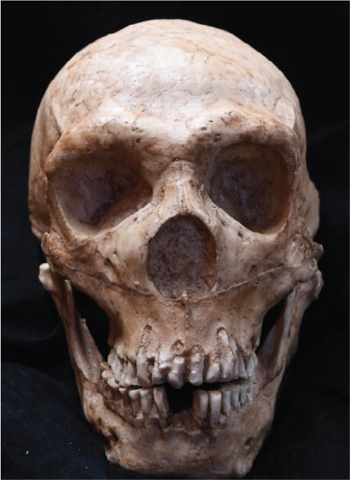
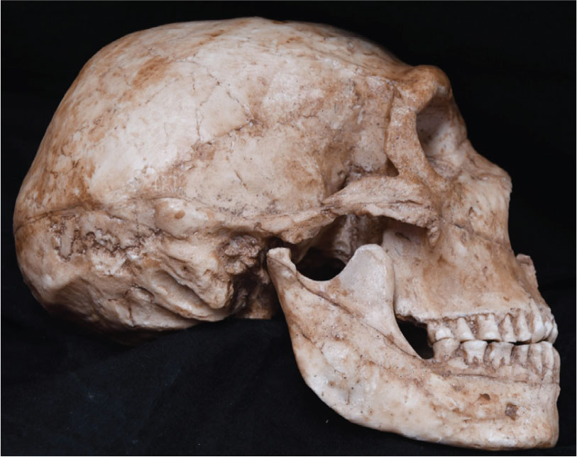
Shanidar 1’s skeleton remains in Iraq, but in the 1970s a mold of the skull was made by the Wenner-Gren Foundation of Anthropological Research. This mold was transferred in 1980 to the Penn Museum, where a cast was made out of the mold. There are very few castings of this skull in the world, so this is a unique opportunity for the Penn Museum to display a pivotal discovery in anthropology.
The Science and Art of Facial Reconstruction
Modern facial reconstruction techniques can be traced back to the work in the late 19th century of German anatomists and physiologists Hermann Welcker and Wilhelm His, who analyzed the relationship between the bone structure of the human skull and the soft tissues of the face. Understanding where muscles and tendons connect beneath layers of skin is crucial to visualizing the contours of a human face.
One aspect of facial reconstruction that might be familiar is the use of depth markers: small pegs of specific length are attached to the skull at particular points to illustrate what thickness the tissue would have been in those places. The work of Wilhelm His, Julius Kollman, and W. Buchly in the late 19th century resulted in a comprehensive table of tissue thicknesses and enough data points to make the accuracy of facial reconstructions exponentially better.
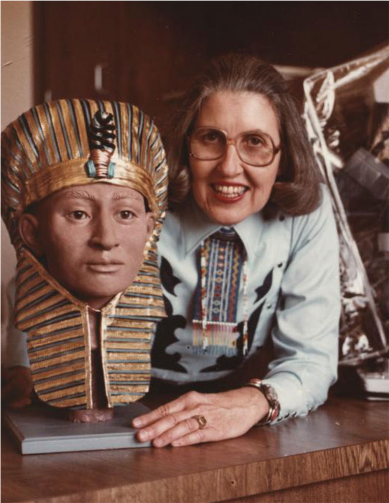
It was not until the 1960s that law enforcement turned to this technique in an effort to identify skeletonized remains. Betty Pat Gatliff (1930–2020) was a pioneer in forensic art. Working with anthropologist Dr. Clyde Snow, she developed the Gatliff/Snow American Tissue Depth Method, which identified features on the skull that should be used as landmarks for building a face. The skull is a map that tells the sculptor where the eyelids converge, where the lips begin, and the approximate length and width of the nose. The tissue depth markers are primarily based on ethnicity, and adjustments are made based on age. Gatliff’s work assisted in identifying victims of serial killer John Wayne Gacy and reproducing the head of President John F. Kennedy, which was used for trajectory tests during the 1978 House investigation into his assassination. Gatliff trained forensic artist Joe Mullins, who taught Kathleen Gallo facial reconstruction.
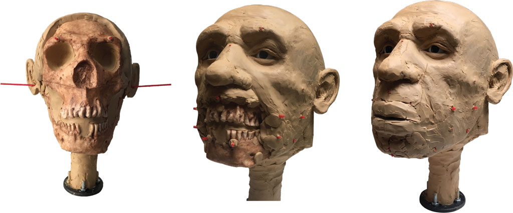
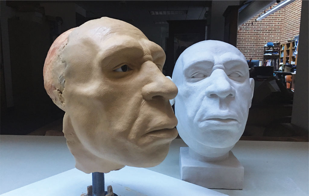
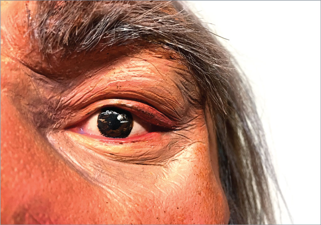
A recognizable face begins to emerge, depth markers still visible and the mouth and chin ready to be built. The eyes in the skull are glass marbles. Once his face is complete, a plaster cast is made for the Museum display.
What Does the “Old Man” of Shanidar Look Like?
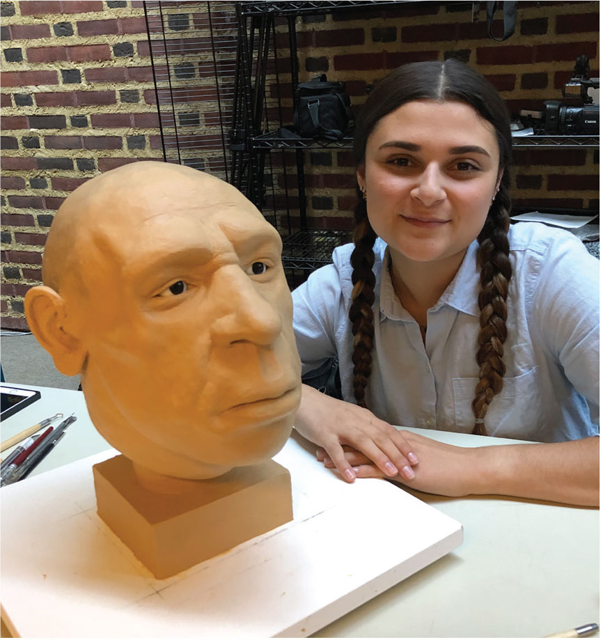
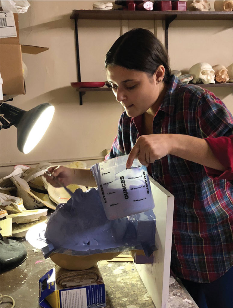
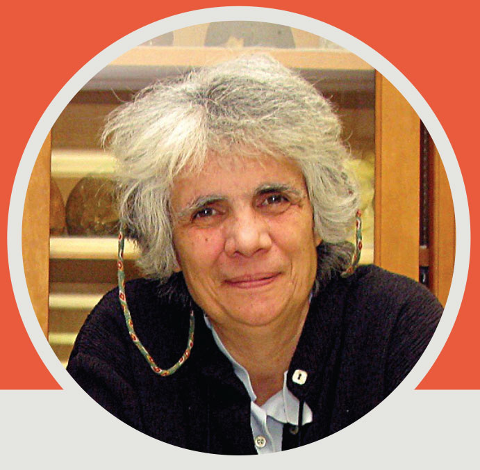
PAFA student Kathleen Gallo, who has provided forensic reconstructions to the Mütter Museum and the Pima County (Arizona) Police, worked under the direction of Dr. Janet Monge to reconstruct the face of Shanidar 1, the “Old Man.” This method of reconstruction involves attaching pegs to the cast indicating different thicknesses of tissue, and using clay to model the shape based on specific points that define the contours of the human face. For example, in the case of a Neandertal, anthropologists know that there is greater muscle mass around the mouth and jaw and larger teeth than in modern humans.
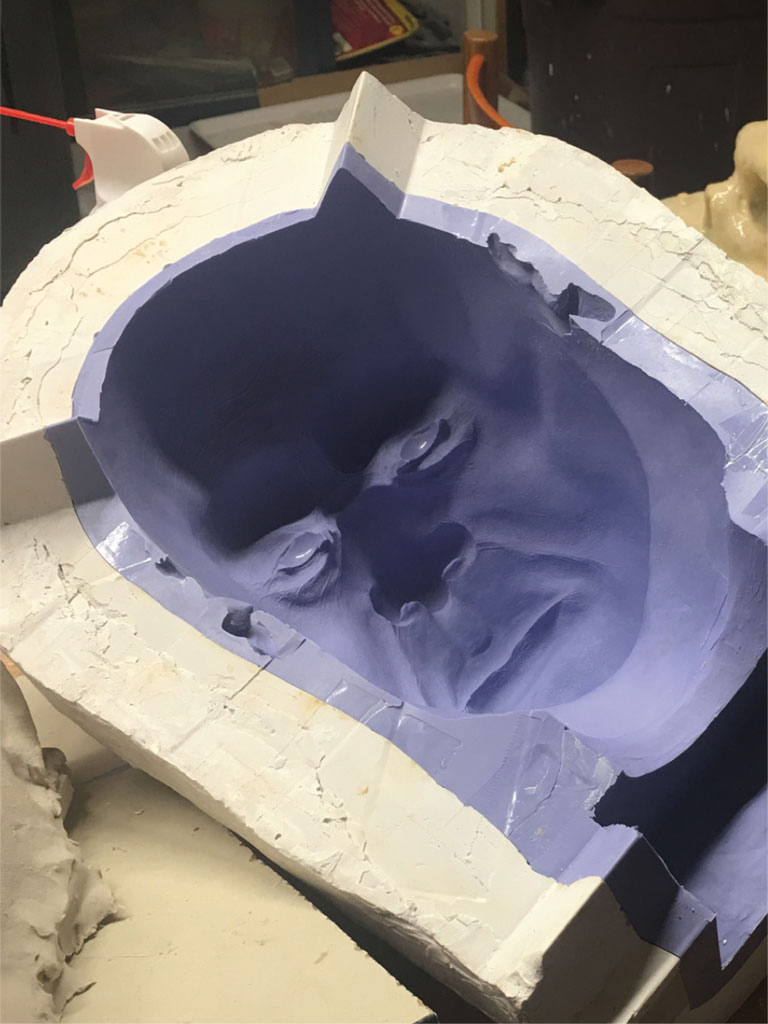
In the next step, the forensic artist reimagines the face, based on the structure of the skull, the position of various features, and what we know about the age and sex of this particular Neandertal. The artist uses oil-based plasticine clay, which will not harden, and adds details to the skin such as pores and wrinkles. She coats the piece with silicone, which will pick up the face in complete and meticulous detail, and a plaster cast is made.
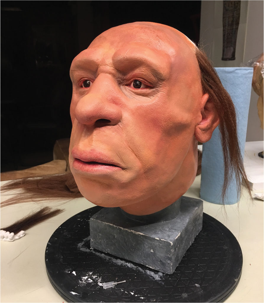
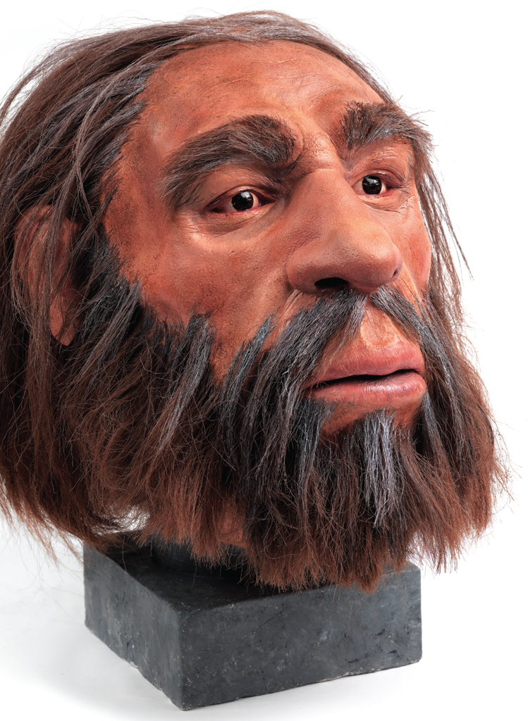
Shanidar 1 is an especially interesting individual with much character in his face. For Janet Monge, he is an example of “how life is embedded essentially in the body.” In recreating his face, Gallo had to take into account features that are the result of his life history: sagging skin, loss of collagen, and even a scar over his left eye. His complexion, that of a man from the Middle East who lived a life in the outdoors, was described by Monge as “ruddy and craggy.” It is known that the gene for red hair present in Europeans came from Neandertals, so an auburn color was chosen for his hair, with some gray to indicate his age. The application of hair and skin details, eyes, and painting skin tone are the final steps that bring to life a distant relative who passed away in a cave tens of thousands of years ago. This work of scientific art, which took about 120 hours to complete, now resides in a case in the Sphinx Gallery.
This project enabled the Penn Museum to engage in a partnership with the Pennsylvania Academy of Fine Arts and a talented student, bringing together the creative and the scientific to provide a powerful experience for Museum visitors. Monge is pleased with the result: “Our visitors will have a good frame of reference when thinking about older individuals and relatives, and how their bodies are really a testament to their life history.” Seeing the face of Shanidar 1 can tell us so much more about his life than we might imagine from his bare bones.
The analysis of Shanidar 1 and his reconstruction illustrate the basis of what makes us human and highlight the commonalities that unite us, which are much more important than our differences.
A Conversation with Janet Monge
Dr. Janet Monge, Associate Curator and Keeper of the Physical Anthropology Section, is a renowned forensic anthropologist well known to Penn Museum members, students, and staff for her engaging lectures and her insightful research in Africa, Europe, and the United States. We sat down with Janet to discuss the reconstruction of a Neandertal head that is now on display in the new Sphinx Gallery.
- In the Sphinx Gallery case, one object represents each of the Museum’s curatorial sections. How does the forensic reconstruction of the Neandertal head represent the work of Physical Anthropology?
- Physical anthropologists are interested in human variation over time and space. We want to answer the big question: what does it mean to be “human”? This question relates to both human biological variation (the differences in each of us) and variation in human behavioral expression (how those differences are seen or expressed in individuals). We also want to understand how “humanness” emerged in our evolutionary lineage.
- Tell us about the reconstruction. Why is it important?
- This is a flesh reconstruction of the fossilized skull of a Neandertal, Shanidar 1, excavated in Iraq and dated to between 60,000 and 45,000 BP (before present). Because the skull is so complete (with an intact cranium and mandible), it is an excellent example of the morphology of an extinct member of the human evolutionary lineage. Thus, this is a critical piece of evidence in the search for human evolutionary history. It gives us insights into the lifeways of very human-like ancestors from the distant past. Since this person was old when he died, he has many of the “scars” of existence, which detail episodes throughout his life. It is not that members of our lineage did not live into older age, but rather that our evidence of older folks is limited.
- Why should this reconstruction matter to a Museum visitor?
- Shanidar 1 illustrates two very important aspects of humanness: living with disability and compassion from community. These two features are key in understanding the unique lifeways of humans. How do we know this? Based on the bones of his skeleton, he had lost his arm many years before death, and we know that his arm was amputated probably right above his elbow joint. Wear on his teeth shows that he was using his mouth as a place to hold things. He also had a scar across his eye which might have compromised his ability to see in that eye and reduced his depth of vision. He would have needed a helper or even a community of helpers to take care of him. In other words, dealing with disability with kindness and compassion has been a deep feature of human evolutionary history.
- How and why was the reconstruction made?
- The reconstruction was made by Kathleen Gallo, a student at the Philadelphia Academy of Fine Arts. She is an expert in putting muscle and flesh onto the bones of humans so that we can get a sense of what people looked like in the past. These very same techniques are used when skeletal materials are found in a forensic context and when we need to know more about the remains to aid in identification of the deceased person. It works in both contexts— evolution and law enforcement.
- How does our understanding of Neandertal lifeways allow us to see similarities between the past and the present?
- The analysis of Shanidar 1 and his reconstruction illustrate the basis of what makes us human and highlight the commonalities that unite us, which are much more important than our differences. Humans are defined by belonging, family, community, connection, and overwhelming kindness and caring— traits we share with Neandertals.
Christina Griffith was, until her recent graduation from the University of Pennsylvania, Associate Editor of Expedition magazine.