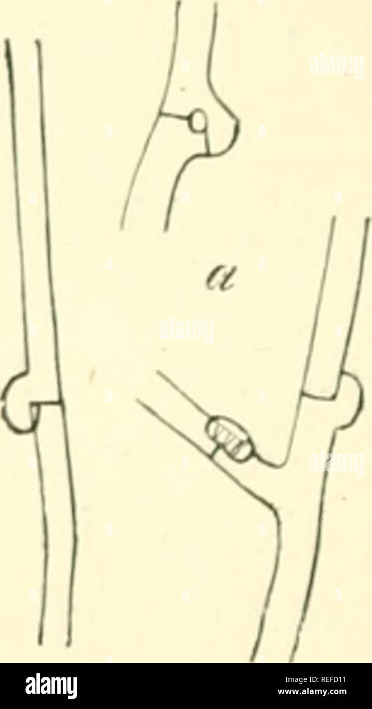
. Comparative morphology and biology of the fungi, mycetozoa and bacteria. Fungi -- Morphology; Bacteria -- Morphology. FIG. i. Germinating gonidia of Nectria (Spicaria) Solani, Reinke ; a developing into an isolated hypha, in the rest the hyphae have coalesced. Magn. 390 times. Fig. 2. Clamp-connections of the my- celium of Hypochnus centrifugns. Tul. M ign. J90 times. following manner :—the lateral wall or the extremity of a branch or of a segment- cell of the branch places itself on another branch or cell, and the membranes of both disappear at the point of contact, so that the cavities and
Image details
Contributor:
The Book Worm / Alamy Stock PhotoImage ID:
REFD11File size:
7.2 MB (103.6 KB Compressed download)Releases:
Model - no | Property - noDo I need a release?Dimensions:
1185 x 2109 px | 20.1 x 35.7 cm | 7.9 x 14.1 inches | 150dpiMore information:
This image is a public domain image, which means either that copyright has expired in the image or the copyright holder has waived their copyright. Alamy charges you a fee for access to the high resolution copy of the image.
This image could have imperfections as it’s either historical or reportage.
. Comparative morphology and biology of the fungi, mycetozoa and bacteria. Fungi -- Morphology; Bacteria -- Morphology. FIG. i. Germinating gonidia of Nectria (Spicaria) Solani, Reinke ; a developing into an isolated hypha, in the rest the hyphae have coalesced. Magn. 390 times. Fig. 2. Clamp-connections of the my- celium of Hypochnus centrifugns. Tul. M ign. J90 times. following manner :—the lateral wall or the extremity of a branch or of a segment- cell of the branch places itself on another branch or cell, and the membranes of both disappear at the point of contact, so that the cavities and protoplasmic contents of the two cells become united into one. Coalescence in this way may take place between the branches of the same hypha, and also between such as are growing together but were originally distinct, being the product of distinct germ-cells. The forms which result from such coalescence are very various; H-shaped cross links or bridges, loops of various form and number, even network of many narrow meshes are found. One curious form must be mentioned here, the clamp-connections (Schnallen- verbindungen), first observed by Hoffmann (Fig. 2). They occur only on hyphae with transverse segmentation, and chiefly in the Basidiomvt etes (many Agaricineae, species ol Polyporus, Typhula, Hypochnus, Cyathus, Hymenogaster, &c). A clamp of this kind when fully formed is usually a nearly semicircular protuberance like a short branch which springs from one cell close to a transverse wall, and is closely applied to the lateral wall of the adjoining cell in such a way that the transverse wall cuts the middle of the plane of contact at a right angle. Sometimes the protuberance not lie close on the lateral wall at all points, but forms an eye-hole. Bivfcld observed the origin of these formations in Coprinus. and found that the protuberance extends itself from the one of the two adjoining cells to the other, and then coalescence takes place, so that the two cells enter into