. Comparative morphology of Fungi. Fungi. 356 COMPARATIVE MORPHOLOGY OF FUNGI In the cavities of Pachyphloeus, this secondary tissue has developed to a pseudoparenchyma of looser structure than the ground tissue. In a cross section through a mature fructification it appears penetrated by two sorts of veins (Fig. 239, 1 and 2). One, the so-called tramal plates (gen- erally darker) or venae internae, which arising in the tissue zone lying under the rind, converge toward the top and bear the hymenial palisade on their surface, and the other, venae externae (generally lighter), which begin at the
Image details
Contributor:
The Book Worm / Alamy Stock PhotoImage ID:
REF6YBFile size:
7.1 MB (428.7 KB Compressed download)Releases:
Model - no | Property - noDo I need a release?Dimensions:
1871 x 1335 px | 31.7 x 22.6 cm | 12.5 x 8.9 inches | 150dpiMore information:
This image is a public domain image, which means either that copyright has expired in the image or the copyright holder has waived their copyright. Alamy charges you a fee for access to the high resolution copy of the image.
This image could have imperfections as it’s either historical or reportage.
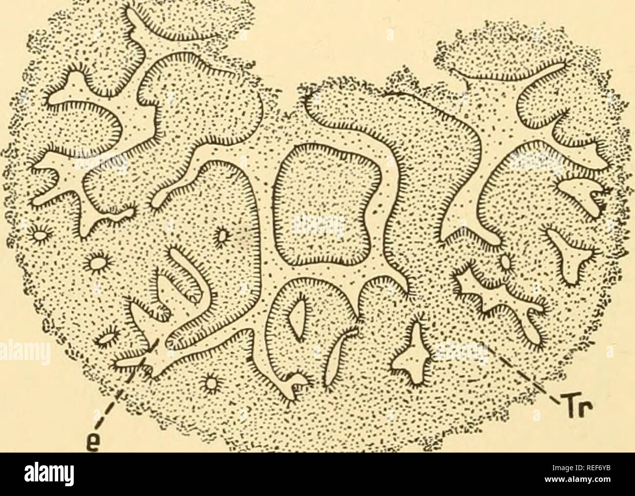
. Comparative morphology of Fungi. Fungi. 356 COMPARATIVE MORPHOLOGY OF FUNGI In the cavities of Pachyphloeus, this secondary tissue has developed to a pseudoparenchyma of looser structure than the ground tissue. In a cross section through a mature fructification it appears penetrated by two sorts of veins (Fig. 239, 1 and 2). One, the so-called tramal plates (gen- erally darker) or venae internae, which arising in the tissue zone lying under the rind, converge toward the top and bear the hymenial palisade on their surface, and the other, venae externae (generally lighter), which begin at the top of the fructification, penetrate its interior and end blindly. Between the two systems of veins there lies a hymenium consisting of parallel paraphyses and asci arranged in a regular palisade with tips directed toward the venae externae. Ontogenetically the venae internae are only the folded parts of the wall of the hollow sphere of the fructifica- tion, such as we have come to know in Hydnotrya, while the venae externae form the originally looser, later pseudoparenchymatous tissue which. â 'ti^'^fi;"^^ â rwf-y-is' '*'â â ;* Fig. 238.âPseudobahamia magnata. Median section of fructification. (X6; after E. Fischer, 1908.) has arisen by the growth of the paraphyses and secondarily has filled the hollow passages in the interior of the fructification. By these venae externae the fructifications of Pachyphloeus, in contrast to those of Hydnotrya, form a compact mass, though ontogenetically heterogeneous, such as we usually meet in Tuber. Apparently in Tuber, the fructifications, as in Hydnotrya and possibly in the above genera, are formed gymnocarpously (Bucholtz, 1897, 1903). Thus, Fig. 239, 3 shows the stage of 1.5 mm. cross section; a flat shell on whose interior project several tramal plates, venae internae. This flat shell is sometimes called ground plate. Apparently it corresponds to the hypothecium of apothecia but is histologically more differentiated and is pen