. Comparative morphology of Fungi. Fungi. 80 COMPARATIVE MORPHOLOGY OF FUNGI In Phytophthora (Fig. 45, 2 to 7), Albugo, Basidiophora, Sclerospora and, in part, Plasmopora (Fig. 48, C), the stage of the germ sac has almost entirely disappeared. Here the mature zoospores swarm directly out of the conidium. In other forms, the formation of swarm spores is suppressed and replaced by the direct development of conidia to a myce- lium. Thus in Plasmopara pygmaea, the undivided content of the conid- ium swells out of the top, rounds off and puts out a germ tube. In Bremia, the conidium germinates dire
Image details
Contributor:
The Book Worm / Alamy Stock PhotoImage ID:
REE45RFile size:
7.1 MB (152.2 KB Compressed download)Releases:
Model - no | Property - noDo I need a release?Dimensions:
1192 x 2096 px | 20.2 x 35.5 cm | 7.9 x 14 inches | 150dpiMore information:
This image is a public domain image, which means either that copyright has expired in the image or the copyright holder has waived their copyright. Alamy charges you a fee for access to the high resolution copy of the image.
This image could have imperfections as it’s either historical or reportage.
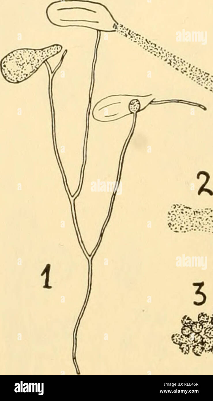
. Comparative morphology of Fungi. Fungi. 80 COMPARATIVE MORPHOLOGY OF FUNGI In Phytophthora (Fig. 45, 2 to 7), Albugo, Basidiophora, Sclerospora and, in part, Plasmopora (Fig. 48, C), the stage of the germ sac has almost entirely disappeared. Here the mature zoospores swarm directly out of the conidium. In other forms, the formation of swarm spores is suppressed and replaced by the direct development of conidia to a myce- lium. Thus in Plasmopara pygmaea, the undivided content of the conid- ium swells out of the top, rounds off and puts out a germ tube. In Bremia, the conidium germinates directly by a germ tube which has grown out of a papilla at the tip; in Peronospora the papilla is lacking and the germ tubes come from any portion of the conidium. Wherever zoospores are formed in the Peronosporaceae they are reni- form and possess two lateral fiagella (Fig. 49, 4; 44, 5); thus they are. $*- " -'riVV;^ ^â¬p -''S>. â¢â >â -, . *->% fs -â¢;; y.'V'-.-.Vv '" -l' .â¢'â >:â > ⺠-;m i Fig. 49.âPythiogeton transversum. 1. Hyphae with three zoosporangia, one of which is discharging. 2. Protoplast has formed an elongate mass on emerging. 3. Zoospore formation. 4. Single swarming zoospore. {After Minden, 1916.) connected directly to the Saprolegnieae of the Achlya type. Those of Pythiogeton swim about for a while (up to two days) then come to rest, surround themselves with a membrane and germinate to a mycelium. Butler (1907) has observed in Pythium diacarpum and Murphy (1922) in Phytophthora infestans the shedding of the membrane, as in Dictyuchus; the zoospores which have found no suitable substrate come to rest and surround themselves with a membrane. After a certain time they again slip out with the same reniform appearance and swarm further. The phenomenon has further been described by Bary for Pythium proliferum and by Atkinson for P. intermedium; ocasionally the bifiagel- late zoospores may split into two uniflagellate ones; th