. Introduction to the study of fungi; their organography, classification, and distribution, for the use of collectors. Fungi. CAPSULAR FUNGI—PYRENOMYCETES 199. tiated into chains of hyaline conidia, which fall away and are capable of germinating and producing a new mycelium. In this condition the parasite is a mould, or Hyphomycete, and was formerly in- cluded under the geniis Oidium, under the supposition that it was a complete and autonomous Fungus. Eecent in- vestigation has shown that this stage of mould is Only the COnidial condition of Fig. 87. — Perithecium and some species of the Erysi
Image details
Contributor:
The Book Worm / Alamy Stock PhotoImage ID:
RDXH4WFile size:
7.2 MB (195.8 KB Compressed download)Releases:
Model - no | Property - noDo I need a release?Dimensions:
1880 x 1330 px | 31.8 x 22.5 cm | 12.5 x 8.9 inches | 150dpiMore information:
This image is a public domain image, which means either that copyright has expired in the image or the copyright holder has waived their copyright. Alamy charges you a fee for access to the high resolution copy of the image.
This image could have imperfections as it’s either historical or reportage.
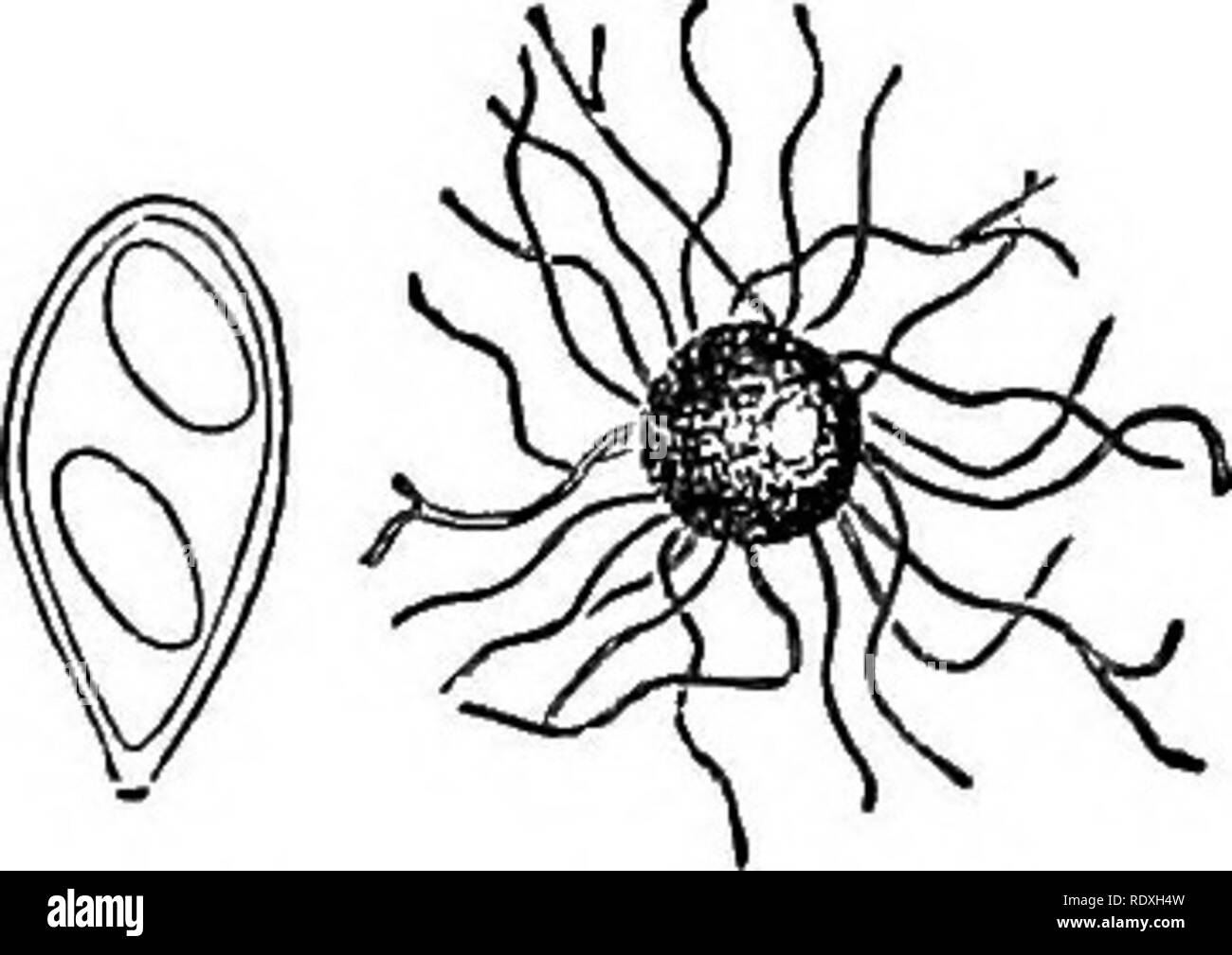
. Introduction to the study of fungi; their organography, classification, and distribution, for the use of collectors. Fungi. CAPSULAR FUNGI—PYRENOMYCETES 199. tiated into chains of hyaline conidia, which fall away and are capable of germinating and producing a new mycelium. In this condition the parasite is a mould, or Hyphomycete, and was formerly in- cluded under the geniis Oidium, under the supposition that it was a complete and autonomous Fungus. Eecent in- vestigation has shown that this stage of mould is Only the COnidial condition of Fig. 87. — Perithecium and some species of the Erysipheae, which ^ ™th sPoridia of succeed the Oidium. In the first in- stance minute spherical yellow bodies appear on the surface of the mycelium, and these gradually enlarge until they become just visible to the naked eye, and acquire a dark brown colour (Fig. 87). These are the perithecia, or membranaceous capsules, attached at the base by a copious mycelium and surrounded by a circlet of free arms, or processes, as appendages, which vary in the different genera. In Erysiphe they are thread-like and fiexuous, of equal diameter throughout, and simple. In Uncinula the arms are hooked or curved at the tips (Fig. 8 8). In Phyllactinia the appendages are straight, and often inflated at the base. In Sphaerotheca they are fiexuous and sometimes vaguely branched. In Podosphaera the appendages are repeatedly forked at the tips, as they are also in Microsphaera (Fig. 89). Internally these globose perithecia are replete with the ascigerous fructification. The asei are nearly globose, or pear-shaped, and contain hyaline elliptic sporidia. In Podosphaera and Sphaerotheca each perithecium en- closes but a single ascus. In the other genera the asci are numerous in each perithecium. The number of sporidia in each ascus varies with the genera, but with the exception of a single genus these sporidia are ovoid and continuous. In the exceptional genus Saccardia. Please note that these images