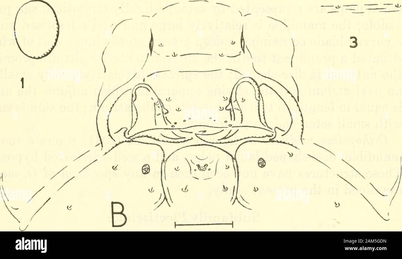
Proceedings of the United States National Museum . Ot m m. Olmm Figure 45.—Head sclerites: a, Collyria calcitratorJGravenhoTSt) (CoUyriinae); b, Orthopelmamediator (Thunberg) (Orthopelmatinae). (1, antenna; 2, spiracle; 3, skin.) 474 PROCEEDINGS OF THE NATIONAL MUSEUM vol. no C. calcitrator is figured by Salt (1931) and a large sclerotized plateis shown underlying the labial area. This plate is the suspensoriumof the hypopharynx (see Short, 1952), and is figured here, althoughin a different position (fig. 45a). The suspensorium is an internalskeletal structuie forming part of the ventral wall
Image details
Contributor:
The Reading Room / Alamy Stock PhotoImage ID:
2AM5GDNFile size:
7.1 MB (158.4 KB Compressed download)Releases:
Model - no | Property - noDo I need a release?Dimensions:
2086 x 1198 px | 35.3 x 20.3 cm | 13.9 x 8 inches | 150dpiMore information:
This image is a public domain image, which means either that copyright has expired in the image or the copyright holder has waived their copyright. Alamy charges you a fee for access to the high resolution copy of the image.
This image could have imperfections as it’s either historical or reportage.
Proceedings of the United States National Museum . Ot m m. Olmm Figure 45.—Head sclerites: a, Collyria calcitratorJGravenhoTSt) (CoUyriinae); b, Orthopelmamediator (Thunberg) (Orthopelmatinae). (1, antenna; 2, spiracle; 3, skin.) 474 PROCEEDINGS OF THE NATIONAL MUSEUM vol. no C. calcitrator is figured by Salt (1931) and a large sclerotized plateis shown underlying the labial area. This plate is the suspensoriumof the hypopharynx (see Short, 1952), and is figured here, althoughin a different position (fig. 45a). The suspensorium is an internalskeletal structuie forming part of the ventral wall of the cibarial partof the stomodaeum, and the position in which it is seen depends onthe position into which it is forced during mounting. Subfamily Orthopelmatinae Figure 45b Orthopelma mediator (Thunberg) was examined. The members ofthis genus are endoparasitic in the cyuipoids Diasirophus on Ruhusand Diplolepis on Rosa. The larval characters resemble those of the Ichneumoninae, althoughthe form of the mandible is different, the hypostoma is more li