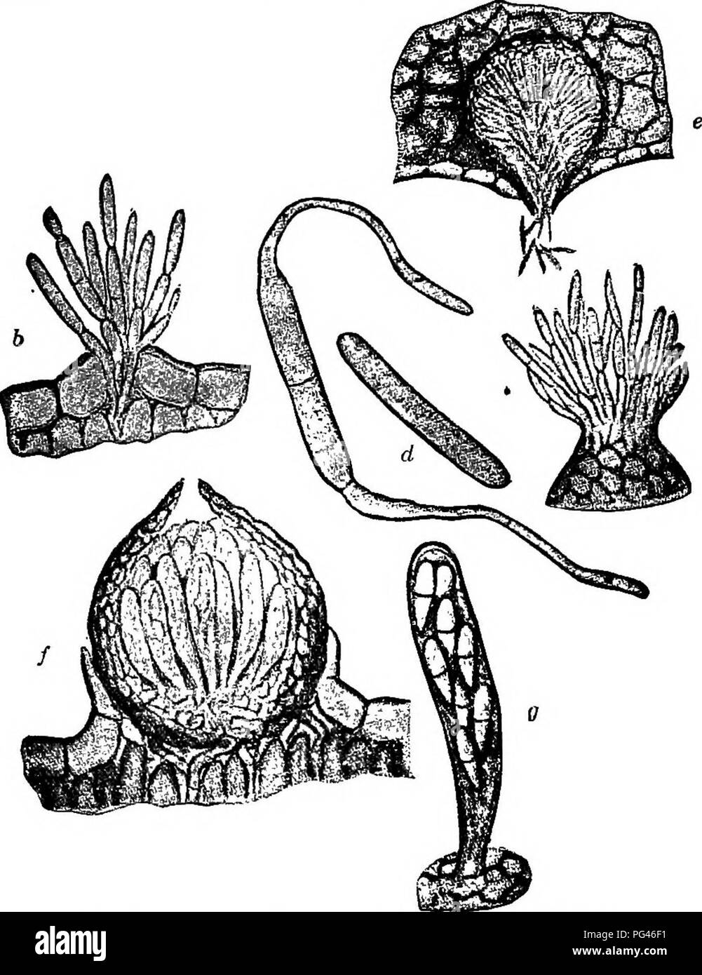
. The fungi which cause plant disease . Plant diseases; Fungi. 244 THE FUNGI WHICH CAUSE PLANT DISEASE M. fragarise (Tul.) Lin.''^ Perithecia on leaves, are produced late in the season, globose, subepidermal, membranous, black, thin-walled; asci few, clavate,. Fig. 179.—Mycosphaerella fragariEe. ft, conidiophores buret- ing through the epidermis; c, arising from apex of a pycnidium; d, summer spores, one germinating; e, s^tion of a spermogonium; /, section of perithecium; g, ascus containing eight two-celled spores. Aft^er Longyear. 8-spored, 4Q ii long; spores hyaline, 2-celled, with acute ti
Image details
Contributor:
Central Historic Books / Alamy Stock PhotoImage ID:
PG46F1File size:
7.2 MB (403.9 KB Compressed download)Releases:
Model - no | Property - noDo I need a release?Dimensions:
1388 x 1801 px | 23.5 x 30.5 cm | 9.3 x 12 inches | 150dpiMore information:
This image is a public domain image, which means either that copyright has expired in the image or the copyright holder has waived their copyright. Alamy charges you a fee for access to the high resolution copy of the image.
This image could have imperfections as it’s either historical or reportage.
. The fungi which cause plant disease . Plant diseases; Fungi. 244 THE FUNGI WHICH CAUSE PLANT DISEASE M. fragarise (Tul.) Lin.''^ Perithecia on leaves, are produced late in the season, globose, subepidermal, membranous, black, thin-walled; asci few, clavate, . Fig. 179.—Mycosphaerella fragariEe. ft, conidiophores buret- ing through the epidermis; c, arising from apex of a pycnidium; d, summer spores, one germinating; e, s^tion of a spermogonium; /, section of perithecium; g, ascus containing eight two-celled spores. Aft^er Longyear. 8-spored, 4Q ii long; spores hyaline, 2-celled, with acute tips, 15 X 3^ fi. Conidia (=Ramularia tulasnei) abundant in early svunmer on reddish spots, stromatic, conidiophores simple; conidia elliptic 20-40 X 3-5 M, 2 to 3-celled. On Fragaria. The life history was first studied in 1863 by the Tulasne brothers under the name Stigmatea. The generic name was changed to Sphaerella in 1882 and later to Mycosphaerella.. Please note that these images are extracted from scanned page images that may have been digitally enhanced for readability - coloration and appearance of these illustrations may not perfectly resemble the original work.. Stevens, Frank Lincoln, 1871-1934. New York : Macmillan