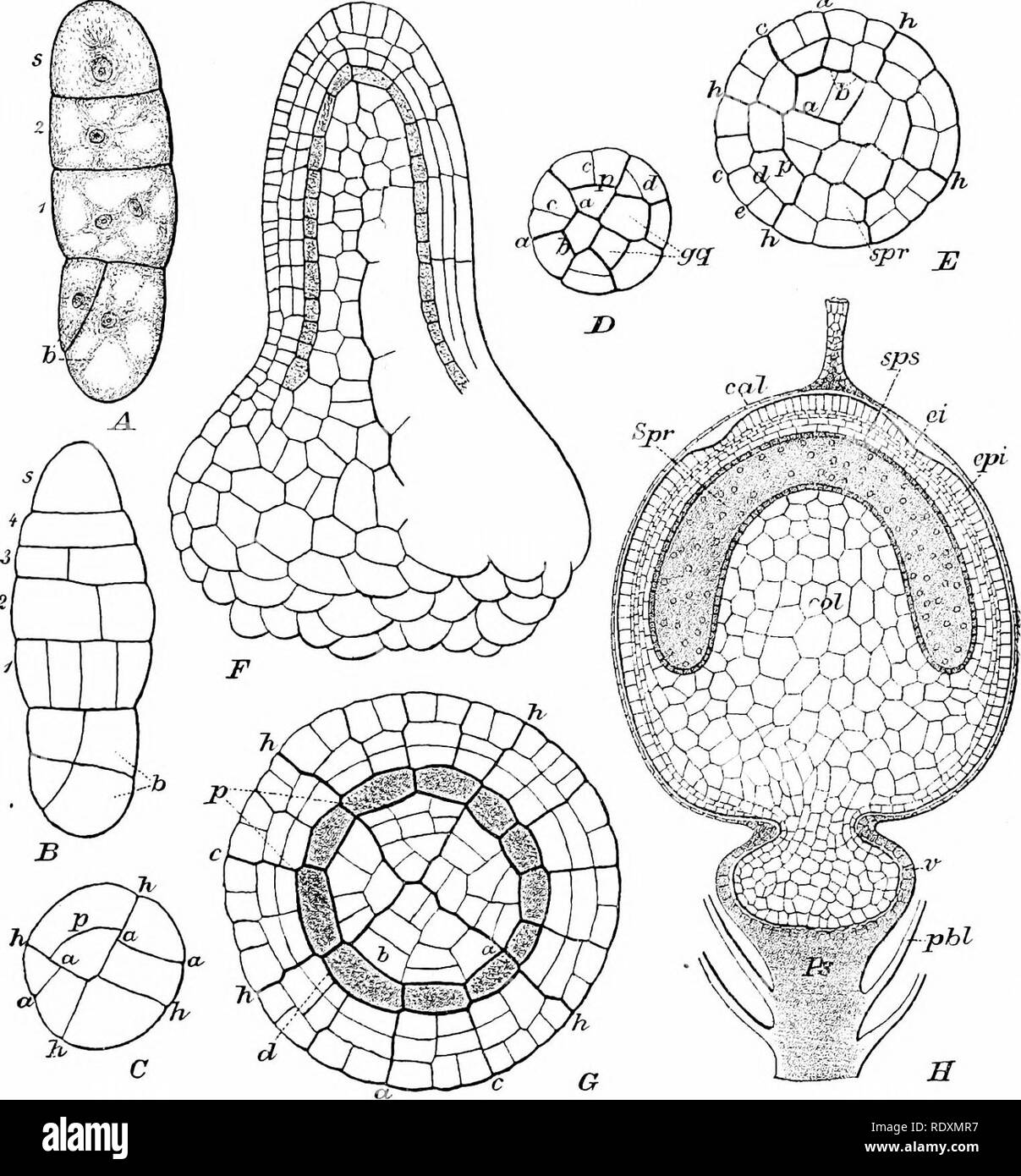
. The origin of a land flora, a theory based upon the facts of alternation. Plant morphology. SPHAGNALES ?73 of tissue with rounded apex (Fig. 133 f, h). The peripheral series of cells, or amphithecium, divides periclinally to give off internally the single. Fig. .32. Development of sporogonium of SpJiagnuin acitiifolium, Ehrh. -4=embryo with four tiers; J = apical cell; />= basal cell with oblique division. j5 = embryo with five tiers. C = optical section of the same embryo; one quadrant is still undivided ; n = aniicmial ; p — periclinal walls; /; = principal walls. D = transverse section
Image details
Contributor:
The Book Worm / Alamy Stock PhotoImage ID:
RDXMR7File size:
7.1 MB (464.3 KB Compressed download)Releases:
Model - no | Property - noDo I need a release?Dimensions:
1523 x 1640 px | 25.8 x 27.8 cm | 10.2 x 10.9 inches | 150dpiMore information:
This image is a public domain image, which means either that copyright has expired in the image or the copyright holder has waived their copyright. Alamy charges you a fee for access to the high resolution copy of the image.
This image could have imperfections as it’s either historical or reportage.
. The origin of a land flora, a theory based upon the facts of alternation. Plant morphology. SPHAGNALES ?73 of tissue with rounded apex (Fig. 133 f, h). The peripheral series of cells, or amphithecium, divides periclinally to give off internally the single. Fig. .32. Development of sporogonium of SpJiagnuin acitiifolium, Ehrh. -4=embryo with four tiers; J = apical cell; />= basal cell with oblique division. j5 = embryo with five tiers. C = optical section of the same embryo; one quadrant is still undivided ; n = aniicmial ; p — periclinal walls; /; = principal walls. D = transverse section of the lower part of an embryo. E = z. rather older stage; ^'^spore-forming layer, /'' — median longitudinal section of a sporogonium showing the bell-shaped sporogenous layer, and the wall covering it externally. 6: = transverse section of a sporogonium of similar age ; lettering as in E. //=median longitudinal section, though a half-ripe sporogonium; <;«/—calypira ; spr = spore-cavity, in which the spore-mother-cells are isolated ; sps = spore-sac ; cpi — epidermis ; Lz'=furrow in wall where the operculum will separate ; /V = pseudopodiuiu ; v — vaginula ; ^<V=perichaetiaI leaves ; £<?/= columella, (After Waldner, from Engler and Prantl.) layer of the archesporium; this appears as a continuous dome closely investing the columella. The external product of the amphithecium forms s. Please note that these images are extracted from scanned page images that may have been digitally enhanced for readability - coloration and appearance of these illustrations may not perfectly resemble the original work.. Bower, F. O. (Frederick Orpen), 1855-1948. London, Macmillan and Co. , Ltd.