The structure & development of the mosses and ferns (Archegoniatae) . t is not until the third set of walls is formed that the separationof endothecium and amphithecium is complete. The nextdivisions (Fig. 95, C) are in the amphithecium, and separateit into two layers. In the endothecium now a series of wallsis formed, almost exactly repeating the first divisions in theoriginal segment (Figs. D, E), and transforming it into a group VII THE BRYINE^ 197 of four central cells and eight peripheral ones. Each of thelatter divides twice by intersecting w^alls, so that a group ofabout sixteen cells (
Image details
Contributor:
The Reading Room / Alamy Stock PhotoImage ID:
2AWXH5PFile size:
7.1 MB (220.1 KB Compressed download)Releases:
Model - no | Property - noDo I need a release?Dimensions:
1921 x 1300 px | 32.5 x 22 cm | 12.8 x 8.7 inches | 150dpiMore information:
This image is a public domain image, which means either that copyright has expired in the image or the copyright holder has waived their copyright. Alamy charges you a fee for access to the high resolution copy of the image.
This image could have imperfections as it’s either historical or reportage.
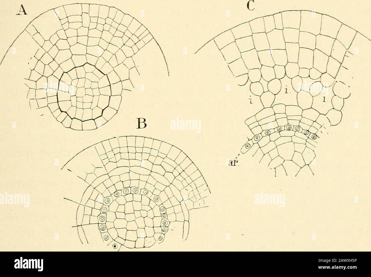
The structure & development of the mosses and ferns (Archegoniatae) . t is not until the third set of walls is formed that the separationof endothecium and amphithecium is complete. The nextdivisions (Fig. 95, C) are in the amphithecium, and separateit into two layers. In the endothecium now a series of wallsis formed, almost exactly repeating the first divisions in theoriginal segment (Figs. D, E), and transforming it into a group VII THE BRYINE^ 197 of four central cells and eight peripheral ones. Each of thelatter divides twice by intersecting w^alls, so that a group ofabout sixteen cells (Fig. 96, A) occupies the middle of theendothecium. The eight peripheral cells divide by radial walls, after which each of these cells is divided by a periclinal wallinto an outer and an inner cell (Fig. 96, B), and the outercells divide rapidly by radial walls and form the archesporium.The single layer of cells immediately within, and therefore sistercells of the primary archesporial ones, is the inner spore-sac.The account of the development of the endothecium here. Fig. 96.- -Three transverse sections of an older sporogonium of F. /lygronetrica, X 400 ;ar, archesporium; /, intercellular spaces. given differs slightly from the account of Kienitz-Gerloff.^ Itwas found first that there was not the absolute constancy inthe number of cells given by him ; thus in Fig. 96, A thereare only fourteen cells in the inner part of the endothecium, and although there are sixteen cells in the outer row theirposition is not perfectly symmetrical. Again the periclinaldivision of the cells of the inner spore-sac takes place laterthan he states is the case. In the eight primary cells of the amphithecium there firstarise periclinal walls that divide each cell into an inner small ^ Kienitz-Gerloff (2). 198 MOSSES AND FERNS chap, vii cell in contact with the endothecium, and an outer larger one.This first division separates the wall of the capsule from theouter spore-sac. The latter next divides by r